Impact Factor ISSN: 1449-2288
- Issue 12; 2026
- Issue 11; 2026
- Issue 10; 2026
- Issue 9; 2026
- Issue 8; 2026
- Volume 22; 2026
- Past Issues
- Advance Articles
- Editorial Board
- Cover Images
- Index & Coverage
- Cover Suggestion
- Special Issues
Introduction
Materials and Methods
Results
Discussion
Supplementary Material
Acknowledgements
References

 Global reach, higher impact
Int J Biol Sci 2026; 22(8):4303-4314. doi:10.7150/ijbs.124330 This issue Cite
Research Paper
Tumor Necrosis Factor Receptor 2 Inhibits HIV-1 Infection by Blocking the Binding of gp120 to CD4
Yang Gao1†, Zhonghao Chen1†, Yibo Chen1, Yang Yang1, Yiru Wang1, Ci Zhu1, Ping Liao1, He Song1,2 ![]() , Xin Chen1,2,3
, Xin Chen1,2,3 ![]()
1. Institute of Chinese Medical Sciences, State Key Laboratory of Mechanism and Quality of Chinese Medicine, University of Macau, Macau SAR, China.
2. Department of Pharmaceutical Sciences, Faculty of Health Sciences, University of Macau, Macau SAR, China.
3. MoE Frontiers Science Center for Precision Oncology, University of Macau, Macau SAR, China.
†These authors contributed equally to this work.
Received 2025-8-27; Accepted 2025-12-13; Published 2026-4-8
Abstract

The HIV-1 envelope glycoprotein gp120 binds to the CD4 molecule and then undergoes conformational changes to interact with the co-receptors CCR5 or CXCR4, resulting in cellular entrance. However, certain types of cells, such as macrophages and CD4+Foxp3+ regulatory T cells (Tregs), have been shown to resist HIV-1 infection despite co-expressing CD4 and co-receptors. In this study, we found that tumor necrosis factor receptor type II (TNFR2) directly binds to gp120, with the binding site on gp120 in proximity to that of CD4. Intriguingly, exogenous TNFR2 had the capacity to inhibit the binding of gp120 to CD4+ T cells. Furthermore, the infection of CD4+CCR5+ cells by pseudoviruses containing the HIV-1 envelope was inhibited by TNFR2 protein. In contrast, TNFR1, which is structurally similar to TNFR2 and shares the same ligand, failed to inhibit the infection of CD4+ T cells by HIV-1 pseudoviruses. This property of TNFR2 may be harnessed in the prevention or treatment of HIV-1 infection and thus warrants future investigation.
Keywords: HIV-1, gp120, TNFR2, TNF, TNFR1, pseudovirus
Introduction
Human Immunodeficiency Virus-1 (HIV-1), the virus that causes AIDS, infects cells by first binding to the CD4 and then to the co-receptors CCR5 or CXCR4. A gradual decrease in CD4+ cells is the characteristic of AIDS, yet many questions relating to the mechanisms underlying this decrease remain unanswered[1]. Among CD4+ cells, several with high co-receptor expression, such as macrophages and regulatory T cells (Tregs), have been found to exhibit reduced susceptibility to HIV-1 infection[2-4]. Previous studies have shown that certain subsets of CD4+ macrophages exhibit relative resistance to HIV-1-induced cytopathic effects and can persist for extended periods during infection[5]. Furthermore, it has been repeatedly reported that, during HIV-1 infection, tumor necrosis factor (TNF) downregulates the expression of CD4 on macrophages, which may contribute to their resistance[2, 6]. However, despite the presence of TNF, effector CD4+ T cells do not exhibit resistance to HIV, whereas CD4+FoxP3+ Tregs are resistant to HIV-1 infection[4, 7, 8]. Interestingly, Tregs could be isolated and expanded in vitro from patients infected with HIV-1, demonstrating that functional Tregs derived from the blood and lymphoid tissues of HIV-1-infected individuals maintain their suppressive capacity, thereby indicating that these cells are not intrinsically impaired in the context of HIV infection[9]. These observations suggest that the infection resistance of Tregs and macrophages may be independent of CD4 expression, and the mechanism remains to be elucidated.
Previous reports have indicated that TNF and TNF receptors can influence HIV-1 infection[10, 11]. For example, it was reported that pretreatment with TNF mutation selective for TNFR2 delays the detection of HIV-1 DNA long terminal repeat sequences in macrophages. It has also been shown that Foxp3, the essential transcription factor for the development and function of Treg cells, markedly inhibits HIV-1 replication when expressed in primary human CD4+ T cells[12, 13]. Previously, compelling evidence has shown that Treg cells predominantly express TNFR2, and the expression of TNFR2 is critical for sustaining Foxp3 expression in Tregs[14, 15]. Therefore, TNFR2 expression and its signaling may be attributable to the resistance of HIV-1 infection by Tregs. Indeed, an in silico molecular docking study suggested that the HIV-1 gp120 can bind to TNFR2[16].
HIV-1 entry into target cells primarily depends on the interaction between gp120 and the CD4 receptor, along with either CCR5 or CXCR4 co-receptors. In addition to these classical entry receptors, several studies have reported that HIV-1 can also bind to other cell surface receptors[17, 18]. However, despite their high affinity for gp120, most of these receptors do not facilitate viral entry and mainly act as attachment factors, capturing viral particles on the cell surface without directly promoting infection. Therefore, experimental validation is required to determine whether TNFR2 indeed binds gp120 and whether such binding affects HIV-1 infection.
In this study, we experimentally verified that the TNFR2 protein binds to the HIV-1 envelope protein gp120 with high affinity. Intriguingly, TNFR2 interrupted the interaction between gp120 and CD4. Moreover, we found that TNFR2 could inhibit the infection of CD4+CCR5+ cells by pseudoviruses containing the HIV-1 envelope. This property of TNFR2 may be therapeutically harnessed and thus merits further investigation.
Materials and Methods
Cell culture
The Jurkat cell line and HEK293T cell line were purchased from American Type Culture Collection (Manassas, USA). Jurkat cells were cultured in RPMI-1640 (Gibco, USA) supplemented with 10% FBS. HEK293T cells were cultured in DMEM (Gibco, USA) supplemented with 10% FBS. Freshly isolated PBMCs were obtained from healthy donors through the Macao Blood Transfusion Service after they provided informed consent approved by the institutional review board. PBMCs were cultured in the T551 medium (Takara, Japan) containing 10% FBS. The incubation was conducted at 37 ℃ in humidified air containing 5% CO2 in a tissue culture flask. The stable cells lines, Jurkat-CCR5, Jurkat-TNFR2-CCR5, 293T-TNFR2, 293T-CD4-CCR5, 293T-TNFR1-CCR5 and 293T-TNFR2-CCR5 were established by lentiviral transduction. All cell lines exhibited transduction efficiencies exceeding 98%.
Reagents
Antibodies were purchased from BD Pharmingen (USA) consisted of PerCP-Cy5.5 anti-human CD4 (Catalog No:566316), APC Streptavidin (Catalog No:554067), PE-Cy7 anti-His (Catalog No: 362620), and BV421 anti-human TNFR2 (Catalog No: 562783) were purchased from Biolegend (USA). The human immunodeficiency virus 1 (HIV-1) (group M, subtype B, Isolate MN) gp120 Protein (DNA sequence: AAC31819.1 (Thr30-Arg513), Catalog No: 40405-V08H), human TNFR2 / CD120b protein (DNA sequence: NP_001057.1 (Met1-Asp257), Catalog No: 10417-H08H) were purchased from Sino Biological (Beijing, China). The human TNFR1 protein (DNA sequence: NP_001056 (Ile 30 - Thr 211), Catalog No: TN1-H5222) was purchased from the ACRO Biosystems (Beijing, China). In addition, TNFR2 protein (DNA sequence: NP_001057 Met1-Gly258) and TNFR1 protein (DNA sequence: NP_001056 Met 1-Thr 211) were also purified and prepared in-house by our laboratory using standard affinity chromatography techniques. CD4 protein (DNA sequence: NP_000607, Catalog No: TP306453) was purchased from OriGene Technologies (Rockville, MD).
Bio-layer interferometry (BLI)
Biotin labeling of human TNFR2 protein was performed according to the manufacturer's instructions using a commercial biotinylation kit, followed by purification with a desalting column (Thermo, USA). The concentration of biotinylated TNFR2 was determined using a BCA protein assay kit (Thermo, USA). Biotinylated TNFR2 was diluted in 1× PBS to a final concentration of 5 µg/mL and immobilized onto streptavidin (SA) biosensors (SARTORIUS, Germany, Catalog No: 18-5017) for 600 seconds. Binding kinetics were assessed by exposing the immobilized TNFR2 to recombinant gp120 protein (diluted in PBST: PBS with 0.02% Tween-20, pH 7.4) at concentrations of 2 μM, 1 μM, 0.5 μM, 0.25 μM, and 0.125 μM for 200 seconds (association phase), followed by dissociation in PBST for 300 seconds. PBST alone was used as a reference control to subtract the background signal. Data were acquired using an Octet® R2 instrument (SARTORIUS, Germany) and analyzed with ForteBio Data Analysis software version 12.0. A global fitting model based on a 1:1 binding interaction was applied to calculate the association rate constant (kon), dissociation rate constant (koff), and dissociation constant (KD = koff/kon).
Co-immunoprecipitation (Co-IP)
HEK293T cells stably expressing TNFR2 were lysed in ice-cold cell lysis buffer (20 mM Tris-HCl, 150 mM NaCl, 1% Triton X-100, 1 mM EDTA, protease inhibitor cocktail, pH 7.4) and incubated on ice for 30 minutes. The lysate was centrifuged at 14,000 rpm for 15 minutes at 4 °C to remove debris. Supernatants were collected and incubated with recombinant gp120 protein (5 µg/mL final concentration) for 2 hours at 4 °C on a rotator. This mixture was then incubated overnight at 4 °C with 30 µL of Dynabeads Protein G (Thermo Fisher Scientific) pre-conjugated with 2 µg of anti-gp120 antibody (Abcam, USA, Catalog No: ab106578) or control rabbit IgG (Cell Signaling Technology, CST, MA, USA, Catalog No: 2729S). Immunocomplexes were collected by magnetic separation and washed three times with ice-cold PBS containing 0.05% Tween-20. The beads were resuspended in 2× SDS loading buffer and boiled at 95 °C for 5 minutes. Proteins were resolved on 10% Bis-Tris SDS-PAGE gels (Invitrogen) and transferred onto PVDF membranes. Membranes were blocked in 3% BSA in TBST (TBS with 0.1% Tween-20) for 1 hour at room temperature, then incubated overnight at 4 °C with primary anti-TNFR2 antibody (1:1000, CST, USA, Catalog No: 3727). After washing, membranes were incubated with HRP-conjugated secondary antibody (1:5000, CST, Catalog No: 7074) for 1 hour at room temperature and developed using ECL chemiluminescent substrate.
Enzyme-linked immunosorbent assay (ELISA)
For gp120-TNFR1/TNFR2 binding assays, 96-well high-binding plates were coated with 100 µL of human TNFR1/TNFR2 (5 µg/mL in 1× CBS (15 mM Na2CO3, 35 mM NaHCO3, pH 9.6) overnight at 4 °C. After washing with buffer (20 mM Tris-HCl, 500 mM NaCl, 0.05% Tween-20, pH 7.5), wells were blocked with 3% BSA in PBS for 1 hour at room temperature. Serial dilutions of gp120 (1 µM, 0.1 µM, 0.01 µM in blocking buffer) were added and incubated for 2 hours at room temperature. After washing, bound gp120 was detected using anti-His-HRP antibody (1:2000, Thermo, Catalog No: MA1-21315-HRP) and developed with TMB substrate. Reactions were stopped with 1 M H₂SO₄ and absorbance was measured at 450 nm. For competition ELISA, 96-well plates were coated with recombinant CD4 protein (5 µg/mL in CBS) overnight at 4 °C. Mixtures of gp120 (1 µM) and TNFR2 (2 µM) were pre-incubated at room temperature for 30 minutes before being added to the CD4-coated wells and incubated for 2 hours. Binding of gp120 was detected as above using anti-His-HRP.
Flow cytometry assay to evaluate gp120 blocking by TNFR2
Jurkat cells and PBMCs were washed and resuspended in FACS buffer (PBS with 2% FBS and 0.1% sodium azide). Cells (1×10⁵ per sample) were incubated with serial dilutions of recombinant TNFR2 protein (800 nM, 400 nM, 200 nM, 100 nM, 50 nM) or TNFR1 (1 µM, 0.6 µM, 0.4 µM) for 1 hour. His-tagged gp120 (5 µg/mL) was then added and incubated for another 1 hour. Cells were washed and stained with PE-Cy7 conjugated anti-His antibody (1:100) for 30 minutes at 4 °C in the dark. For PBMCs, anti-human CD4- PerCP-Cy5.5 antibody (1:100) was used to identify CD4⁺ T cells. After final washes, samples were analyzed using a BD LSRFortessa flow cytometer. Data analysis was performed using FlowJo software.
HIV-1 pseudovirus infection
The HIV-1 pseudovirus from a CCR5-tropic (R5) strain containing HIV-Env protein encapsulated with green fluorescent protein (GFP)-encoding RNA was obtained from Fubio Biotechnology (Suzhou, Jiangsu, China; Catalog No: FNV5011G). Different concentrations of TNFR2 protein or TNFR1 protein were prepared and incubated with the virus for one hour at 37 °C. Following the incubation period, appropriate target cells were prepared for infection. For infection of 293T-CD4-CCR5 cells, cells were seeded in 24-well plates and maintained under optimal conditions. The pre-treated pseudoviruses were added at a multiplicity of infection (MOI) of 5 or 10. After 16 hours, GFP expression was analyzed by flow cytometry to evaluate infection efficiency. For infection of 293T-CCR5, 293T-TNFR2-CCR5,293T-TNFR1-CCR5 cells, cells were seeded in 24-well plates and maintained under optimal conditions. The pseudoviruses were added at a multiplicity of infection (MOI) of 10. After 16 hours, GFP expression was analyzed by flow cytometry to evaluate infection efficiency.
For Jurkat-CCR5 cell and Jurkat-TNFR2-CCR5 infection, cells were directly infected without prior activation. The virus-protein mixture was added at an MOI of 50, also in the presence of polybrene (1 µg/mL). After 48 hours of incubation, the percentage of GFP-positive cells was measured by flow cytometry to determine infection efficiency.
The complex structure prediction based on AlphaFold3
Utilized AlphaFold3 to predict the structure of the protein complex[19]. Collected the amino acid sequences of TNFR2 (PDB ID: 3ALQ) and gp120 (PDB ID: 4RZ8) from the UniProt database and performed multiple sequence alignments using HHblits. These alignments were then input into AlphaFold3, which was configured for multichain complex predictions. The model generated several candidate structures, which were assessed using the pLDDT scoring system to determine their reliability. The highest-scoring structure was selected as the final model. Visualization and further analysis were conducted using PyMOL[20-22].
Statistical analysis
All data were presented as means ± SEM, and the statistical analysis was performed by t test or one-way ANOVA test by using GraphPad Prism 10.0. The P value < 0.05 was considered to be statistically significant.
Results
HIV-1 envelope protein gp120 interacts with TNFR2
We examined the interaction between the extracellular domain of TNFR2 (residues 1-257) and the gp120 core (residues 30-513) using an Enzyme-Linked Immunosorbent Assay (ELISA). The results showed that at a concentration of 0.1 μM, gp120 had the capacity to bind to immobilized TNFR2 (Fig. 1A), and this binding occurred in a concentration-dependent manner (Fig. 1B), demonstrating a direct interaction between the two proteins. To rule out non-specific binding, we also performed ELISA using uncoated (blank) wells and observed no detectable gp120 signal under the same conditions (Fig. S1A).
The HIV-1 gp120 protein directly interacts with the TNFR2 protein. (A) The binding interaction between gp120 and TNFR2 was assessed using an ELISA assay with varying concentrations of gp120 (1 μM, 0.1 μM, 0.01 μM, blank). The results of color development from the TMB substrate. (B) The statistical analysis of the final OD450 measurements. (C) Co-immunoprecipitation analysis was used to validate the interaction between gp120 and TNFR2. The IgG group was included as a control to ensure the specificity of the experiment. (D) Detection of gp120 and TNFR2 binding using bio-layer interferometry, with five concentrations of gp120 (2 μM, 1 μM, 0.5 μM, 0.25 μM, 0.125 μM) to determine the affinity. Kd values were determined from the BLI results, providing a quantitative assessment of the equilibrium binding affinity between gp120 and TNFR2. One-way ANOVA was used to analyze group differences. Significance levels: * p < 0.05, ** p < 0.01, *** p < 0.001, **** p < 0.0001, ns no significant difference.
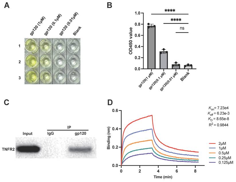
For further verification, a co-immunoprecipitation (Co-IP) assay was performed to study protein-protein interaction between TNFR2 and gp120. To this end, gp120 was incubated with lysates of TNFR2-overexpressing cells, followed by pulling down the protein complexes with a gp120-specific antibody which conjugated to magnetic beads. The results showed that TNFR2 was present in the immunoprecipitate, indicating a direct physical interaction between gp120 and TNFR2 (Fig. 1C).
The binding of gp120 to TNFR2 was further evaluated using bio-layer interferometry (BLI) to measure biomolecular interactions in real time. Biotinylated human TNFR2 protein was immobilized on a streptavidin (SA) sensor tip and exposed to a gradient of gp120 concentrations, ranging from 0.125 µM to 2 µM. With 5 µg/ml of immobilized TNFR2-Biotin, gp120 could bind to TNFR2 with high affinity (Fig. 1D). As the concentrations of gp120 increased, the response signal correspondingly increased, indicating specific and concentration-dependent binding to TNFR2. BLI analysis of the binding kinetics revealed a binding affinity (Kd) of 86.5 nM for the gp120-TNFR2 interaction, indicative of a high affinity and a strong, specific binding interaction.
TNFR2 interrupts the binding of gp120 to CD4
We further examined the effect of TNFR2 on the interaction between gp120 and CD4. To visualize the competition between TNFR2 and CD4 for binding to gp120, AlphaFold3 was used to predict the complex structure of TNFR2 and gp120[19]. The resulting model exhibited high confidence in the overall complex structure, providing a detailed and reliable representation of the interaction (Fig. S2A). The model showed that TNFR2 binds to gp120 at a specific epitope crucial for the interaction between CD4 and gp120. The interaction of the surface between gp120 and TNFR2 was analyzed in detail, showing that it buries a total area of 1,139.5 Ų. The structural analysis revealed a specific interaction interface between TNFR2 and gp120, involving several key amino acid residues. On the TNFR2 side, residues W62, Y65, Y68, Q72, L73, and V75 contribute to the binding, while on gp120, residues L71, T72, T74, M426, W427, and Y435 are involved. Hydrophobic interactions, with key residues forming hydrogen bonds, salt bridges, and hydrophobic contacts that stabilize the gp120-TNFR2 complex (Fig. S2B-S2C), with the CD4 binding epitope (Fig. S2D). This overlap indicates that TNFR2's binding to gp120 could sterically block the binding of CD4 and potentially disrupt the CD4-mediated HIV-1 entry process. Compared to the docking model previously reported[16], the AF3-predicted complex shows a similar overall binding orientation but highlights additional interactions near the CD4-binding region of gp120. These observations indicate that TNFR2 and CD4 likely compete for the same binding site on gp120.
To verify this hypothesis, we investigated the competitive binding between TNFR2 and CD4 for their interaction with gp120. CD4 was immobilized on the surface of a plate and then incubated with gp120 in the presence or absence of TNFR2 protein. Remarkably, the addition of TNFR2 protein resulted in a significant reduction in gp120 binding to CD4, with an inhibition rate reaching up to 70% (Fig. 2A). This finding demonstrates that TNFR2 competes with CD4 for binding to gp120, which supports our hypothesis. To further verify it at the cellular level, we conducted experiments using the Jurkat cell line, a commonly used cell model in the study of HIV-1 infection[23]. The binding of the gp120 protein to Jurkat cells was quantified by detecting the His tag on the gp120 protein. The results showed that in the absence of TNFR2, approximately 75% of the cells exhibited gp120 binding. In contrast, the addition of TNFR2 protein resulted in a 65% reduction in gp120 binding to the cells; only ~10% of cells exhibited gp120 association (Fig. 2B-2D). To validate the specificity of this inhibitory effect, we used an unrelated control protein in parallel. No inhibition of gp120 binding was observed with this control, supporting the specificity of the blocking effect to TNFR2 (Fig. S3A). The inhibition of gp120 binding to the cells by TNFR2 appeared to be dose-dependent, and IC50 was 106.9 nM (Fig. 2E-2F). Almost 200 nM TNFR2 can basically inhibit the binding of gp120 to Jurkat cells to the greatest extent. To further test whether TNFR2 engages gp120 at or near the CD4-binding site, we performed a competition experiment with the broadly neutralizing antibody VRC01, which targets the CD4 epitope. As a positive control, VRC01 efficiently blocked gp120-CD4 binding in Jurkat cells (Fig. S3B). We then assessed gp120-TNFR2 binding on TNFR2-expressing 293T cells after pre-incubation of gp120 with VRC01 (Fig. S3C). Flow cytometry showed a marked reduction in gp120 binding (decreased MFI) in the VRC01 condition relative to gp120 alone, indicating that VRC01 impairs gp120-TNFR2 engagement. These data support that TNFR2 recognizes an epitope overlapping or immediately adjacent to the CD4-binding site.
The TNFR2 protein blocks the binding of gp120 to CD4. (A) ELISA was used to assess whether TNFR2(2 µM) can inhibit the interaction between gp120 and immobilized CD4 on the plate. The final OD450 measurement statistics are shown. (B) Flow cytometry was utilized to evaluate cell-bound His-tag, indicating the binding of gp120-His to the Jurkat cells, and also assessed if TNFR2 (1 µM) can block gp120-cell binding. (C) The percentage of positive cells was determined from flow cytometry analysis results. (D) The statistical analysis of mean fluorescence intensity. (E) Further examination assessed whether the blocking effect of TNFR2 (50 nM, 100 nM, 200 nM, 400 nM, 800 nM) exhibited dose-dependency. (F) The half-maximal inhibitory concentration (IC50) was calculated through the dose-dependent inhibition curve. (G) Flow cytometry was utilized to detect the binding of gp120 to human peripheral blood mononuclear cells and to evaluate the effect of TNFR2. (H) The statistical analysis of the double positive cells. One-way ANOVA was used to analyze group differences. Significance levels: * p < 0.05, ** p < 0.01, *** p < 0.001, **** p < 0.0001, ns no significant difference.
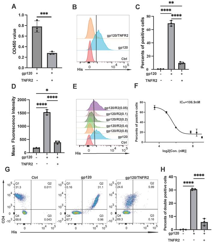
To verify this in primary human cells, PBMCs from healthy donors were treated with gp120, with or without TNFR2. We confirmed that gp120 exclusively binds to CD4 cells in PBMCs (Fig. 2G). The binding of gp120 to the cells was potently inhibited by TNFR2, and 200 nM of TNFR2 resulted in 80% inhibition of gp120 binding (Fig. 2G-2H). Together, our data demonstrate that TNFR2 could potently block the binding of gp120 to CD4 at both the cellular and molecular levels.
TNFR2 inhibits HIV pseudovirus infection in CD4+CCR5+ cells
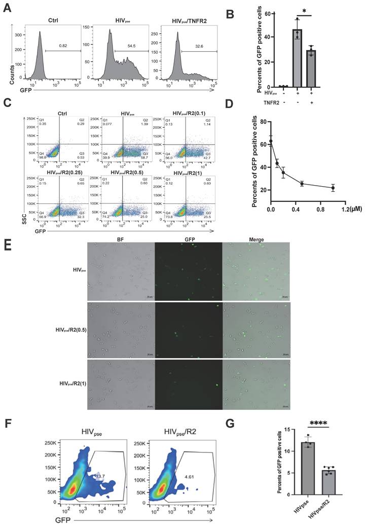
Building on our earlier observations, we sought to determine whether TNFR2 could inhibit the cellular infection of HIV-1 by blocking the binding of gp120 to CD4. To explore this possibility, we utilized a pseudovirus-based assay system. HIV-1 pseudoviruses were generated by co-transfecting HEK-293T cells with plasmids encoding Gag-Pol, the HIV-1 Env of interest, and a reporter gene (GFP). The components self-assemble into replication-incompetent particles that package the reporter and undergo only a single cycle of infection. These particles mimic Env-mediated receptor engagement and membrane fusion, enabling quantitative assessment of entry inhibition.
For this experiment, HEK293T-CD4-CCR5 cells were exposed to these pseudoviruses, in which the expression of the CD4 and CCR5 receptors reached approximately 99% (Fig. S4A). The green fluorescence signal was used as an indicator of viral infection efficiency, providing a quantitative way to measure infection rates. Notably, treatment with exogenous TNFR2 protein at 1 µM concentration significantly decreased the infection rate of HIV-1 pseudoviruses. Specifically, the infection rate was reduced by 20% compared to untreated control cells (Fig. 3A-3B). Furthermore, the inhibitory effect of TNFR2 on pseudovirus infection showed a dose-dependent relationship, with higher concentrations of TNFR2 leading to greater reductions in infection efficiency (Fig. 3C-3D). These results suggest that TNFR2 can help reduce HIV-1 pseudovirus infection by blocking the binding of gp120 to CD4. This conclusion is further supported by the observed reduction in infection efficiency with higher doses of TNFR2, emphasizing its role as a competitive inhibitor in this context (Fig. 3E). To further explore the antiviral role of TNFR2 in T cells, we repeated the infection experiments using Jurkat cells overexpressing CCR5 as the target cells. CD4-CCR5 cells, pre-treatment of the virus with TNFR2 protein also resulted in a noticeable decrease in infection efficiency in these CCR5⁺ Jurkat cells, supporting the broader inhibitory effect of TNFR2 on HIV-1 entry across different cell types (Fig. 3F-3G).
The TNFR2 protein can inhibit HIV pseudovirus infection of CD4+CCR5+ cells. (A) HIV pseudovirus carrying green fluorescent protein genes was used to infect CD4+CCR5+293T cells (MOI=10). The flow cytometry was used to analyze the rate of GFP-positive cells to illustrate the virus infection efficiency. (B) The statistical analysis for the ratio of GFP-positive cells in each group. (C) Further examination assessed whether the blocking effect of TNFR2 (0.1 µM、0.25 µM、0.5 µM、1 µM) exhibited dose-dependency. (D) The statistical analysis for the dose-dependent effects. (E) Infected CD4+CCR5+ 293T cells were observed by fluorescence microscopy on the 12h post infection. (F) Flow cytometry analysis of Jurkat-CCR5 cells infected with HIV-1 pseudovirus, with or without pre-incubation of the virus with 10 µg/mL TNFR2 protein. (G) Quantification of infection efficiency. One-way ANOVA was used to analyze group differences. Significance levels: * p < 0.05, ** p < 0.01, *** p < 0.001, **** p < 0.0001, ns no significant difference.
Collectively, these results suggest a promising potential for TNFR2 in therapeutic strategies targeting the interaction between gp120 and CD4 to prevent HIV-1 infection.
Transmembrane TNFR2 expression inhibits HIV-1 pseudovirus infection
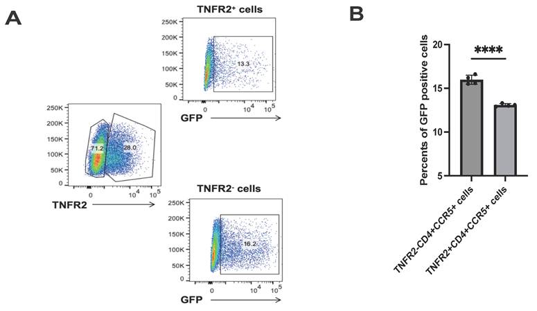
After establishing that soluble TNFR2 protein can inhibit HIV-1 infection by blocking the interaction between gp120 and CD4, we then examined whether transmembrane TNFR2 expression on host cells could have a similar influence on viral susceptibility. We used a Jurkat cell model overexpressing CCR5, where only a subset of cells expressed TNFR2. Following HIV-1 pseudovirus infection, flow cytometry analysis revealed that TNFR2⁺ cells showed a 17.9% reduction in GFP-positive cells compared to TNFR2⁻ cells (Fig. 4A-4B). These findings directly demonstrate that transmembrane TNFR2 inhibits HIV-1 infection, emphasizing its function as a protective factor in cells.
TNFR2 expression inversely correlates with HIV-1 pseudovirus infection. (A) CCR5⁺ Jurkat cells with partial TNFR2 overexpression (~30% TNFR2⁺) were infected with GFP-expressing HIV-1 pseudovirus. GFP expression was analyzed in TNFR2⁺ and TNFR2⁻ subpopulations by flow cytometry. (B) Quantification of the percentage of GFP⁺ cells in TNFR2⁺ and TNFR2⁻ populations to evaluate pseudovirus infection efficiency. Significance levels: * p < 0.05, ** p < 0.01, *** p < 0.001, **** p < 0.0001, ns no significant difference.
TNFR1 affects the binding of gp120 to CD4 but promotes HIV-1 infection
Due to the structural similarities between TNFR1 and TNFR2, as well as their shared ligand TNF, we extended our investigation to evaluate whether TNFR1 interacts with gp120. We first performed ELISA assays and confirmed that recombinant TNFR1 protein directly binds to gp120 in a concentration-dependent manner, similar to TNFR2 (Fig. 5A). Specifically, when Jurkat cells were treated with TNFR1, we observed a significant reduction in the binding of gp120 to these cells (Fig. 5B-5C). A similar trend was observed in primary lymphocytes, where the presence of TNFR1 markedly reduced gp120 binding (Fig. 5D- 5E).
TNFR1 impacts the binding of gp120 to CD4 but promotes the virus infection. (A) The binding interaction between gp120 and TNFR1 was assessed using an ELISA assay with varying concentrations of gp120 (5 μM, 0.5 μM, 0.05 μM, blank). (B) Flow cytometry was utilized to evaluate cell-bound His-tag, indicating the binding of gp120 to the Jurkat cells, and also assess if TNFR1 (0.4µM, 0.6µM, 1µM) can block gp120-cell binding. (C) The percentage of positive cells was determined from flow cytometry analysis results. (D) Flow cytometry was utilized to detect the binding of gp120 to human peripheral blood mononuclear cells and to evaluate the blocking effect of TNFR1. (E) The statistical analysis of the double-positive cells. (F) HIV pseudovirus carrying green fluorescent protein genes was used to infect CD4+CCR5+293T cells (MOI=5, MOI=10). Flow cytometry was used to analyze the rate of GFP-positive cells, illustrating the virus infection efficiency. (G) The percentage of GFP-positive cells was determined from flow cytometry analysis results. (H) His-tagged gp120 was pre-incubated with recombinant TNFR1 or TNFR2 and applied to CCR5⁺CD4⁻ 293T cells. (I) CD4⁻CCR5⁺ 293T, TNFR1+CD4⁻CCR5⁺ 293T, and TNFR2+CD4⁻CCR5⁺ 293T cells were infected with GFP-expressing HIV-1 pseudovirus. Infection efficiency was assessed by flow cytometry based on the percentage of GFP⁺ cells. Significance levels: * p < 0.05, ** p < 0.01, *** p < 0.001, **** p < 0.0001, ns no significant difference.
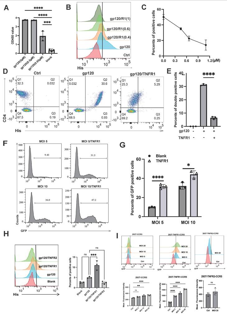
Interestingly, when we further investigated the role of TNFR1 in the context of HIV-1 infection, we uncovered a surprising contrast. While we initially hypothesized that TNFR1, like TNFR2, might act to inhibit HIV-1 infection by blocking the gp120-CD4 interaction, our results showed the opposite. In experiments using HIV-1 enveloped pseudoviruses, TNFR1 was found to facilitate viral infection rather than inhibit it (Fig. 5F-5G). This unexpected finding suggests that TNFR1 may play a distinct role in promoting the entry or replication of HIV-1 pseudoviruses. These results highlight a fundamental difference in the functional roles of TNFR1 and TNFR2, with TNFR1 potentially enhancing HIV-1 infectivity under certain conditions.
These contrasting outcomes may result from the distinct conformational changes in gp120 induced by TNFR1 versus TNFR2. To directly examine whether TNFR1 binding to gp120 facilitates CCR5 engagement, we performed flow cytometry-based binding assays. His-tagged gp120 was pre-incubated with recombinant TNFR1 or TNFR2 and applied to CCR5⁺CD4⁻ 293T cells. TNFR1 significantly increased gp120 binding to the cells, confirming that TNFR1 promotes gp120-CCR5 association (Fig. 5H). We further investigated whether TNFR1 could promote viral infection without CD4. We used engineered cell lines that express CCR5 alone (as a control), or both CCR5 and either TNFR1 or TNFR2 (Fig. 5I). Notably, TNFR1—but not TNFR2—was able to promote HIV-1 pseudovirus infection under these CD4-deficient conditions. These results suggest that TNFR1 may act as a cofactor by inducing conformational changes in gp120 that expose or stabilize CCR5-binding sites, thereby enhancing viral entry even in the absence of canonical CD4 engagement.
Discussion
In this study, we present compelling evidence that the TNFR2 protein plays a crucial role in modulating the entry of HIV-1 into CD4 cells, serving as an effective inhibitor of viral infection. Our findings suggest that this inhibitory effect is primarily mediated through the competitive binding between TNFR2 and CD4 to the HIV-1 envelope protein gp120, which is essential for viral entry. By binding to gp120, TNFR2 outcompetes the CD4, preventing the necessary conformational changes in gp120 required for its subsequent interaction with the coreceptor CCR5. This disruption in the viral entry process effectively blocks the fusion of the virus with the host cell membrane, thereby impeding HIV-1 infection and reducing viral replication within the target cells. The mechanistic underpinning of this process is complex but intriguing. It appears that TNFR2 acts not merely as a passive competitor for gp120 binding but as a dynamic regulator of viral entry. By preventing the interaction between gp120 and CD4, TNFR2 interferes with the early stages of HIV-1 infection. This disruption in the viral lifecycle is a critical step in preventing HIV-1 from successfully establishing infection in immune cells.
Our findings are consistent with prior studies demonstrating the impact of TNF mutant proteins on HIV-1 infection. Specifically, pretreating macrophage cells with a TNF mutant protein that selectively binds to TNFR2 resulted in a significant reduction in HIV-1 virus entry into the cells[11]. We believe that their findings are based on TNF mutants activating TNFR2 signaling, leading to upregulation of TNFR2 expression on the cell surface, thereby inhibiting HIV-1 infection. We propose that the therapeutic potential of these TNF mutants lies not only in their ability to bind TNFR2 but in their capacity to activate TNFR2 signaling, leading to an upregulation of TNFR2 expression on the cell surface. The increased availability of TNFR2 on the cell membrane could, in turn, create an environment that is less permissive to HIV-1 infection, as the heightened receptor density disrupts the viral entry process.
Our previous studies have demonstrated that TNFR2 is highly expressed on Tregs and plays a crucial role in maintaining their immunosuppressive function[14]. Tregs, known for their essential role in immune homeostasis, rely on TNFR2 signaling for their survival and suppressive activity[24-26]. When the HIV virus approaches Treg cells, TNFR2 acts as a binding partner for the virus, potentially preventing viral entry and mitigating the impact of infection on these critical immune regulators. This interaction suggests a mechanism that limits HIV's ability to compromise Tregs, thereby preserving immune suppressive function.
In this study, we found that both TNFR1 and TNFR2 are capable of competitively binding to the HIV-1 envelope protein gp120, thereby interfering with its interaction with the CD4 receptor. This competitive binding may block the initial step of viral attachment to host cells, providing a novel mechanistic insight into the regulation of HIV-1 entry. Previous studies have shown that the structure of gp120 is highly dependent on its binding state, and certain proteins or molecules can induce conformational changes in gp120 that affect viral fusion and entry[27]. Interestingly, TNFR1 and TNFR2 exhibited markedly different effects on HIV-1 pseudovirus infection. TNFR2 showed a strong inhibitory effect, while TNFR1 appeared to promote infection. These contrasting outcomes may be attributed to the distinct conformational changes induced in gp120 upon binding with TNFR1 or TNFR2. It has also been reported that gp120 can expose or conceal different functional domains—such as the V3 loop or fusion peptide—depending on the nature of its interacting partner, thus affecting the virus's infectivity[28]. Based on our observations, we speculate that TNFR2 binds to a region of gp120 that sterically hinders CD4 attachment or induces an unfavorable conformation for subsequent fusion events, thereby blocking viral entry. In contrast, TNFR1 may bind to a different epitope or interact with gp120 in a way that partially exposes fusion-relevant structures, inadvertently facilitating viral entry under certain conditions. These findings not only highlight the unique roles of TNFR1 and TNFR2 in the context of HIV infection but also provide a theoretical basis for developing antiviral strategies that leverage the inhibitory potential of TNFR2.
In conclusion, our study provides crucial insights into the role of TNF receptors in the dynamics of HIV-1 infection, shedding light on the complex interplay between HIV viral entry and host immune responses. Although biosafety constraints prevented us from conducting live virus experiments, our pseudovirus-based assays still provided strong theoretical support for these findings. We demonstrate that TNFR2 plays a critical and unique role in inhibiting HIV-1 entry into host cells, offering a potential mechanism by which the immune system can limit HIV viral propagation.
Our research also provides a rationale for exploring TNFR2-based fusions as potential modulators of HIV-1 infection. By modifying the TNFR2 protein, we aim to investigate strategies that may enhance the host's ability to restrict HIV-1 entry and replication. While further studies are needed to fully assess their efficacy and safety, this approach could contribute to the development of novel interventions to limit viral proliferation and support immune function in the context of HIV-1 infection. In summary, our study provides a fresh perspective on understanding the mechanisms of HIV-1 infection. By elucidating the functions of TNF receptors, we hope to make meaningful contributions in tackling the global challenge of HIV/AIDS.
Supplementary Material
Supplementary figures.
Acknowledgements
This project has been funded by The Science and Technology Development Fund, Macau SAR (File no. 0002/2025/NRP, 0008/2025/EQP, 0007/2022/AKP, 0001/2025/AKP and 0049/2025/AIJ), University of Macau (File No. MYRG-GRG2024-00300-ICMS-UMDF, MYRG-GRG2025-00176-ICMS, CPG2026-00018-ICMS).
Authorship
Contribution: XC and HS provided project oversight for experimental design, data management, data analysis and interpretation, and methods development; YG and ZC helped with workflow development, experiments, data processing, management, and analysis; and all authors wrote the manuscript and provided feedback.
Competing Interests
The authors have declared that no competing interest exists.
References
1. Capon DJ, Ward RH. The CD4-gp120 interaction and AIDS pathogenesis. Annu Rev Immunol. 1991;9:649-78
2. Hewson TJ, Logie JJ, Simmonds P, Howie SE. A CCR5-dependent novel mechanism for type 1 HIV gp120 induced loss of macrophage cell surface CD4. J Immunol. 2001;166:4835-42
3. Vallejo A, Abad-Fernández M, Moreno S, Moreno A, Pérez-Elías MJ, Dronda F. et al. High levels of CD4⁺ CTLA-4⁺ Treg cells and CCR5 density in HIV-1-infected patients with visceral leishmaniasis. Eur J Clin Microbiol Infect Dis. 2015;34:267-75
4. Nilsson J, Boasso A, Velilla PA, Zhang R, Vaccari M, Franchini G. et al. HIV-1-driven regulatory T-cell accumulation in lymphoid tissues is associated with disease progression in HIV/AIDS. Blood. 2006;108:3808-17
5. Le Douce V, Herbein G, Rohr O, Schwartz C. Molecular mechanisms of HIV-1 persistence in the monocyte-macrophage lineage. Retrovirology. 2010;7:32
6. Karsten V, Gordon S, Kirn A, Herbein G. HIV-1 envelope glycoprotein gp120 down-regulates CD4 expression in primary human macrophages through induction of endogenous tumour necrosis factor-alpha. Immunology. 1996;88:55-60
7. Holmes D, Jiang Q, Zhang L, Su L. Foxp3 and Treg cells in HIV-1 infection and immuno-pathogenesis. Immunol Res. 2008;41:248-66
8. López-Abente J, Correa-Rocha R, Pion M. Functional Mechanisms of Treg in the Context of HIV Infection and the Janus Face of Immune Suppression. Front Immunol. 2016;7:192
9. Angin M, Kwon DS, Streeck H, Wen F, King M, Rezai A. et al. Preserved function of regulatory T cells in chronic HIV-1 infection despite decreased numbers in blood and tissue. J Infect Dis. 2012;205:1495-500
10. Herbein G, Montaner LJ, Gordon S. Tumor necrosis factor alpha inhibits entry of human immunodeficiency virus type 1 into primary human macrophages: a selective role for the 75-kilodalton receptor. J Virol. 1996;70:7388-97
11. Herbein G, Gordon S. 55- and 75-kilodalton tumor necrosis factor receptors mediate distinct actions in regard to human immunodeficiency virus type 1 replication in primary human macrophages. J Virol. 1997;71:4150-6
12. Ohkura N, Sakaguchi S. Transcriptional and epigenetic basis of Treg cell development and function: its genetic anomalies or variations in autoimmune diseases. Cell Res. 2020;30:465-74
13. Selliah N, Zhang M, White S, Zoltick P, Sawaya BE, Finkel TH. et al. FOXP3 inhibits HIV-1 infection of CD4 T-cells via inhibition of LTR transcriptional activity. Virology. 2008;381:161-7
14. Chen X, Bäumel M, Männel DN, Howard OM, Oppenheim JJ. Interaction of TNF with TNF receptor type 2 promotes expansion and function of mouse CD4+CD25+ T regulatory cells. J Immunol. 2007;179:154-61
15. Chen X, Wu X, Zhou Q, Howard OM, Netea MG, Oppenheim JJ. TNFR2 is critical for the stabilization of the CD4+Foxp3+ regulatory T. cell phenotype in the inflammatory environment. J Immunol. 2013;190:1076-84
16. Alves NMP, de Moura RR, Bernardo LC, Agrelli A, de Oliveira A, da Silva NP. et al. In silico analysis of molecular interactions between HIV-1 glycoprotein gp120 and TNF receptors. Infect Genet Evol. 2021;92:104837
17. Ouellet M, Mercier S, Pelletier I, Bounou S, Roy J, Hirabayashi J. et al. Galectin-1 acts as a soluble host factor that promotes HIV-1 infectivity through stabilization of virus attachment to host cells. J Immunol. 2005;174:4120-6
18. Ruffin N, Gea-Mallorquí E, Brouiller F, Jouve M, Silvin A, See P. et al. Constitutive Siglec-1 expression confers susceptibility to HIV-1 infection of human dendritic cell precursors. Proc Natl Acad Sci U S A. 2019;116:21685-93
19. Abramson J, Adler J, Dunger J, Evans R, Green T, Pritzel A. et al. Accurate structure prediction of biomolecular interactions with AlphaFold 3. Nature. 2024;630:493-500
20. Schrodinger, LLC. The AxPyMOL Molecular Graphics Plugin for Microsoft PowerPoint, Version 1.8. 2015
21. Schrodinger, LLC. The JyMOL Molecular Graphics Development Component, Version 1.8. 2015
22. Schrodinger, LLC. The PyMOL Molecular Graphics System, Version 1.8. 2015
23. Wang X, Gao Y, Tan J, Devadas K, Ragupathy V, Takeda K. et al. HIV-1 and HIV-2 infections induce autophagy in Jurkat and CD4+ T cells. Cell Signal. 2012;24:1414-9
24. Zhang X, Lao M, Xu J, Duan Y, Yang H, Li M. et al. Combination cancer immunotherapy targeting TNFR2 and PD-1/PD-L1 signaling reduces immunosuppressive effects in the microenvironment of pancreatic tumors. J Immunother Cancer. 2022 10
25. Yan F, Du R, Wei F, Zhao H, Yu J, Wang C. et al. Expression of TNFR2 by regulatory T cells in peripheral blood is correlated with clinical pathology of lung cancer patients. Cancer Immunol Immunother. 2015;64:1475-85
26. Whiteside TL. Clinical Impact of Regulatory T cells (Treg) in Cancer and HIV. Cancer Microenviron. 2015;8:201-7
27. Narumi T, Ochiai C, Yoshimura K, Harada S, Tanaka T, Nomura W. et al. CD4 mimics targeting the HIV entry mechanism and their hybrid molecules with a CXCR4 antagonist. Bioorg Med Chem Lett. 2010;20:5853-8
28. Laumaea A, Marchitto L, Ding S, Beaudoin-Bussières G, Prévost J, Gasser R. et al. Small CD4 mimetics sensitize HIV-1-infected macrophages to antibody-dependent cellular cytotoxicity. Cell Rep. 2023;42:111983
Author contact
![]() Corresponding authors: Xin Chen; xchenedu.mo, He Song; hesongedu.mo. Institute of Chinese Medical Sciences, State Key Laboratory of Quality Research in Chinese Medicine, University of Macau, Macau, 999078, P.R. China.
Corresponding authors: Xin Chen; xchenedu.mo, He Song; hesongedu.mo. Institute of Chinese Medical Sciences, State Key Laboratory of Quality Research in Chinese Medicine, University of Macau, Macau, 999078, P.R. China.