Impact Factor ISSN: 1449-2288

- Issue 12; 2026
- Issue 11; 2026
- Issue 10; 2026
- Issue 9; 2026
- Issue 8; 2026
- Volume 22; 2026
- Past Issues
- Advance Articles
- Editorial Board
- Cover Images
- Index & Coverage
- Cover Suggestion
- Special Issues
Introduction
1. Classical pathophysiological...
2. Gut microbiome-associated...
3. Gut microbiota...
4. Gut microbiota...
5. Systemic effects of...
6. Microbiome-modulating...
7. Conclusions and perspectives
Acknowledgements
References

 Global reach, higher impact


Int J Biol Sci 2026; 22(12):6709-6734. doi:10.7150/ijbs.134169 This issue Cite
Review
From Host-Microbiome Symbiosis to Clinical Translation: A Gut Microbiome Perspective on Radiation Enteritis
Jieru Zhuang1,2,3,4, Yun Zhong1,2,3,4, Zhongyao Lin5, Yongjun Lu5, Hui Wang1,2,3,4, Lingming Chen6, Keli Yang1,2,3,4, Delin Tan1,2,3,4, Yunyi Qiu1,2,3,4, Yuanxin Zhang1,2,3,4  , Huaiming Wang1,2,3,4 , Zhenhuang Ge5,7
1. Department of General Surgery (Colorectal surgery), The Sixth Affiliated Hospital, Sun Yat-sen University, Guangzhou, 510655, China.
2. Guangdong Provincial Key Laboratory of Colorectal and Pelvic Floor Diseases, The Sixth Affiliated Hospital, Sun Yat-sen University, China.
3. Key Laboratory of Human Microbiome and Chronic Diseases (Sun Yat-sen University), Ministry of Education, China.
4. Biomedical Innovation Center, The Sixth Affiliated Hospital, Sun Yat-sen University, China.
5. Run Ze Laboratory for Gastrointestinal Microbiome Study, School of Life Sciences, Sun Yat-sen University, Guangzhou, 510275, China.
6. Institute of Laboratory Medicine, School of Medical Technology, Guangdong Medical University, Dongguan, 523808, China.
7. Guangdong Provincial Key Laboratory of Diabetology, Guangzhou Key Laboratory of Mechanistic and Translational Obesity Research, The Third Affiliated Hospital of Sun Yat-sen University, Guangzhou, 510630, China.
Received 2026-3-10; Accepted 2026-6-9; Published 2026-7-20
Abstract
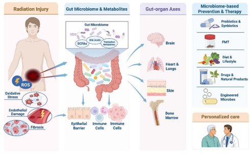
Radiotherapy is an essential component of multimodal treatment for solid tumors, and more than half of patients with cancer receive radiation during their disease course. Because of the unique anatomical and physiological features of the intestine, radiation enteritis (RE) remains a common and clinically challenging complication of abdominal and pelvic irradiation, with limited effective treatment options. In this review, we re-examine RE from a host-microbiome perspective. We summarize classical pathophysiological mechanisms and discuss how radiotherapy reshapes gut microbial composition and metabolism. We also highlight the roles of microbial metabolites, including short-chain fatty acids, bile acids and tryptophan derivatives, in barrier repair, immune homeostasis and stem-cell regeneration. Finally, we discuss microbiome heterogeneity across disease phases, tumor types and host factors, as well as microbiota-mediated gut-brain, gut-cardiopulmonary, gut-skin and gut-bone-marrow axes involved in systemic radiation injury. We further outline microbiome-based strategies for individualized risk stratification and early prediction, and recent advances and limitations of probiotics and synbiotics, fecal microbiota transplantation, dietary and lifestyle interventions, drugs and natural products, engineered microbes and novel delivery systems, highlighting the gut microbiome as a promising entry point to improve prevention and treatment of RE and systemic radiation toxicity.
Keywords: radiation enteritis, gut microbiota, microbial metabolites, host-microbiome interaction, gut-organ axis, microbiome-based therapy
Introduction
Radiotherapy is a cornerstone of cancer management and is widely deployed across a broad spectrum of malignancies worldwide. It is estimated that more than 50% of patients with cancer receive radiotherapy at some point during their treatment, whether with curative or palliative intent. Despite continued advances in precision radiotherapy, gastrointestinal injury remains a frequent and challenging complication in the treatment of abdominal and pelvic tumors, owing to the unique anatomical and physiological milieu of the bowel. Radiation enteritis (RE) affects up to 80% of patients undergoing pelvic or abdominal irradiation, leading to substantial deterioration in quality of life and, in some cases, necessitating treatment interruption or dose reduction, with potential compromise of oncological control.
RE is typically categorized into acute and chronic forms according to timing and pathophysiology. Acute RE emerges within weeks of initiating radiotherapy and is characterized by diarrhea, abdominal pain, nausea and tenesmus, reflecting direct epithelial injury and an acute inflammatory response. By contrast, chronic RE arises months to years after completion of treatment and is marked by intestinal fibrosis, endothelial proliferation and microvascular dysfunction, with clinical sequelae including persistent abdominal pain, altered bowel habits and intermittent bleeding[1]. Approximately 10-20% of patients with acute RE progress to a chronic phenotype; however, the mechanisms underpinning this transition remain incompletely defined.
The gut microbiome is increasingly implicated in the onset, progression and outcomes of RE. Radiotherapy profoundly alters microbial diversity and composition, typically with depletion of taxa traditionally associated with short-chain fatty acid (SCFA) production and barrier maintenance, such as members of Lachnospiraceae and the genus Roseburia, alongside expansion of potentially pathogenic or inflammation-associated lineages, including Enterobacteriaceae and Streptococcaceae[2]. Notably, these shifts are not merely consequences of radiation injury; they actively shape the host response to irradiation by influencing mucosal inflammation, epithelial barrier integrity and tissue repair[3]. On this basis, microbiome-directed interventions have become a salient target for the prevention and treatment of RE. Beyond local intestinal effects, the gut microbiome communicates with multiple host systems through the gut-brain, gut-cardiopulmonary, gut-skin and gut-bone-marrow axes. These interactions may contribute to the systemic effects of radiotherapy. This systems-level perspective broadens understanding of radiation-associated complications and may inform the development of integrated therapeutic strategies.
In this review, we synthesize current evidence on RE across clinical features, pathophysiology, microbiome alterations, therapeutic approaches and systemic effects, with particular emphasis on the central role of the gut microbiome and related translational advances. By integrating contemporary clinical practice with insights from microbiology and radiobiology, we aim to inform precision prevention, early diagnosis and individualized management of RE, and to outline future research priorities and challenges. To facilitate critical interpretation of this heterogeneous literature, we applied a tiered translational evidence framework throughout the review. Evidence was considered strong when clinical observations were accompanied by mechanistic validation in experimental models; moderate when clinical associations were biologically consistent with mechanisms demonstrated in independent preclinical studies; potentially translational when preclinical-only findings involved microorganisms, metabolites or pathways shared between humans and mice; and weak or exploratory when preclinical-only findings centered on mouse-specific taxa or mechanisms without clear human counterparts. This framework was used to distinguish clinically supported associations from mechanistic hypotheses and to highlight current limitations, inconsistencies and areas requiring prospective validation.
1. Classical pathophysiological mechanisms
Radiation-induced intestinal injury is a common complication among patients receiving abdominal or pelvic radiotherapy, arising from both direct and indirect effects of ionizing radiation on cells and tissues.
1.1 Radiation-induced oxidative injury and cell death
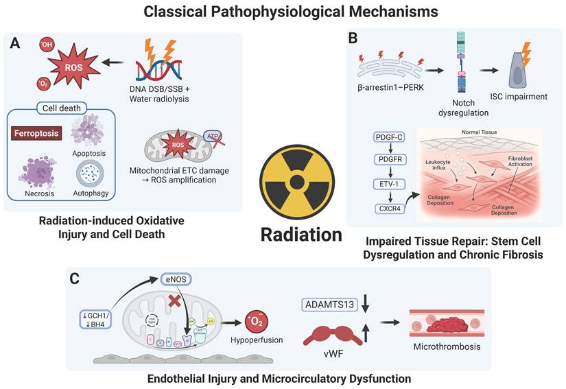
The direct effects of ionizing radiation primarily involve the ionization of DNA, resulting in single- and double-strand breaks and base damage. If unrepaired, these lesions activate cell-cycle checkpoints and apoptotic signaling pathways. Indirect effects occur via radiolysis of water, generating abundant reactive oxygen species (ROS), including hydroxyl radicals (•OH), superoxide anions (O2•-) and hydrogen peroxide (H2O2). These molecules oxidatively damage cellular constituents such as lipid membranes, proteins and nucleic acids. Excessive ROS also disrupt the mitochondrial electron transport chain, impair ATP synthesis and perturb energy metabolism, further amplifying ROS accumulation and creating a vicious cycle of oxidative injury[4]. Oxidative stress not only damages cellular architecture directly but also activates stress-responsive signaling, including ATM/ATR-p53 pathways and the NF-κB inflammatory cascade[5].
Radiation-induced cell death is not monolithic; rather, it encompasses a network of death programs, including apoptosis, ferroptosis, necrosis and autophagy, which collectively drive tissue injury and organ dysfunction. Recent animal studies indicate a pivotal role for ferroptosis in radiation-induced intestinal damage. In Göttingen minipigs, ionizing radiation markedly reduced glutathione peroxidase 4 (GPX4) protein and GPX4 mRNA expression (by approximately 70-83% and 90-94%, respectively), a hallmark of ferroptosis[6]. Complementary mouse studies show that the ferroptosis inhibitor Ferrostatin-1 mitigates ROS generation, suppresses both ferroptosis and apoptosis, significantly improves survival after lethal irradiation and restores intestinal structural and functional integrity[7].
Notably, studies have revealed that microbes play a significant role in radiation-induced oxidative damage and cell death, as demonstrated by in vitro and murine research on a novel “theft-ferroptosis” mechanism. Pseudomonas aeruginosa secretes a bacterial 15-lipoxygenase that catalyzes host phosphatidylethanolamine to form 15-hydroperoxy-eicosatetraenoyl-phosphatidylethanolamine (15-HpETE-PE), while concomitantly releasing phospholipases that blunt endogenous anti-ferroptotic defenses. In mice, this synergy markedly exacerbates radiation-induced intestinal injury, shortening survival after total-body irradiation from 15 to 4 days[8]. Collectively, these data reveal complex microbiome-radiation interactions (Figure 1A).
Classical pathophysiological mechanisms of RE. (A) Radiation-induced oxidative injury and cell death. (B) Impaired tissue repair associated with stem-cell dysregulation and chronic fibrosis. (C) Endothelial injury and microcirculatory dysfunction. ROS, reactive oxygen species; SSB/DSB, single-/double-strand breaks; ETC, electron transport chain; ATP, adenosine triphosphate; ISC, intestinal stem cell; TCA, tricarboxylic acid (cycle); ADAMTS13, a disintegrin and metalloproteinase with thrombospondin motifs 13; PERK, protein kinase R-like ER kinase; PDGF-C, platelet-derived growth factor-C; PDGFR, platelet-derived growth factor receptor; ETV1, ETS variant 1; CXCR4, C-X-C chemokine receptor 4; eNOS, endothelial nitric oxide synthase; GCH1, GTP cyclohydrolase 1; BH4, tetrahydrobiopterin; vWF, von Willebrand factor.
1.2 Repair of impaired tissue: stem cell dysregulation and chronic fibrosis
The clinical burden of intestinal radiation injury stems not only from acute epithelial loss and barrier disruption, but also from chronic long-term repair damage. Two interlinked processes contribute to chronic disease: dysfunction of intestinal stem cells (ISCs) and a pathological inflammation-fibrosis cascade[9].
Endoplasmic reticulum (ER) stress is a key mediator of radiation-induced stem cell dysfunction, constraining ISC proliferation and differentiation. In mouse models, β-arrestin-1 (βarr1) is markedly upregulated after irradiation. βarr1 directly binds and suppresses protein kinase R-like ER kinase (PERK)-mediated ER stress signaling, modulating downstream Notch activity and ultimately limiting ISC proliferative capacity and regenerative potential. In knockout mouse models, genetic ablation of β-arrestin 1 (βarr1) enhanced ISC proliferation and accelerated crypt regeneration. It also mitigated acute gastrointestinal syndrome and prolonged survival after lethal-dose irradiation[10]. When stem cell function is compromised and repair is delayed, unresolved acute inflammation evolves into chronic inflammation and fibrosis—the defining pathology of chronic RE. In mice, platelet-derived growth factor C (PDGF-C) engages PDGF receptors to activate an ETV1-driven CXCR4 signaling axis, sustaining immune-cell infiltration and fibroblast activation, with consequent collagen overdeposition and tissue fibrosis[11]. These mechanisms interlock to form a self-amplifying vicious cycle: impaired stem-cell function blunts epithelial regeneration and delays repair, while persistent inflammation and fibrogenesis erode the stem-cell niche—including vascular perfusion and stromal support—thereby deepening regenerative failure. This failed-regeneration-chronic inflammation-fibrosis axis may represent an important mechanism underlying the transition from acute to chronic RE (Figure 1B).
1.3 Endothelial injury and microcirculatory dysfunction
Endothelial damage is an early, prominent event in RE. Experimental studies show that ionizing radiation downregulates GTP cyclohydrolase 1 (Gch1), limiting tetrahydrobiopterin (BH4) synthesis and precipitating uncoupling of endothelial nitric oxide synthase (eNOS). Under physiological conditions, BH4 enables eNOS to generate nitric oxide (NO), which confers vasoprotection; when BH4 is deficient, eNOS instead produces superoxide, culminating in endothelial dysfunction and intestinal microischaemia. Clinically, patients exhibit a 45.9% reduction in plasma BH4 after radiotherapy, with intestinal perfusion falling to approximately 37% of baseline, indicating that endothelial dysfunction and microcirculatory compromise are key pathophysiological substrates of RE[12].
Imbalance of the von Willebrand factor (vWF)/ADAMTS13 axis further aggravates vascular injury. Clinical observations indicate that radiotherapy increases prothrombotic vWF while relatively reducing its sole physiological cleaving protease, ADAMTS13, thereby tipping the hemostatic balance towards thrombosis. Using mice with platelet- and endothelium-specific deletion of vWF, vWF was shown to promote radiation-induced intestinal injury. Importantly, exogenous recombinant human ADAMTS13 restored vWF/ADAMTS13 equilibrium, attenuated inflammation and oxidative stress, and protected the intestine from radiation damage, nominating this axis as a potential biomarker and therapeutic target for radiation-induced gut injury[13] (Figure 1C).
In summary, decades of work have elucidated canonical mechanisms of RE, encompassing direct cytotoxic effects, oxidative stress, multiple cell death programs, vascular injury, ER stress and an inflammation-fibrosis cascade. However, the translation of these established mechanisms into durable and effective therapies remains limited. Single-target interventions (for example, antioxidants, ferroptosis inhibitors, vasoprotective agents) show promise in animal models but have struggled to halt a disease driven by intertwined pathological processes in humans. Traditional paradigms also fail to explain the marked inter-individual variability under similar radiation regimens or predict why only 10-20% of acute cases progress to chronic disease, and these limitations are further compounded by the lack of reliable biomarkers. Moreover, a predominant focus on local intestinal injury has underappreciated systemic effects of radiotherapy and their bidirectional interplay with gut pathology. Crucially, the intestine has been viewed largely as a host organ, with insufficient consideration of the gut microbiome as a “hidden organ.” Emerging concepts, such as the “theft-ferroptosis” mechanism, show that microbes are not passive bystanders but active participants and modulators whose dysbiosis may link disparate pathological threads. These insights argue for a paradigm shift: from a host-centric view to a holistic host-microbiome interaction framework to explain individual variability, predict treatment responses, clarify acute-chronic transitions and enable rational, multi-target interventions.
2. Gut microbiome-associated regulatory network in the pathogenesis of RE
Limitations of single-target mechanistic studies underscore the need for a systems biology perspective. With advances in high-throughput sequencing, the gut microbiome has emerged as a potential modulator and biomarker of RE, linking environment, diet, immunity and metabolism. Microbiome-focused research offers a new framework to explain inter-individual variability, predict treatment responses and design personalized interventions. Crucially, the tractability of the microbiome and its metabolites provides actionable targets for clinical translation.
2.1 Impact of radiotherapy on gut microbiome composition
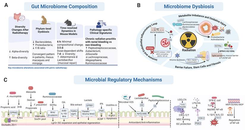
As the largest microbial ecosystem in the human body, the gut microbiome has been increasingly associated with, and may contribute to, the onset, progression and outcomes of RE. Multiple clinical studies using high-throughput 16S rRNA gene sequencing have shown that radiotherapy significantly alters microbial diversity and community structure. Diversity exhibits a dual pattern. Patients with RE show a marked reduction in alpha diversity (Simpson and Shannon indices), alongside a significant increase in beta diversity, indicating both loss of taxa and restructuring of the overall community[14]. Phylum-level dysbiosis shows cross-species consistency. The most characteristic change is a consistent decrease in Bacteroidetes coupled with an expansion of Proteobacteria. In patients receiving radiotherapy, 16S rRNA gene profiling indicates a decline in the Firmicutes/Bacteroidetes (F/B) ratio after treatment, correlating with dysbiosis and tissue injury; however, the F/B ratio should be interpreted only as a broad ecological descriptor rather than a standalone predictive biomarker[15]. Studies in two large animal models (rhesus macaques and minipigs) further confirm that, despite baseline inter-species differences, radiation induces highly convergent phylum-level shifts: decreased Bacteroidetes and increased Proteobacteria, whereas changes in Firmicutes vary by irradiation regimen and host species[16]. Time-resolved and pathology-specific shifts furnish candidate clinical biomarkers. In mouse irradiation models, microbiome dynamics are distinctly phase-dependent: during the prodromal phase (6 hours post-irradiation), changes are minimal; in the critical phase (3.5 days), composition closely tracks radiation dose; by the recovery phase (7 days), overall diversity declines, but the relative abundance of genera such as Akkermansia and Lactobacillus increases, potentially contributing to mucosal repair[17]. Clinically, patients with chronic radiation proctitis and rectal bleeding show distinct microbiome signatures compared with non-bleeding counterparts: enrichment of Peptostreptococcaceae, Eubacterium and Allisonella, with reduced Lachnospiraceae, Megasphaera and Ruminococcaceae—features that may serve as microbial markers for bleeding risk stratification and disease monitoring[18] (Figure 2A).
Gut microbiome as a central regulatory network in RE. (A) Changes in the gut microbiome after irradiation. (B) Radiation-induced dysbiosis sustains barrier failure, inflammation and impaired tissue repair. (C) Commensal microbes modulate post-irradiation gut barrier, survival and inflammatory circuits. 3-HB, 3-hydroxybutyrate; ACBP, acyl-CoA-binding protein; AhR, aryl hydrocarbon receptor; ARE, antioxidant response element; F/B ratio, Firmicutes/Bacteroidetes ratio; COX-2, cyclooxygenase-2; CXCL12, C-X-C motif chemokine ligand 12; E. coli, Escherichia coli; GPR43, G protein-coupled receptor 43; GPR81, G protein-coupled receptor 81; GPBAR1/TGR5, G protein-coupled bile acid receptor 1; H₂S, hydrogen sulfide; I3A, indole-3-aldehyde; I3C, indole-3-carbinol; IA, indoleacrylic acid; IL-1β, interleukin-1 beta; IL-6, interleukin-6; IL-10, interleukin-10; IPA, indole-3-propionic acid; ISC, intestinal stem cell; LGG, Lactobacillus rhamnosus GG; LGR5, leucine-rich repeat-containing G protein-coupled receptor 5; LPS, lipopolysaccharide; LTA, lipoteichoic acid; MAPK, mitogen-activated protein kinase; MDP, muramyl dipeptide; MSC, mesenchymal stem cell; MUC2, mucin 2; MyD88, myeloid differentiation primary response 88; NF-κB, nuclear factor kappa-B; NOD2, nucleotide-binding oligomerization domain-containing protein 2; NOX2, NADPH oxidase 2; Nrf2, nuclear factor erythroid 2-related factor 2; PGE2, prostaglandin E2; ROS, reactive oxygen species; SCFAs, short-chain fatty acids; SFB, segmented filamentous bacteria; TLR, Toll-like receptor; TLR2, Toll-like receptor 2; TLR4, Toll-like receptor 4; YAP1, Yes-associated protein 1; ZO-1, zonula occludens-1.
However, direct comparisons across studies should be made with caution. Existing cohorts differ substantially in patient population, tumor type, irradiation site and treatment strategy, including cervical, prostate, rectal, endometrial and anal cancers treated with radiotherapy alone, concurrent chemoradiotherapy, postoperative adjuvant radiotherapy or radionuclide therapy. Radiation field, intestinal dose exposure, concurrent chemotherapy, antibiotic use, diet and comorbidities can all markedly reshape the gut microbiome. These variables may partly explain why the direction of change in specific taxa is not always consistent across studies. For example, Faecalibacterium, Roseburia and some Clostridia are generally regarded as SCFA producers with barrier-protective functions, yet in some clinical cohorts these taxa or related enterotypes have been associated with more severe radiation-induced gastrointestinal toxicity. Similarly, reported changes in the F/B ratio vary across studies, and this ratio alone has limited predictive value for RE. These inconsistencies suggest that the effects of putatively protective or deleterious taxa are context dependent and should not be extrapolated without considering tumor type, radiation dose, sampling time point, sequencing approach, diet and medication exposure.
Collectively, these human and animal data indicate that radiation induces characteristic, phase-dependent patterns of gut dysbiosis that actively shape the onset, progression and clinical manifestations of RE, while simultaneously offering candidate microbial biomarkers for risk stratification and longitudinal disease monitoring.
2.2 Microbiome dysbiosis tightly links to pathophysiological mechanisms
Under radiation-induced oxidative injury and cell-death programs, both animal and clinical studies consistently report reduced microbial diversity, depletion of beneficial and SCFA-producing members, a decrease in the F/B ratio, and expansion of pathobionts (for example, Proteobacteria and Actinobacteria). These shifts may increase endotoxin burden and activate inflammatory pathways, thereby potentially establishing a self-reinforcing “radiation-oxidative injury-barrier disruption-dysbiosis” positive feedback loop[19]. Notably, Akkermansia exhibits a biphasic, niche- and dose-dependent behavior: several studies show its depletion, and that supplementation or upregulation of its metabolite propionate can strengthen tight junctions and the mucus layer via GPR43, thereby attenuating radiation-induced intestinal injury; conversely, in acute high-dose models, Akkermansia has been reported to bloom post-irradiation, deplete mucin, and promote pathogenic adhesion and inflammation, thereby exacerbating injury[20]. In parallel, survivor-type models suggest that taxa enriched after irradiation (for example, Lachnospiraceae and Enterococcaceae) and their metabolites (propionate; indole pathway products such as I3A and kynurenic acid) mitigate DNA damage and ROS and promote hematopoiesis and mucosal repair. The tryptophan metabolite indole-3-propionic acid (IPA) confers systemic radioprotection via the pregnane X receptor (PXR)/acyl-CoA-binding protein (ACBP) axis; these interactions unite microbes, metabolites, redox homeostasis, and cell-death programs into an integrated network[21].
In the setting of endothelial injury and microcirculatory dysfunction, barrier breakdown and translocation of bacterial products into the bloodstream may activate Toll-like receptor 4 (TLR4)-dependent NADPH oxidase-ROS production, eNOS uncoupling, and MAPK/NF-κB signaling, which together perpetuate endothelial dysfunction, microcirculatory derangement, dysbiosis, and endotoxemia/inflammation, although direct validation of this pathway in RE remains limited[22]. Microbial metabolites are both targets and modulators within this loop: SCFAs can restore eNOS and mitochondrial function in endothelial cells, lower ROS and dampen inflammation, thereby offering a means to interrupt the cycle; additionally, the commensal-derived SCFA valerate (pentanoate) confers systemic and gastrointestinal radioprotection in vivo and reshapes community composition[23].
With respect to impaired repair, stem-cell dysregulation and chronic fibrosis, SCFAs act via GPR41, GPR43 and GPR109A and through histone deacetylase (HDAC) inhibition to reinforce tight junctions and the mucus barrier, and to restrain TLR/NF-κB amplification, thereby alleviating mucosal inflammation and promoting barrier reconstruction. Dietary fiber elevates SCFAs and other co-metabolites, which can attenuate gastrointestinal toxicity from radiotherapy while potentially enhancing anti-tumor efficacy[24]. The microbiota also directly influences epithelial stem-cell lineages: Lactobacillus promote ISC proliferation and barrier repair via the IL-22/STAT3 axis. Beyond bacteria, phage (virome) dysbiosis can suppress regeneration by overactivating RIG-I/Notch signaling within ISCs. Fecal virome transplantation from healthy donors partially restores stem-cell proliferation and differentiation, implicating multiple microbial compartments in the regulation of post-irradiation repair[25]. Collectively, the failed repair-stem cell dysregulation-chronic fibrosis axis is coupled to microbiome/virome disruption, metabolite depletion, and inflammatory amplification, defining a targetable network amenable to dietary fiber, probiotics/synbiotics, microbiota/virome transplantation, and metabolite supplementation[26]. It is worth noting that the roles of the gut mycobiome and broader virome in RE remain poorly defined, with current evidence remaining limited, preliminary and largely indirect, thereby highlighting the need for future studies integrating bacterial, fungal and viral microbiome datasets to better delineate host-microbiome interaction networks in RE.
Accordingly, dysbiosis should not be regarded solely as a downstream consequence of radiation injury; rather, it may contribute to the amplification and persistence of pathology: (1) Metabolite imbalance weakens endogenous protection. Post-irradiation loss of protective metabolites produced by beneficial taxa diminishes intrinsic anti-inflammatory, antioxidant and reparative capacity. (2) Immune dysregulation fuels an inflammatory vicious cycle. Overgrowth of pathobionts (for example, Proteobacteria) and their cell-wall components chronically activate TLR-MyD88-NF-κB signaling, escalating inflammation and reinforcing a loop of “dysbiosis-heightened inflammation-tissue injury-further dysbiosis”[27,28]. (3) Barrier disruption promotes bacterial translocation. Depletion of beneficial genera (for example, Lactobacillus, Bifidobacterium) thins the protective mucus layer; dysbiosis-driven signals downregulate tight-junction proteins (ZO-1, occludin), increasing permeability and facilitating translocation of bacteria and toxins, which triggers local and systemic inflammation[29]. Radiation also perturbs the physical barrier to skew microbe-immune crosstalk: loss of REGγ reduces goblet cells and thins the mucus layer post-irradiation, enabling closer bacterial-epithelial contact and exacerbating inflammation. It is a phenotype closely tied to compositional shifts, particularly loss of probiotics such as Lactobacillus and Bifidobacterium[30]. Moreover, radiation-induced NF-κB activation upregulates miR-221/222 and suppresses Syndecan-1, leading to ZO-1 and occludin downregulation and barrier failure. Dysbiosis magnifies these changes, reflected by elevated circulating soluble Syndecan-1 and inflammatory cytokines (TNF-α, IL-1β) in patients with RE[29]. It should be emphasized that most clinical microbiome studies in RE are based on associations between fecal sequencing profiles and symptom scores or toxicity grades. Antibiotic depletion, fecal microbiota transplantation, specific strain supplementation and metabolite rescue experiments in animals provide stronger evidence for causality. Accordingly, clinical causality remains to be established through prospective, mechanism-guided interventional studies (Figure 2B).
2.3 Insights into microbiome-based regulatory mechanisms: emerging perspectives
Microbial metabolites play pivotal roles in the progression of RE. These small molecules act across multiple signaling axes to coordinate barrier function, inflammatory responses and stem-cell regeneration, forming a multifaceted interactive network. The SCFA-GPR axis is a meaningful regulator of barrier integrity. SCFAs, the principal products of microbial fermentation of dietary fiber, are central to gut homeostasis. Radiotherapy significantly reduces SCFA production; notably, depletion of mucosa-associated, SCFA-producing genera correlates with radiation injury of the anterior rectal mucosa[31]. Propionate and butyrate exert particularly salient protective effects. Akkermansia muciniphila protects the gut against irradiation by secreting propionate, which engages G-protein-coupled receptor 43 (GPR43) on intestinal epithelial cells, enhances histone acetylation and upregulates tight-junction proteins (occludin, ZO-1) and mucin (MUC2), thereby strengthening epithelial barrier integrity[20]. In patients, the abundance of A. muciniphila inversely correlates with the duration of diarrhea; supplementation improves survival and preserves intestinal architecture in mice[32]. Recent work also indicates that Faecalibaculum rodentium is a radioprotective keystone species whose depletion coincides with reduced butyrate and increased ionizing radiation sensitivity. Mechanistically, butyrate curtails ERK-driven nuclear translocation of PKM2, thereby attenuating p53/BAX-dependent apoptosis in hematopoietic stem and progenitor cells (HSPCs)[33]. 3-hydroxybutyrate (3HB) represents a second protective route. 3HB ameliorates radiation proctopathy by blocking GPR43-mediated IL-6 signaling. As an important source of 3HB, A. muciniphila declines after irradiation, with 3HB levels inversely associated with IL-6 expression; supplementation with 3HB or A. muciniphila lowers GPR43 and IL-6 expression and improves clinical symptoms and histopathology[34].
Additional microbiota-derived factors fortify the barrier. Dextran produced by Weissella increases propionate and promotes mucosal barrier function[35]. Prophylactic administration of Faecalibacterium prausnitzii protects the colonic barrier by maintaining the Sox-9+ stem/progenitor pool and stimulating crypt epithelial proliferation[36]. Lactobacillus rhamnosus GG (LGG) safeguards the gut via dual mechanisms: suppression of JAK2/STAT3 signaling to reduce pro-inflammatory cytokines, and activation of a TLR2/COX-2-dependent pathway that drives COX-2+ mesenchymal stem cells to home towards the ISCs niche, creating a protective microenvironment[37,38].
The Wnt signaling network governs ISC dynamics. The Wnt/β-catenin pathway is essential for ISC self-renewal and differentiation and is finely tuned by microbial metabolites. SCFAs produced by the Ruminococcaceae family activate Wnt/β-catenin signaling, enhance Lgr5+ ISC expansion and promote epithelial regeneration[39,40]. The microbial indole-3-aldehyde (I3A) activates the aryl hydrocarbon receptor (AhR)/IL-10/Wnt axis to stimulate epithelial proliferation, mitigate mucosal injury and maintain barrier integrity[41]. As an environmental sensor, AhR constrains Wnt-β-catenin signal strength by modulating RNF43 and ZNRF3, thereby preserving ISC homeostasis and barrier function[42]. By contrast, under pathological conditions FOXQ1 upregulates SIRT1 and promotes β-catenin nuclear translocation, heightening stemness and radioresistance in colorectal cancer cells while reshaping the microbiota, illustrating the pathogenicity of Wnt pathway dysregulation[43]. The bile acid-TGR5 axis underpins metabolism and regeneration. Untargeted metabolomics reveals significant post-irradiation reductions in secondary bile acids (for example, deoxycholic acid and lithocholic acid), which possess anti-inflammatory properties; their loss may worsen radiation-induced inflammation[44]. Supplementation with bile extract or lithocholic acid promotes ISC proliferation and intestinal regeneration by modulating G-protein-coupled bile acid receptor 1 (TGR5) and Yes-associated protein 1 (YAP1)[45]. Lactate provides an additional regenerative cue. By engaging GPR81, lactate induces Paneth and stromal cells to produce Wnt3, accelerating ISC-mediated epithelial development; pre-feeding lactate protects mice from chemotherapy- and radiation-induced intestinal injury[46].
Beyond these major axes, several specialized metabolites confer added protection. Indole-3-propionic acid (IPA) activates PXR to sustain ACBP expression, thereby protecting the gut from radiation injury[47]. Urolithin A (UroA) has shown radioprotective activity by inhibiting p53-mediated apoptosis and remodeling the gut microbiota; in an immunosuppressive tumor microenvironment, UroA reduces tumor-associated macrophages and enhances the efficacy of radiotherapy[48]. The Nrf2 antioxidant pathway is involved in countering radiation-induced oxidative injury. Microbiota-derived hydrogen sulphide (H2S) directly activates the Keap1/Nrf2/ARE cytoprotective axis, upregulating antioxidant proteins and glutathione to mitigate damage in normal tissues. Acid-producing taxa such as Lactobacillus acidophilus catalyze the conversion of dietary indole-3-carbinol (I3C) into bioactive anticancer compounds that lessen radiation injury via Nrf2-dependent mechanisms. Indoleacrylic acid (IA) produced by commensal Peptostreptococcus likewise suppresses inflammation and strengthens the barrier through Nrf2-ARE activation[49]. KEGG analyses further implicate multiple microbiome-related metabolic routes in RE, including arginine, arachidonic acid and linoleic acid metabolism[50].
The gut microbiota also engages host immunity through multiple pattern-recognition receptors (PRRs). Under physiological conditions, beneficial microbes elicit TLR-mediated protective responses that confer radioprotection. LGG releases lipoteichoic acid (LTA), a TLR2 agonist that binds TLR2 on subepithelial macrophages, triggering MyD88-dependent signaling and robust CXCL12 secretion. The resulting chemokine gradient actively recruits mesenchymal stem cells to the crypt niche, thereby shielding ISCs from radiation injury[38,51]. Lipopolysaccharide (LPS) and hyaluronan provide radioprotection via TLR4-COX-2 activation, boosting prostaglandin E2 (PGE2) production and protecting crypt cells from radiation-induced apoptosis[27]. When irradiation drives dysbiosis, TLR signaling shifts towards inflammation. Radiation-induced overgrowth of segmented filamentous bacteria (SFB) and Escherichia coli activates TLR-MyD88-NF-κB signaling, enhancing production of IL-1β, IL-6 and IL-23. This promotes Th17 polarization and accumulation; Th17-derived IL-17 and granulocyte-macrophage colony-stimulating factor (GM-CSF) in turn expand and activate neutrophils, foster oxidative stress and exacerbate dysbiosis, closing a vicious cycle[28]. The microbiota can also modulate RE via NOD2: muramyl dipeptide (MDP) and N-acetylglucosamine-MDP, generated by Enterococcus peptidoglycan hydrolases, engage NOD2 to activate NF-κB and regulate inflammation[52].
Taken together, these findings suggest that microbiome-related signaling in RE is better understood as a hierarchical, phenotype-linked network rather than a set of equivalent pathways. Based primarily on preclinical evidence, the SCFA-GPR/HDAC axis represents the most direct microbiome-dependent barrier-protective mechanism, maintaining tight-junction integrity, limiting mucosal inflammation and supporting intestinal and hematopoietic stem/progenitor cell survival. A second major module is the microbial metabolite-regulated regenerative axis, which includes Wnt/β-catenin, AhR/IL-10/Wnt, bile acid-TGR5/YAP1 and lactate-GPR81-Wnt3 signaling and collectively governs ISC maintenance, epithelial regeneration and post-irradiation repair. In contrast, TLR-MyD88-NF-κB signaling is best positioned as a downstream inflammatory amplifier that becomes prominent when irradiation-induced barrier failure and dysbiosis increase exposure to microbial products, thereby connecting acute mucosal inflammation with persistent immune activation. Other pathways, including Nrf2, PXR/ACBP, cGAS-STING and metabolite-specific mechanisms, should be viewed as context-dependent modifiers or therapeutic entry points that intersect with these core processes. This hierarchy also helps align mechanisms with clinical phenotypes: SCFAs depletion and barrier failure are most closely associated with acute diarrhea and mucosal injury; impaired regenerative signaling may contribute to delayed repair and acute-to-chronic transition; and sustained inflammatory amplification together with pro-fibrotic remodeling is more relevant to chronic bleeding, fibrosis and stricture formation (Table 1 and Figure 2C).
Gut bacteria and microbial metabolites involved in RE progression
| Bacteria | Metabolite | Effect | Mechanism | Model | Refs. |
|---|---|---|---|---|---|
| Akkermansia muciniphila | Propionate | Protective (barrier) | Strengthens epithelial barrier (ZO-1, occludin and MUC2) via GPR43 activation. | Murine models; pelvic RT patient cohorts. | 20, 32 |
| Akkermansia muciniphila | 3-Hydroxybutyrate (3HB) | Protective (anti-inflammatory) | Blocks GPR43-mediated IL-6 signaling in rectal mucosa, lowering GPR43 and IL-6 expression. | Human cohorts; murine models. | 34 |
| Faecalibaculum rodentium | Butyrate | Protective (intestinal & hematopoietic) | Prevents ERK-dependent PKM2 nuclear translocation and reduces p53/BAX-mediated apoptosis in HSPCs. | Murine models. | 33 |
| Faecalibacterium prausnitzii | - | Radioprotective | Maintains the Sox-9⁺ stem/progenitor pool, stimulates crypt epithelial proliferation. | Rat models (male Sprague-Dawley rats). | 36 |
| - | Indole-3-propionic acid (IPA) | Protective (systemic) | Gut microbiota-derived IPA activates PXR-ACBP axis and enhances antioxidant and metabolic homeostasis. | Murine models. | 21, 47 |
| - | Indole-3-aldehyde (I3A) | Protective (barrier & regeneration) | Activates AhR/IL-10/Wnt signaling to promote epithelial proliferation and ISC-mediated regeneration. | Murine models. | 41 |
| Peptostreptococcus | Indoleacrylic acid (IA) | Protective (anti-inflammatory, barrier) | Activates Nrf2-ARE signaling and upregulates antioxidant and barrier-related genes. | In vitro systems and human cohorts. | 49 |
| - | Urolithin A (UroA) | Protective (gut), Sensitizing (tumor) | Inhibits p53-mediated apoptosis, remodels gut microbiota and reduces tumor-associated macrophages. | Murine models. | 48 |
| - | Secondary bile acids / bile extract | Protective (ISC-driven regeneration) | Activates epithelial TGR5-YAP1 signaling to promote ISC proliferation and mucosal regeneration. | Murine models. | 44, 45 |
| - | Lactate | Protective (epithelial development) | Engages GPR81 on Paneth and stromal cells to induce Wnt3 and drive Lgr5⁺ ISC-mediated regeneration. | Murine models. | 46 |
| - | H₂S | Protective (antioxidant) | Activates the Keap1/Nrf2/ARE cytoprotective axis to enhance antioxidant defenses. | In vitro systems. | 49 |
| Weissella cibaria | Dextran and propionate | Protective (mucosal barrier) | Dextran increases propionate, reshapes gut microbiota and strengthens the mucus layer and tight junctions. | Murine models. | 35 |
| Lactobacillus rhamnosus | Lipoteichoic acid (LTA) | Protective (crypt/ISC protection) | Activates TLR2-MyD88 signaling and induces CXCL12 to recruit COX-2⁺ mesenchymal stem cells. | Murine models. | 37, 38, 51 |
Nevertheless, most mechanistic studies are based on mouse or rat models of acute, high-dose irradiation, including total abdominal irradiation, total-body irradiation, and localized intestinal irradiation. Although these models are valuable for elucidating epithelial injury, inflammatory activation, and microbe-metabolite interactions, they differ substantially from clinical settings that involve fractionated radiotherapy, intensity-modulated radiotherapy, or concurrent chemoradiotherapy. Therefore, findings from animal studies should be interpreted primarily as mechanistic insights, and validation in human cohorts remains essential.
3. Gut microbiota characteristics in RE across different tumor types
The gut microbiome in RE is not a homogeneous entity. Microbial composition and inferred function in RE vary substantially with primary tumor type, anatomical location and radiation regimen, and these tumor- and treatment-specific microbiome configurations are closely linked to differences in the clinical presentation and severity of RE, providing a basis for personalized risk stratification and precise prevention strategies (Figure 3).
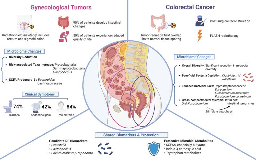
Tumor-specific landscapes of RE. In colorectal cancer, radiation-associated dysbiosis features loss of SCFA producers and enrichment of anaerobic, pro-fibrotic and Fusobacterium-dominated communities linked to symptoms, fibrosis and reduced radiosensitivity. In gynecological tumors, the gut microbiome exhibits reduced diversity with Proteobacteria and Gammaproteobacteria increases and Bacteroides decreases; baseline Coprococcus enrichment identifies patients at higher enteritis risk.
3.1 Colorectal tumors
Colorectal tumor-associated RE has unique anatomical and treatment characteristics. In standard neoadjuvant regimens for rectal cancer, pelvic radiation fields routinely encompass the mesorectum, presacral and internal iliac nodes, and, in T4 or distal tumors, extend anteriorly to cover the external iliac or even inguinal basins, which tends to bring additional small bowel and the anal canal into the high-dose region[53]. Because the tumor often overlaps the irradiation field, sparing normal tissue is technically challenging. Consequently, pelvic radiotherapy in colorectal tumors may promote epithelial injury, barrier disruption, inflammatory activation, and local microbial remodeling.
Existing evidence indicates that patients with colorectal cancer (CRC) already exhibit marked intestinal dysbiosis at the time of diagnosis, characterized by reduced overall microbial diversity and richness, enrichment of pro-inflammatory and opportunistic pathogenic taxa, and depletion of butyrate-producing “beneficial” bacteria. Neoadjuvant radiochemotherapy (NRCT) does not substantially alter the global diversity or overall structure of the gut microbiota, but can decrease the relative abundance of potentially pathogenic genera such as Fusobacterium, Escherichia and Klebsiella, while increasing Bifidobacterium, a shift that is considered relatively favorable[54]. Analyses of clinical specimens suggest that the intratumoral microbiome may have predictive value for radiotherapeutic response. Specifically, the presence of Fusobacterium canifelinum has been associated with unfavorable prognosis, whereas microbiota-directed interventions, such as antibiotics or probiotics, may enhance radiosensitivity by modulating tumor-associated microbial communities[55]. In murine models, oral inoculation with Fusobacterium nucleatum followed by irradiation allows this species to translocate from the oral cavity to CRC lesions and attenuate the efficacy of radiotherapy, whereas metronidazole effectively suppresses F. nucleatum colonization and restores radiosensitivity. These findings highlight the critical role of the oral-gut microbiota axis in shaping responses to radiation therapy and suggest that optimizing oral hygiene and oral microbiome status may represent an important component of comprehensive management in CRC patients undergoing radiotherapy[56].
3.2 Gynecological tumors
Radiotherapy for gynecological malignancies has distinct anatomical and physical characteristics: target positioning is markedly influenced by bladder and rectal filling, and pelvic organs such as the uterus and cervix can undergo substantial displacement and deformation, in contrast to the relatively fixed targets seen in most thoracic and head-and-neck tumors. At the same time, gynecological pelvic irradiation typically involves large fields and high doses, and, combined with generally long patient survival, this results in a high incidence of multisystem chronic toxicities[57]. Moreover, fields for cervical and endometrial cancer inevitably include the rectum and sigmoid colon[58].
Even with intensity-modulated radiotherapy (IMRT), about 90% of patients experience persistent intestinal changes and 50% report marked deterioration in quality of life[14]. During cervical cancer radiotherapy, the incidence of diarrhea and abdominal pain rises weekly, peaking at treatment end (diarrhea 74%, abdominal pain 42%)[59]. Nutritional risk escalates throughout treatment: by completion, up to 84% exhibit deficiency, with significant declines in body weight, skeletal muscle mass and serum albumin[60]. In older women treated for endometrial cancer, risks of constipation (HR=2.27), abdominal pain (HR=2.94) and fecal incontinence (HR=1.96) exceed those in cancer-free peers and persist for at least 5 years[61]. High-throughput sequencing shows marked loss of gut microbial diversity during therapy, with increased Proteobacteria and Gammaproteobacteria and decreased Bacteroides. Pre-treatment microbiota can predict enteritis risk; for example, higher baseline abundance of Coprococcus associates with severe RE[14]. Diversity tightly relates to chemoradiation toxicity: the Shannon index fell from baseline (2.9±0.5) to week 5 (2.49±0.7), and patients with higher diversity generally experienced fewer gastrointestinal symptoms[62].
Although reduced diversity and depletion of SCFA-producing bacteria are shared features, the gut microbiota associated with RE displays tumor-type-specific compositional and functional profiles shaped by the underlying malignancy and radiation strategy, indicating that we should take tumor-type specificity into account rather than focusing solely on inflammation itself. These cancer-specific microbiome signatures highlight the need for tumor-type-specific microbial risk assessment and tailored intervention strategies.
4. Gut microbiota characteristics in acute and chronic RE
The gut microbiota characteristics in RE not only vary depending on the tumor type and corresponding radiation regimen but are also closely associated with distinct stages of RE progression. RE can be clearly divided into acute and chronic phases based on timing and pathophysiology. According to expert consensuses and clinical guidelines from various countries, including the United States, Europe, and Australia, as well as the 2021 Chinese expert consensus, a 3-month interval after radiotherapy is commonly used in clinical practice to distinguish acute from chronic RE. This cutoff has also been widely adopted in clinical and translational studies. The two differ fundamentally in clinical phenotype, histological underpinnings and putative microbiome signatures. Crucially, deciphering the roles of multiple factors, including the gut microbiome, in the transition from acute to chronic disease is key to preventing long-term severe complications (Figure 4).
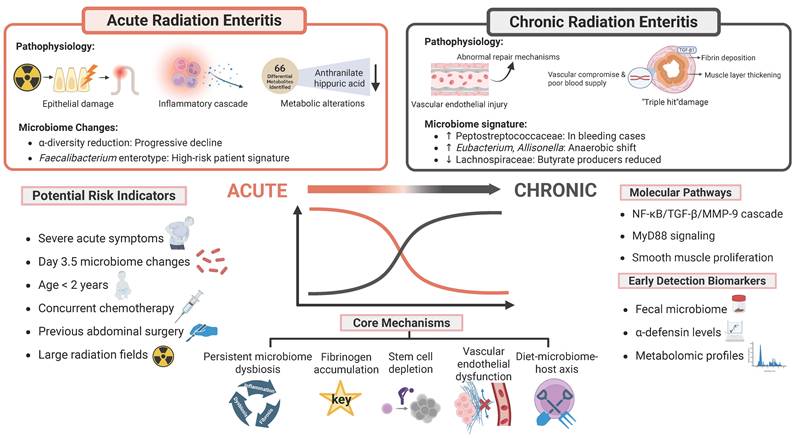
Features of acute and chronic RE and mechanisms of transition. The figure illustrates microbiome-driven transitions from acute inflammatory dysbiosis to chronic, anaerobic, pro-fibrotic community states. Core mechanisms link acute injury to chronic disease through sustained dysbiosis, impaired regeneration, fibrosis, vascular dysfunction and disrupted diet-microbiome-host metabolism. Representative interventions are shown on both sides. α-diversity, alpha diversity; MMP-9, matrix metalloproteinase-9; MyD88, myeloid differentiation primary response 88; NF-κB, nuclear factor kappa B; TGF-β, transforming growth factor beta.
4.1 Features of acute phase
Acute RE typically emerges during radiotherapy or within weeks after completion. Pathologically, ionizing radiation inflicts direct, rapid injury on the mucosa—most prominently on crypt stem cells with high mitotic rates. This damage drives extensive apoptosis of clonogenic crypt cells, disrupts the epithelial renewal cycle and triggers downstream cascades. Histology shows villous atrophy, blunting and fusion; crypt disarray or loss; marked depletion of goblet cells; submucosal oedema; and diffuse inflammatory infiltrates dominated by neutrophils. The resultant physical barrier breakdown increases intestinal permeability and elicits a strong innate immune response with surges of pro-inflammatory cytokines such as IL-1β, IL-6 and TNF-α[63-65]. Clinically, patients develop diarrhea, abdominal pain, nausea, vomiting and tenesmus, with severity closely linked to cumulative dose and often peaking at treatment end. In a cervical-cancer cohort, diarrhea rose from 28% in week 2 of radiotherapy to 74% by treatment completion[59]. Time-series analyses identify week 4 post-radiotherapy as a critical inflection point: levels of arachidonic acid and other metabolites reach a nadir and then rebound, consistent with the onset of gut self-regulatory responses[59]. In parallel, nutritional status deteriorates; by treatment end, up to 84% of patients exhibit malnutrition with significant declines in body weight, skeletal muscle mass and serum albumin[60].
The microbiome shifts sharply in the acute phase, with reduced diversity and dysbiosis. Changes scale with radiation dose: at a critical window around day 3.5 post-exposure, the relative abundance of Proteobacteria and the genus cluster Escherichia-Shigella increases dose-dependently, while beneficial Lactobacillus murinus decreases[17]. We speculate that such dysbiosis likely arises on the background of radiation-induced epithelial and mucus barrier disruption, which facilitates overgrowth and mucosal adhesion of facultative pathobionts and enhances exposure of pattern-recognition receptors to luminal pathogen-associated molecular patterns, thereby amplifying mucosal inflammation and further impairing barrier integrity. As many of these depleted taxa are SCFA producers that support epithelial energy supply and tight-junction maintenance, their early loss is expected to weaken barrier repair and promote a pro-inflammatory milieu. Serum metabolomics in an acute rat model identified 66 significantly altered metabolites, with anthranilic acid and hippuric acid showing the most pronounced decreases; the lipid metabolite linoleic acid emerged as a candidate biomarker of high nutritional risk[44,60]. Clinically, fecal microbiome profiling shows promise as a biodosimeter for radiation injury. Thus, acute RE should be viewed not merely as a process of microbial compositional change, but as a reciprocal loop in which radiation-induced barrier disruption drives dysbiosis, while dysbiosis further aggravates mucosal inflammation and barrier failure. Although direct mechanistic evidence for microbiome-mediated pathways in acute RE is still limited, based on the above findings we hypothesize that acute-phase barrier disruption and dysbiosis are mechanistically linked and together shape the inflammatory and metabolic landscape that determines progression versus resolution of injury in later phases.
4.2 Features of chronic phase
In contrast, chronic RE is a delayed injury that can present months to up to three decades after therapy. Its pathological core is aberrant repair of early damage, culminating in progressive ischemic vasculopathy and tissue fibrosis[31,66]. Among patients receiving pelvic radiotherapy, incidence is approximately 5-20%[18]. TGF-β1, a key pro-fibrotic mediator, is activated in cascade to drive fibroblast proliferation and extracellular-matrix deposition[67], accompanied by intimal hyperplasia and microvascular remodeling. This establishes a characteristic “triple hit” of the bowel wall, comprising excess fibrin deposition, pathological thickening of the muscularis and compromised vascular supply[66], which together define the histological basis of chronic injury. These substrates produce a broad clinical spectrum dominated by altered bowel habits, persistent abdominal pain and intermittent bleeding. Unlike the inflammation-driven symptoms of the acute phase, chronic manifestations reflect permanent architectural change; roughly 50% of long-term survivors develop some form of chronic gastrointestinal dysfunction[31]. With advancing fibrosis, about 10% progress to strictures, fistulas or perforation, often requiring surgery[66]. Symptom timing shows a bimodal pattern: in some, symptoms track continuously with treatment, whereas in others they arise after a 6- to 24-month latency, underscoring the complex dynamics of fibrogenesis[67].
Microbiome studies reveal deep ecological shifts in chronic disease that likely participate in maintenance and progression rather than being mere sequelae of radiation. This distinction may influence reparative capacity: long-term depletion of classical butyrate-producing taxa, including Clostridium cluster IV, Roseburia and Phascolarctobacterium, correlates positively with progression of intestinal fibrosis and loss of butyrate (which serves as both a primary energy source for colonocytes and an anti-inflammatory and epigenetic modulator) may contribute to the inflammation-fibrosis cycle by permitting sustained TGF-β/SMAD-dependent myofibroblast activation and collagen deposition[68]. Deeper mucosal profiling shows significant reductions in homeostatic cytokines that sustain barrier integrity, including IL-7, IL-12/IL-23p40, IL-15 and IL-16[31], which we speculate reflects disruption of microbial ecological niches following acute barrier injury, leading to persistent immune dysregulation that in turn fuels chronic inflammation and progressive fibrosis.
4.3 Hallmarks of the acute-to-chronic transition
The transition from acute to chronic RE arises from interlocking pathophysiological processes. In the acute phase, abrupt epithelial barrier loss, mucosal cytokine shifts and dose-dependent dysbiosis create a highly inflamed and metabolically perturbed luminal environment; early expansion of facultative pathobionts and depletion of SCFA producers such as classical butyrate-producing taxa may weaken barrier repair and prolong immune activation. Persistent dysbiosis may be an important contributor to a sustained low-grade inflammatory milieu that is increasingly recognized as a legacy of these early ecological disturbances rather than a de novo phenomenon. At the molecular level, fibrinogen accumulation in intestinal tissues is pivotal; its deficiency markedly attenuates both early and delayed injury and reduces abnormal smooth-muscle proliferation and TGF-β expression[69]. Beyond TGF-β signaling, Wnt/β-catenin dysregulation impairs stem-cell renewal, and persistent NF-κB activation maintains a chronically inflamed microenvironment. Vascular injury is likewise key: radiation-induced endothelial damage leads to microvascular occlusion and impaired angiogenesis, producing ischemic tissue injury that amplifies fibrosis. Critically, repetitive acute insults deplete the intestinal stem-cell pool, limiting long-term epithelial regeneration[26].
Integrating microbial and host factors, we hypothesize that early barrier failure and microbiome perturbation may increase the risk of chronic disease. These changes may promote a feed-forward loop involving dysbiosis, cytokine dysregulation and pro-fibrotic signaling, including TGF-β/SMAD and NF-κB pathways. Over time, this process may contribute to fibrosis and vascular remodeling. For risk stratification, the severity of acute symptoms strongly predicts chronic progression; all patients who later developed delayed RE had previously exhibited pronounced acute reactions[70]. Microbiome signatures can also serve as predictors, especially changes at the critical ~3.5-day timepoint post-irradiation rather than at 6 hours (prodrome)[17]. Additional high-risk groups include patients with significant comorbidities and those receiving high doses and/or large irradiation fields. In children, younger age (particularly under 2 years), prior abdominal surgery and concurrent chemoradiation, especially with anthracyclines (for example, doxorubicin), increase risk[70].
Technical differences further complicate interpretation of phase-specific microbiome signatures. Existing studies differ in sequencing platform, 16S rRNA variable region, bioinformatic workflow and taxonomic database. Most rely on 16S rRNA sequencing, which often provides genus-level or coarser taxonomic resolution and has limited capacity to resolve strain-level variation or functional genes. Functional inference using tools such as PICRUSt can generate useful hypotheses but cannot replace shotgun metagenomics, metatranscriptomics, metabolomics or culture-based validation. These limitations should be considered when interpreting acute- and chronic-phase biomarkers or comparing microbial signatures across studies.
5. Systemic effects of radiotherapy: microbiome-mediated inter-organ crosstalk
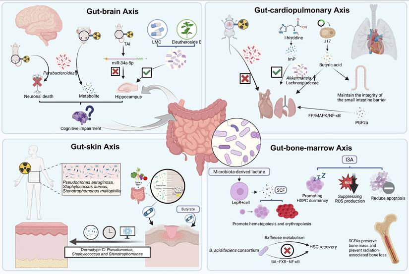
The impact of the gut microbiome in RE in response to radiotherapy on the host does not represent an isolated event; through the gut-organ axes, which constitute a multilayered communication network, it systemically influences distant organ function. Here we outline the roles of the gut-brain, gut-cardiopulmonary, gut-skin, and gut-bone-marrow axes in microbiota-mediated radiation-associated injury. Because evidence specifically linking gut-organ axes to RE remains limited, this section presents the gut-organ axis framework as a conceptual and mechanistic extension derived from broader studies of radiation-associated systemic injury (Figure 5).
Microbiome-mediated gut-organ axes underlying systemic effects of radiotherapy. Radiotherapy elicits systemic effects via microbiome-mediated gut-organ axes, revealing actionable targets. Gut-brain axis: After irradiation, disruption of the gut-brain axis links microbial shifts with miR-34a-5p/BDNF alterations and candidate neuroprotective interventions. Gut-cardiopulmonary axis: A gut-cardiopulmonary axis emerges whereby microbial metabolites and engineered strains modulate barrier integrity, immune balance and cardiopulmonary injury. Gut-skin axis: In radiation-induced dermatitis, distinct microbiome “dermotypes” marked by opportunistic taxa correlate with chronicity and delayed healing. Gut-bone marrow axis: Microbial metabolites and host mediators regulate post-irradiation hematopoiesis by promoting HSPC dormancy, survival and supportive niche signaling. TAI, total abdominal irradiation; BDNF, brain-derived neurotrophic factor; EE, Eleutheroside E; LMC, Lactobacillus microcapsules; ImP, imidazole propionate; HSPC, hematopoietic stem and progenitor cell; HSC, hematopoietic stem cell; LepR⁺, leptin receptor-positive; SCF, stem cell factor; TNF, tumor necrosis factor; RID, radiation-induced dermatitis.
5.1 Gut-brain axis
The gut-brain axis denotes bidirectional communication between the central nervous system and the gastrointestinal tract, integrating the central nervous system, enteric and autonomic nervous systems, neuroendocrine, neuroimmune pathways and the gut microbiota with its metabolites[71]. Cognitive impairment after radiotherapy reflects not only direct cranial exposure but also distant effects mediated by the gut-brain axis, although current evidence comes almost entirely from preclinical rodent studies and varies considerably in mechanistic depth. In a mouse model with causal validation, total abdominal irradiation (TAI) impairs cognition through the miR-34a-5p/BDNF axis. TAI significantly elevates miR-34a-5p in the small intestine and peripheral blood while lowering hippocampal BDNF, resulting in cognitive deficits. Tail-vein delivery of a miR-34a-5p antagonist restores hippocampal BDNF and cognition and reverses dysbiosis, notably rebalancing Bacteroidetes and Firmicutes. Fecal microbiota transplantation (FMT) further confirms that microbiota changes directly contribute to cognitive impairment[72]. In contrast, other reports remain largely observational and have not yet been causally verified. In a rat model, a single 6 Gy dose markedly altered the microbiota, particularly increasing the genera Parabacteroides, Sutterella and Desulfovibrio, and induced neuronal death, reduced neuronal maturity and astrocyte activation, ultimately affecting exploratory behavior in rats. Although this regimen had limited impact on memory, hippocampal plasticity markers such as Nmda2 decreased, revealing a pathway by which pelvic irradiation influences neuronal plasticity via the gut-brain axis[73]. Conversely, cranial irradiation can perturb distant gut ecology and systemic metabolism, underscoring the bidirectional nature of this communication. In a mouse model, after 20 Gy X-ray cranial exposure, gut community structure shifts; at 2 weeks, Muribaculum and Parabacteroides increase, whereas by 4 weeks Akkermansia and Helicobacter decline. Brain irradiation can affect the intestinal flora and metabolome far away from the irradiation site through the gut-brain axis, and there are common pathways for metabolic changes in different organs, especially arginine and proline metabolism[74].
Radioprotective strategies targeting this axis are emerging, though evidence to date is limited to mouse models. Eleutheroside E (EE) pretreatment mitigated radiation-induced cognitive dysfunction by remodeling the microbiota (increasing Lactobacillus, decreasing Helicobacter), modulating neurotransmitters and activating PKA/CREB/BDNF signaling[75]. Lactobacillus microcapsules (LMC) likewise alleviated radiation-related brain injury by improving the intestinal milieu, enriching Lachnospiraceae, Blautia and Lactobacillus, lowering inflammatory cytokines, ameliorating hippocampal pathology, reducing anxiety and enhancing memory[76].
From the perspective of RE risk, the most important implication of brain irradiation studies is not that cranial radiation directly causes RE, but that cranial exposure can remotely remodel gut microbial communities. Several altered taxa have known links to intestinal barrier homeostasis. In particular, the decline of Akkermansia after cranial irradiation overlaps with observations from radiation-induced intestinal injury and radiation proctopathy, in which Akkermansia-derived metabolites support tight-junction integrity, mucus production and anti-inflammatory signaling. By contrast, changes in Parabacteroides, Muribaculum and Helicobacter are more context-dependent and should be interpreted cautiously. These findings suggest that cranial irradiation may generate a gut microbial state with potential relevance to intestinal inflammatory susceptibility, although direct evidence for cranial-irradiation-induced enteritis is still lacking.
5.2 Gut-cardiopulmonary axis
The gut-cardiopulmonary axis centers on the microbiota and its metabolites and links the gut with the cardiovascular and respiratory systems via immune, metabolic, neuroendocrine and barrier pathways. Shared features include dysbiosis and increased permeability driving systemic inflammation and endothelial dysfunction; circulating microbial metabolites shaping myocardial/vascular and pulmonary immune milieus; and reciprocal remodeling of gut ecology by cardiorespiratory disease through low perfusion, hypoxia, medications and diet[77,78]. Thoracic irradiation has also been shown to remodel the gut microbiota itself. Chest irradiation alters gut microbial composition in mice, and gut microbiota-derived prostaglandin F2α (PGF2α) can penetrate the intestinal barrier, enter the circulation and activate the FP/MAPK/NF-κB signaling pathway to mitigate lung injury[79]. Thoracic irradiation is further associated with a decrease in Firmicutes and an increase in Bacteroidetes[80], and, in related models, gut microbiota attenuate acute LPS-induced lung damage via the TLR4/NF-κB pathway[81].
Specific microbiota-derived metabolites confer radioprotection to the heart and lungs. Oral L-histidine prevents radiation-induced structural injury and collagen accumulation in the heart and lungs while preserving microbial balance, notably increasing Akkermansia and Lactobacillus; ImP acts downstream to inhibit pyroptotic cell-death programs in lung cells[82]. From the perspective of intestinal inflammatory risk, these findings are relevant because thoracic irradiation, although primarily causing lung or heart injury, may also generate gut microbial perturbations that overlap with enteritis-associated dysbiosis. In particular, depletion of Firmicutes may indicate a reduced capacity for SCFA-production, whereas changes in Bacteroidetes may reflect broader ecological imbalance. Conversely, preservation or enrichment of Akkermansia and Lactobacillus during radioprotective interventions suggests that microbial configurations associated with cardiopulmonary protection may also favor intestinal barrier integrity.
Taken together, these studies highlight a gut-cardiopulmonary axis in the context of radiotherapy and support microbiome-targeted radioprotective strategies. However, the available evidence is currently derived mainly from preclinical animal experiments. Therefore, it should not be interpreted as proof that thoracic irradiation directly causes RE. Rather, these findings indicate that extra-abdominal irradiation can induce gut microbial changes that overlap with microbial patterns implicated in intestinal inflammation, thereby suggesting a potential, hypothesis-generating link between thoracic irradiation-induced dysbiosis and intestinal inflammatory susceptibility.
5.3 Gut-skin axis
The gut-skin axis is a bidirectional network anchored by the gut microbiota and its metabolites, connecting gut and skin through immune-inflammatory, metabolic-endocrine, neuroendocrine and barrier-integrity pathways. Eubiosis supports skin barrier function and immune tolerance, whereas dysbiosis and increased gut permeability can drive systemic inflammation and endothelial/keratinocyte dysfunction, contributing to atopic dermatitis, psoriasis and acne; conversely, the skin microenvironment and dermatological therapies may also remodel the gut ecosystem[83]. Radiation-induced dermatitis (RID) is closely linked to local skin microbial dysbiosis and may also be influenced by the broader gut-skin axis. Radiotherapy significantly reshapes the skin microbiome; RID is associated with reduced microbial diversity, and microbial diversity has been reported to correlate positively with wound healing[84]. Compositionally, RID lesions are often dominated by opportunistic or inflammation-associated taxa such as Klebsiella, Staphylococcus or Pseudomonas; the co-occurrence of Pseudomonas aeruginosa, Staphylococcus aureus and Stenotrophomonas maltophilia correlates with chronicity, particularly in diabetes[84]. A microbiome-based “skin-type” classifier identifies a type C pattern (dominated by Pseudomonas, Staphylococcus and Stenotrophomonas) that has been associated with delayed healing and may aid risk stratification[84]. Some of these taxa, particularly Klebsiella, have established roles as intestinal pathobionts in Inflammatory Bowel Disease-like inflammation; however, the available evidence mainly concerns local skin microbiome alterations and does not demonstrate that skin irradiation induces gut dysbiosis, enteritis, or RE.
Gut-derived SCFAs are key mediators of epithelial barrier integrity, including in the skin. Dietary-fiber-derived SCFAs, especially butyrate, promote keratinocyte differentiation and barrier-protein expression. Orally administered butyrate rapidly reaches skin, enhances mitochondrial fatty-acid β-oxidation in keratinocytes and improves barrier function[85]. However, most mechanistic insights and interventional data, particularly regarding SCFAs and butyrate supplementation, are derived from preclinical animal models, and extrapolation to humans should be undertaken with caution. Thus, compared with the brain, cardiopulmonary, and hematopoietic axes, the gut-skin axis currently provides the most indirect and speculative evidence for a link with RE. It mainly highlights the broader principle that radiation can disrupt barrier-associated microbiomes and that gut-derived metabolites may influence epithelial repair, rather than establishing skin irradiation as a proven route to RE.
5.4 Gut-bone marrow axis
The gut-bone marrow axis positions the intestinal microbiota as a hub whose circulating signals reach the marrow niche, where stromal and immune cells sense and regulate HSPC quiescence, proliferation and lineage decisions. In turn, infection, inflammation, hypoxia, medications and diet remodel gut communities and metabolism to influence marrow homeostasis and emergency hematopoiesis[86]. Through a gut-hematopoietic-skeletal axis, microbiota and their metabolites have been reported to afford radioprotection to marrow and bone. Microbial metabolites bidirectionally tune hematopoiesis. I3A, a tryptophan-derived microbial metabolite, enhances HSPC dormancy, suppresses reactive oxygen species and reduces apoptosis, thereby ameliorating radiation-induced hematopoietic injury[87]. Conversely, a Bacteroides acidifaciens-dominant consortium impairs HSC recovery via raffinose metabolism and the bile acid-FXR-NF-κB axis; the FXR inhibitor ursodeoxycholic acid (UDCA) reverses this effect[88]. Dietary fiber and its metabolites are radioprotective. A colon-retentive inulin gel increases beneficial taxa and SCFAs, boosts anti-inflammatory mediators, preserves barrier integrity and protects hematopoiesis[89]. Microbially derived lactate activates LepR+ marrow stromal cells to secrete stem cell factor (SCF), promoting hematopoiesis and erythropoiesis; a single oral administration of lactate-producing bacteria yields hematopoietic benefits lasting up to 16 weeks[90]. SCFAs also preserve bone mass and prevent radiation-associated bone loss: propionate and butyrate inhibit osteoclast differentiation and resorption and induce metabolic reprogramming; short-term propionate supplementation reduces bone-resorption markers in healthy humans[91]. However, direct evidence demonstrating that bone marrow irradiation reciprocally drives gut dysbiosis and contributes to RE pathogenesis is still lacking. Therefore, the gut-bone marrow axis in the specific context of RE should currently be regarded as a hypothesis-generating concept.
In summary, building on the well-established roles of the gut microbiota in multi-organ-related diseases through gut-X axes[92], we propose a potential framework in which radiotherapy, in the context of gut-multi-organ axes, may drive intestinal inflammation by perturbing the gut microbiota. Gut-derived metabolites can further reach distant organs to modulate stem-cell behavior and tissue metabolism, forming a cross-organ network with radioprotective potential. Given the current paucity of direct evidence linking gut-organ axes to RE, this section synthesizes broader literature on radiation-associated injury and the microbiota to provide a conceptual and mechanistic framework that may guide future investigations and inform microbiota-targeted strategies for RE prevention and management (Table 2).
Microbiota- and metabolite-mediated gut-organ axes implicated in radiation-induced injury and protection
| Axis | Microbiota / metabolite / intervention | Change / role in radiotherapy context | Radiation-related outcome (distant organ) | Key mechanisms / pathways (keywords) | Model | Refs. |
|---|---|---|---|---|---|---|
| Gut-brain | miR-34a-5p / BDNF axis | miR-34a-5p increase; hippocampal BDNF decrease | Cognitive impairment, memory deficits | Microbiota-dependent regulation of miR-34a-5p and neurotrophic signaling | Murine models. | 72 |
| Parabacteroides, Sutterella, Desulfovibrio | Increased after pelvic irradiation | Neuronal death, reduced neuronal maturity, altered exploratory behavior | Gut-brain signaling, astrocyte activation, reduced hippocampal plasticity markers | Murine models. | 73 | |
| Muribaculum, Parabacteroides | Increased at early timepoints after focal cranial irradiation | Associated with changes in behavior and metabolism | Reshaped gut community, altered arginine and proline metabolism | Murine models. | 74 | |
| Akkermansia, Helicobacter | Decreased at later timepoints after cranial irradiation | Linked to sustained metabolic and neural alterations | Fecal, serum and cortical metabolites, including tryptophan pathway | Murine models. | 74 | |
| Eleutheroside E | Oral pretreatment before irradiation | Attenuates radiation-induced cognitive dysfunction | Lactobacillus increase, Helicobacter reduction, PKA/CREB/BDNF activation | Murine models. | 75 | |
| Lactobacillus microcapsules | Probiotic supplementation after irradiation | Improves hippocampal pathology, reduces anxiety-like behavior, enhances memory | Enriches Lachnospiraceae, Blautia, Lactobacillus; lowers inflammatory cytokines; improves intestinal milieu | Murine models. | 76 | |
| Gut-cardio pulmonary | L-histidine / imidazole propionate (ImP) | Oral L-histidine; microbial conversion to ImP | Protects heart and lung from structural injury and collagen accumulation. | Akkermansia and Lactobacillus increase, ImP inhibit the pyroptotic lung cell death | Murine models. | 82 |
| Gut microbiota-derived prostaglandin F2α (PGF2α) | Chest irradiation remodels gut microbiota and promotes circulating PGF2α signaling | Mitigates radiation-induced lung injury | PGF2α crosses the intestinal barrier and activates FP/MAPK/NF-κB signaling | Murine models. | 79 | |
| Gut-skin | “Type C” skin microbiome | Characteristic pattern in a subset of RID patients | Predicts chronic or delayed-healing RID, especially in diabetes | Pathobiont overgrowth and persistent local inflammation | Human cohorts. | 84 |
| SCFAs, especially butyrate | Reduced with low-fiber diet; restored by dietary fiber or supplementation | Improved skin-barrier integrity; potential mitigation of RID | Butyrate enhances mitochondrial β-oxidation and keratinocyte differentiation | Murine models. | 85 | |
| Gut-bone marrow | Indole-3-aldehyde | Tryptophan-derived microbial metabolite | Ameliorates radiation-induced hematopoietic injury | Enhances HSPC dormancy, reduces reactive oxygen species and apoptosis | Murine models. | 87 |
| Bacteroides acidifaciens-dominant consortium | Overrepresentation after specific microbial colonization | Impairs HSC recovery after irradiation | Activates bile acid-FXR-NF-κB axis, damages HSC niche | Human cohorts; murine models. | 88 | |
| Ursodeoxycholic acid (UDCA) | FXR inhibitor given after irradiation | Reverses consortium-induced HSC impairment | Blocks FXR-NF-κB signaling; restores hematopoietic recovery | Human cohorts; murine models. | 88 | |
| Colon-retentive inulin gel | Fermentable fiber targeted to the colon | Protects hematopoiesis and intestinal barrier after irradiation | Increases beneficial taxa and SCFAs and boosts anti-inflammatory mediators | Murine models. | 89 | |
| Lactate-producing bacteria | Single oral administration; increased systemic lactate | Long-lasting enhancement of hematopoiesis and erythropoiesis | Activates LepR-positive marrow stromal cells to secrete stem cell factor (SCF) | Murine models. | 90 | |
| Propionate and butyrate (SCFAs) | Supplementation or high-fiber diets | Preserve bone mass; prevent radiation-associated bone loss | Inhibits osteoclast differentiation, induces metabolic reprogramming and lowers bone resorption markers | Human cohorts; murine models. | 91 |
6. Microbiome-modulating strategies and clinical translation
6.1 Individualized risk stratification and precision prophylaxis
Individualized risk assessment underpins precision prevention of RE. By integrating clinical parameters, microbiome features and multi-omics data, high-risk patients can be identified pre-radiotherapy and targeted preventive strategies deployed.
Baseline gut composition has shown predictive potential. In a prospective cervical-cancer cohort, pre-treatment enterotyping had strong prognostic value: a Faecalibacterium-predominant enterotype conferred a 4.4-fold higher risk of severe acute RE[50], indicating that simple enterotyping can rapidly flag high-risk individuals in practice. In prostate cancer, the MICLIDE decision-tree model, built on six genera (Faecalibacterium, Bacteroides, Parabacteroides, Alistipes, Prevotella and Phascolarctobacterium), effectively stratified gastrointestinal toxicity risk and warrants further prospective validation before clinical implementation[93]. Targeted biomarkers further enhance performance: combined quantification of the bacterium Erysipelatoclostridium and its metabolite ptilosteroid A achieved superior prediction (AUC = 0.875), outperforming standalone clinical parameters[94].
Dynamic monitoring adds actionable value. Large-animal studies corroborate dose-response biology, showing linear relationships between radiation dose and the abundance of key genera such as Prevotella, Lactobacillus and Elusimicrobium/Treponema[16,95]. Multi-omics integration markedly boosts predictive accuracy: microbiome-metabolome analyses not only identify risk-linked taxa but also reveal functional mechanisms, for example the central regulatory role of the S24-7 family within intestinal metabolic networks[95]. Radiomics further refines prognostication: combining radiomics with clinical factors increased AUC from 0.6641 to 0.6855, while interpretable SHAP-based models preserved accuracy and provided transparent attribution[96]. An integrated radiomics nomogram that fuses dose-volume and image-derived features accurately predicts severe radiation proctitis in post-operative cervical-cancer patients, informing individualized planning. Risk-tiered prophylaxis can then be implemented. Low-risk patients generally require standard supportive care. For intermediate-risk patients, evidence-supported probiotics or synbiotic formulations, such as those containing LGG and Bifidobacterium longum, may be considered in selected clinical contexts. High-risk patients merit multimodal interventions: prophylactic anti-inflammatory therapy (for example, triamcinolone acetonide 40 mg intramuscularly on days 1, 11 and 21, which reduces gastrointestinal and genitourinary toxicity during pelvic radiotherapy)[97], intensified nutritional support, combined probiotic-prebiotic therapy and consideration of advanced radiotherapy techniques. Finally, imbalance of the vWF/ADAMTS13 axis has emerged as a potential preventive target[13] (Figure 6A).
Integrated therapeutic strategies and clinical applications. (A) Personalized risk assessment integrates clinical factors, microbiome signatures and multi-omics/AI models to stratify radiotherapy-related outcomes. (B) Emerging therapeutic strategies span pharmacologic agents, ecological microbiome remodeling, diet-lifestyle interventions, and advanced delivery platforms targeting radiation-related toxicity. cGAS-STING, cyclic GMP-AMP synthase-stimulator of interferon genes.
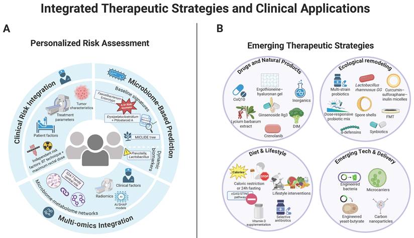
6.2 Clinical strategies for microbiome intervention
Although diverse microbiome-modulating strategies have shown intestinal protective effects in experimental models, their clinical translation remains constrained by several factors. First, most interventional evidence still comes from animal studies, whereas randomized controlled trials in patients receiving radiotherapy remain limited. Second, strains, doses, timing and treatment duration differ widely across studies, and no standardized regimen has yet been established. Finally, for patients with cancer, any radioprotective strategy must demonstrate that it does not simultaneously protect tumor cells or compromise the antitumor efficacy of radiotherapy; this issue has not been systematically addressed in many studies. These considerations should guide the interpretation of the intervention studies summarized below.
6.2.1 Ecological remodeling
Multi-strain probiotics outperform single strains in radioprotection. A consortium comprising Bifidobacterium longum BL21, Lactobacillus paracasei LC86 and L. plantarum Lp90 synergistically mitigates radiation-induced intestinal injury by promoting intestinal stem-cell proliferation and differentiation, strengthening barrier integrity and modulating oxidative stress and inflammation[98]. Orally administered composite probiotics show a clear dose-response in attenuating murine radiation enteropathy; during the post-irradiation critical window they suppress pathogenic taxa such as the Escherichia-Shigella cluster and increase taxa generally associated with SCFA production, such as Lachnospiraceae[17]. Spore-shell biomaterials offer a live-microbe-free alternative resilient to gastric acid and irradiation. Spore shells derived from Bacillus species (for example, Bacillus subtilis and B. coagulans) efficiently alleviate radiation-induced gut injury, improve survival and restore microbial homeostasis in mice[99]. Host defense peptides also contribute: intestinal α-defensins facilitate recovery by reshaping the microbiome and fecal metabolome[100].
Synbiotics leverage prebiotic-probiotic synergy, but most supporting data currently come from preclinical studies. Clinically, in a randomized controlled trial in prostate cancer, a synbiotic containing Lactobacillus reuteri plus dietary fiber significantly alleviated acute radiation proctitis symptoms, especially tenesmus and defecatory urgency during urination, improving quality of life, with peak benefit at weeks 2-3[101]. By contrast, the following findings are derived from mouse models and other preclinical work. Inulin hydrogel combined with multi-strain probiotics significantly improves survival in irradiated mice, outperforming either component alone; as a prebiotic carrier, inulin prolongs probiotic gut residence and cooperatively enhances SCFA production[102]. For prevention of radiation-induced colonic fibrosis, inulin promotes expansion of context-dependent SCFA-producing or fermentative taxa, including Ruminococcaceae, Bacteroides acidifaciens and Clostridium papyrosolvens, reducing collagen deposition and α-SMA expression[103]. A colon-retentive inulin gel similarly protected both the hematopoietic system and the gastrointestinal tract in irradiated mice, in parallel with increased SCFAs output and elevated anti-inflammatory cytokines IL-22 and IL-10, in a microbiota-dependent manner[89]. In mouse models, konjac glucomannan (KGM), another prebiotic, fosters beneficial taxa and SCFAs and confers protection against both total- and abdominal-irradiation injury[104]. Multifunctional oral curcumin-thione-inulin conjugate micelles integrate direct antioxidant activity (radical scavenging) with inulin-mediated dysbiosis correction, yielding multilevel protection[105]. Together, these data suggest that inulin- and KGM-based prebiotics and synbiotics are promising candidates, but their efficacy and safety in patients receiving radiotherapy remain to be defined.
FMT is being explored as a microbiome-based intervention. FMT ameliorates murine RE by restoring microbiota-mediated tryptophan metabolism, increasing production of I3A and related metabolites to dampen oxidative stress and inflammation[106]. Microbiota harvested from “elite survivor” mice that live through high-dose irradiation provide pronounced radioprotection; Lachnospiraceae and Enterococcaceae were identified as key protective families, with propionate and tryptophan metabolites underpinning durable benefits[21]. Sex-matched FMT significantly improves survival, gastrointestinal function and epithelial integrity after irradiation, and combines favorably with bone-marrow transplantation[107]. Targeted, composition-guided consortia are advancing; inocula enriched for Lachnospiraceae protect ISCs and preserve barrier integrity.
6.2.2 Pharmacological and natural-product interventions
Natural products, including plant-derived compounds and fungus-derived bioactive polysaccharides, have attracted interest as potential protectants against radiation-induced intestinal injury, although most supporting evidence remains preclinical. Clinically, epigallocatechin-3-gallate (EGCG) protects via a novel gut microbiota/D-tagatose/AMPK axis, preserving mucosal architecture and promoting regeneration. In pelvic-cancer patients, 400 mg/day EGCG reduced the incidence of grade ≥2 RE from 65% to 24.3%[108]. By contrast, the following data are derived from rat or mouse models. Ginsenoside Rg3 suppresses the TLR4/MyD88/NF-κB inflammatory cascade while rebalancing the gut microbiota. At a high dose (80 mg/kg), Rg3 achieved efficacy comparable to dexamethasone and exhibited a greater capacity to remodel microbial communities[65]. The cruciferous-vegetable metabolite 3,3′-diindolylmethane (DIM) confers marked protection against TAI-induced gut injury by enhancing survival and function of Lgr5+ ISCs and progeny, activating the Nrf2 antioxidant pathway to mitigate oxidative stress, and restoring radiation-induced dysbiosis[109]. Lycium barbarum (goji) extract alleviates radiation injury through multi-pathway mechanisms and increases beneficial taxa such as Turicibacter and Akkermansia[110]. The vanillin-derived small molecule VND3207 precisely tunes the p53/NOXA axis to sustain DNA repair without triggering excessive apoptosis, while correcting dysbiosis; it significantly improved survival after lethal-dose irradiation in mice[111]. Fungus-derived polysaccharides may represent a promising class of natural radioprotective agents, and Tremella fuciformis polysaccharide TFP-23 has been reported to mitigate radiation-induced intestinal injury in mice by reducing colon shortening and crypt loss. This protection was associated with partial restoration of radiation-disrupted gut microbiota, including increased microbial diversity, reduced Proteobacteria expansion and enrichment of beneficial taxa such as Akkermansia, Bacteroides, Faecalibacterium and Oscillospirales, although further validation is still required[112].
Conventional drugs also provide benefit. In patients with chronic RE, a pentoxifylline-vitamin E regimen improved symptoms of chronic RE by transcriptionally downregulating TGF-β1, reducing SOMA scores by 41% and 80% at 6 and 18 months, respectively[67]. In rat models, coenzyme Q10 attenuates inflammation and fibrosis by inhibiting NF-κB/TGF-β/MMP-9 signaling and increases the relative abundance of Bacteroidetes, Akkermansia and SCFA producers[64]. In mouse models, inorganic agents have also been reported to afford additional protection. Selenium nanoparticles (SeNPs) ameliorate radiation colitis by modulating the cGAS-STING pathway, suppressing X-ray-induced excess ROS and DNA damage, and reshaping the inflammatory immune microenvironment[113]. Similarly, in mouse models, dietary nitrate prevents whole-body-irradiation-induced colonic injury by activating the NO3-/NO2-/NO pathway, limiting oxidative stress and maintaining microbiome homeostasis[114].
Epigenetic and cell-death pathway modulators offer complementary strategies, but current mechanistic evidence comes predominantly from animal models. The histone deacetylase inhibitor trichostatin A (TSA) mitigates gastrointestinal toxicity via epigenetic and anti-inflammatory effects and shifts the microbiome by increasing Verrucomicrobia and Lactobacillus[115]. Ferrostatin-1, a specific ferroptosis inhibitor, suppresses both apoptosis and ferroptosis after irradiation, through inhibition of p53-mediated apoptotic signaling and restoration of microbial balance, thereby improving survival[7]. The HSP90 inhibitor NVP-AUY922 downregulates epoxide hydrolase 1 (EPHX1), curbing radiation-induced intestinal inflammation and remodeling gut communities[116]. The PDGF-C signaling pathway is a key regulator of radiation proctopathy; its inhibitor crenolanib attenuates colorectal tissue injury by blocking the PDGF-C/PDGFRs/ETV1/CXCR4 axis[11].
Other notable interventions include an ergothioneine-hyaluronic acid gel, which reduces apoptosis and inflammation while improving microbial balance[117]; in rat models, combined supplementation with β-hydroxy-β-methylbutyrate (HMB), L-glutamine and L-arginine to promote protein synthesis, suppress proteolysis and reinforce epithelial structure and function[63]; and in mouse models, hydrogen-rich water, which exerts antioxidant and anti-inflammatory effects via MyD88 signaling while stabilizing the gut microbiota[118].
6.2.3 Diet and lifestyle interventions
Dietary and lifestyle measures provide non-invasive, readily implementable strategies for mitigating radiation-induced intestinal injury. Exercise is a simple, effective lever: low-intensity walking (1 m per minute, 35 minutes daily) increased the relative abundance of A. muciniphila and attenuated radiation enteropathy without compromising tumor control[119].
Dietary restriction, either 30% caloric restriction or a 24-hour fast, ameliorates acute intestinal injury by suppressing cGAS/STING activation[120]. These strategies exhibit sex-dependent protective effects: 30% caloric restriction in females primarily reduces pro-inflammatory taxa (for example, Helicobacter and Desulfovibrionaceae), whereas in males it enriches SCFA producers (for example, Faecalibaculum and Lactobacillus), thereby improving barrier function[121]. By contrast, dietary methionine supplementation exacerbates ionizing-radiation-induced gastrointestinal syndrome, accelerating the expansion of pathogenic Burkholderiales and thus warrants caution[122]. High-fiber intake increases Bacteroides acidifaciens and can significantly enhance tumor radiosensitivity[123]. Fiber supplementation can both potentiate tumor response and mitigate gut toxicity: psyllium plus resistant starch delivered superior radiosensitization, whereas psyllium plus inulin afforded better protection against acute enterotoxicity[124].
Circadian alignment also shapes radiosensitivity. Disruption of circadian rhythms induces dysbiosis and weakens radiotolerance, whereas mice maintained under a standard 12-hour dark/12-hour light cycle display greater resistance to radiation injury[125]. Vitamin D supplementation protects by preserving barrier integrity and modulating microbiota composition[126]. Selective antibiotic strategies (for example, vancomycin) that deplete butyrate-producing bacteria may, in certain settings, both reduce intestinal toxicity and enhance the anti-tumor efficacy of radiotherapy[127].
6.2.4 Emerging technologies and delivery systems
Engineered microbes are being investigated for both radiosensitization and radioprotection. A tumor-targeting probiotic, Escherichia coli Nissle 1917, markedly increases catalase secretion within the tumor microenvironment (TME), catalyzing tumor-derived H2O2 into O2. By relieving hypoxia and improving oxygen availability, this approach enhances radiation-induced ROS generation and achieves radiosensitization in mice[128]. An IL-22-secreting engineered probiotic promotes direct intestinal epithelial repair and restores bone-marrow hematopoiesis; notably, administration as late as 24 hours post-irradiation significantly improves survival, offering a practical window for delayed intervention[129]. A butyrate-engineered yeast releases butyrate to restore GPR109A expression, protecting the intestine and, via the gut-heart axis, mitigating radiation-induced cardiac injury while activating the cardioprotective genes Nppa and Sgcg, illustrating the multi-organ protective potential of engineered strains[130].
Targeted delivery systems can substantially amplify microbial and metabolic therapies. An oral micro-carrier based on the microalga Spirulina efficiently delivers amifostine (AMF), protecting the small intestine from radiation injury and stabilizing the gut microbiota, particularly by increasing beneficial taxa such as Lactobacillus, Prevotella and Alloprevotella, thereby providing broad radioprotection[131]. A metal-organic framework (MOF) platform constructed from zinc ions and 2-methylimidazole provides pH-responsive, intestinal release of sitagliptin alongside Zn2+, affording multifaceted protection through a microbiota-SCFAs-tight junction axis[132]. Clinically approved carbon nanoparticles (CNSI), administered orally, leverage potent free-radical scavenging and intestinal targeting to preserve crypt stem cells and maintain microbial balance, with notable enrichment of probiotic Lactobacillus and the order Bifidobacteriales[133].
From a radiation-physics perspective, modern ultra-high-dose-rate (FLASH) radiotherapy, by virtue of its unique temporal pulse structure, spares normal tissues. Implemented as a single pulse with a mean dose rate ≥ 280 Gy/s, FLASH significantly attenuates damage to the intestinal microbiota and mucosal tissues[134], providing a complementary physical modality to protect the gut ecosystem (Figure 6B).
6.3 Clinical implementation of integrated therapeutic strategies
Management of RE should be tailored to disease phase. In the acute phase (during radiotherapy to within 3 months after completion), priorities are diarrhea control and preservation of epithelial barrier integrity. In the chronic phase (beyond 3 months), the focus shifts to antifibrotic measures and quality-of-life optimization. Interventions such as nicotinamide mononucleotide (NMN) and inulin modulate the gut microbiota—particularly increasing Bacteroidetes, Akkermansia and short-chain fatty-acid producers—and thereby indirectly mitigate fibrogenesis[68,103]. For severe cases refractory to conservative therapy, anatomically precise resection of diseased bowel remains an effective salvage option; in one series, no recurrence was observed over a median 42-month follow-up[66].
From a translational perspective, clinical confounders remain a major challenge. Although clinical studies have controlled for antibiotic use during fecal sample collection, many patients with locally advanced or metastatic cancer receive cytotoxic chemotherapy and/or undergo surgical resection, both of which can substantially perturb intestinal ecology, mucosal immunity, intestinal anatomy and microbial niches. These factors may interact with radiation-induced epithelial injury and dysbiosis, thereby complicating causal attribution of microbiome alterations to radiotherapy or RE alone. In addition, inter-individual variability related to age, sex, diet, geography, lifestyle, and host genetic background can influence baseline microbiome composition and function and may partly explain the heterogeneity of microbial signatures and clinical RE phenotypes observed across cohorts. Therefore, such heterogeneity should be interpreted in a balanced manner, with explicit consideration of how clinical management and patient-level factors may modulate both the microbiota and the risk of RE. Taken together, these clinical constraints underscore the gap between controlled research settings and everyday oncology practice and must be carefully considered when designing and interpreting microbiome-based interventions.
In parallel with these clinical constraints, much of the available evidence still comes from preclinical studies, particularly mouse models, and should therefore be regarded primarily as mechanistic and conceptual insight rather than definitive clinical proof. Nevertheless, these studies collectively highlight that host factors, including age, sex, genetic background, metabolic state/comorbidities and circadian status, substantially influence the onset and trajectory of RE. Risk-assessment frameworks are coalescing from clinical phenotyping to microbiome readouts and multi-omics integration. Microbiome modulation is evolving from conventional probiotics to precision, engineered live biotherapeutics. Layered, multi-target strategies offer promising avenues for the prevention and treatment of RE. As microbiome science and enabling technologies continue to advance, RE management may become increasingly precise and effective, with meaningful improvements in patient outcomes and quality of life. However, despite substantial mechanistic progress in animal studies, interspecies differences in host-microbiome composition, metabolism and treatment response mean that not all preclinical findings will be reproducible in humans. Accordingly, these strategies should be interpreted with caution and will require rigorous, well-designed clinical trials before they can be translated into routine practice.
7. Conclusions and perspectives
Research on RE is moving toward multidimensional integration, with three priorities emerging. First, multi-omics integration will deepen understanding of host-microbiome crosstalk[95]. In this context, future studies should integrate bacterial, fungal, and viral microbiome data to better define the host-microbiome interaction network in RE. Prospective clinical studies are needed to validate microbiome biomarkers[135]. Second, in translational medicine, individualized risk-assessment models based on radiomics and artificial intelligence are advancing; radioprotective microbes and metabolites identified from radiation survivors provide actionable targets for next-generation protectants[21]. Third, on the clinical front, systems-level regulation along the gut-multi-organ axis, together with rigorous assessment of the long-term efficacy and safety of microbial interventions, warrants in-depth investigation. Clinical practice should adopt systematic, personalized strategies: perform pre-radiotherapy microbiome profiling to identify high-risk patients; implement tumor-specific management and stepwise regimens tailored to the acute and chronic phases; deploy evidence-based microbial interventions; and integrate nutritional support that recognizes the microbiome as a key transducer of host nutrient signaling[136]. Multidisciplinary teamwork is essential to optimize outcomes.
Overall, RE has evolved from a concept of purely local inflammation to a systemic disease involving complex cell-death programs, oxidative stress, immune dysregulation and microbiome imbalance. Its clinical manifestations exhibit clear dependence on tumor type, anatomical site and host factors. The gut microbiome appears to contribute to the initiation, progression and resolution of RE, with effects extending from the intestinal compartment to multi-system involvement[8]. Dynamic equilibria between the microbiome and specific metabolites, including SCFAs, bile acids and tryptophan derivatives, provide critical radioprotective targets. Diagnostics and risk assessment are shifting from single-parameter approaches to personalized, multi-omics models, while therapy is moving from symptomatic care to mechanism-based, multi-target interventions. In the era of precision medicine, management is converging on a “predictive, preventive, personalized and participatory” continuum; integrating microbiome-modulating, individualized strategies will continue to drive clinical innovation, with the goal of delivering precise protection and optimal prognosis for every patient.
Acknowledgements
This work was supported by the Guang Dong Cheung Kong Philanthropy Foundation (E2018096 to YJL), National Natural Science Foundation of China (32500039 to ZHG; 32370057 to YJL; 82302537 to LMC; 82573330 to HMW), Key Technologies R&D Program of Guangdong Province (2024B1111150001 to YJL), Key laboratory start-up project (Sixth Affiliated Hospital of Sun Yat-sen University) (2023WST02 to YJL), Guangdong Basic and Applied Basic Research Foundation (2024A1515012253 to ZHG), Dongguan Science and Technology of Social Development Program (No.20231800936102 to HMW), Doctoral Initial Funding of Guangdong Medical University (4SG23186G to LMC). Supported by National Key Clinical Discipline. All figures were created with BioRender.com under publication licenses.
Author contributions
JRZ conceived and drafted the manuscript. JRZ and ZHG drew the figures. YJL, HW and LMC discussed the concepts of the manuscript. YZ, ZYL, DLT and YXZ revised figure legends. ZHG and HMW provided valuable discussion and revised the manuscript. All authors have read and approved the article.
AI usage statement
The study conception, data organization, manuscript structure, and initial draft were independently completed by the authors. During the preparation and revision of this manuscript, ChatGPT was used only for language polishing and readability improvement, such as grammar correction, wording refinement, and sentence clarity. The authors independently developed the manuscript content, scientific arguments, conclusions, and references. ChatGPT was not used to generate scientific content, analyze literature, produce data, or serve as a source of scientific evidence. All AI-assisted edits were carefully reviewed and approved by the authors, who take full responsibility for the accuracy, integrity, and final content of the manuscript.
Competing Interests
The authors have declared that no competing interest exists.
References
1. Yu Z, Xu C, Song B, Zhang S, Chen C, Li C. et al. Tissue fibrosis induced by radiotherapy: current understanding of the molecular mechanisms, diagnosis and therapeutic advances. J Transl Med. 2023Oct9;21(1):708 doi:10.1186/s12967-023-04554-0 PubMed PMID: 37814303; PubMed Central PMCID: PMC10563272
2. Chenxiang D, Jing Z, Xiaojian Y, Hua X, Xi Y. Interplay between the abdominopelvic radiotherapy and gut microbiota. Front Microbiol. 2025;16:1692179 doi:10.3389/fmicb.2025.1692179 PubMed PMID: 41459231; PubMed Central PMCID: PMC12742314
3. Lu Q, Liang Y, Tian S, Jin J, Zhao Y, Fan H. Radiation-Induced Intestinal Injury: Injury Mechanism and Potential Treatment Strategies. Toxics. 2023Dec10;11(12):1011 doi:10.3390/toxics11121011 PubMed PMID: 38133412; PubMed Central PMCID: PMC10747544
4. Srinivas US, Tan BWQ, Vellayappan BA, Jeyasekharan AD. ROS and the DNA damage response in cancer. Redox Biol. 2019Jul;25:101084 doi:10.1016/j.redox.2018.101084 PubMed PMID: 30612957; PubMed Central PMCID: PMC6859528
5. Wang K, Tepper JE. Radiation therapy-associated toxicity: Etiology, management, and prevention. CA Cancer J Clin. 2021Sep;71(5):437-54 doi:10.3322/caac.21689 PubMed PMID: 34255347
6. Horseman T, Rittase WB, Slaven JE, Bradfield DT, Frank AM, Anderson JA. et al. Ferroptosis, Inflammation, and Microbiome Alterations in the Intestine in the Göttingen Minipig Model of Hematopoietic-Acute Radiation Syndrome. Int J Mol Sci. 2024Apr20;25(8):4535 doi:10.3390/ijms25084535 PubMed PMID: 38674120; PubMed Central PMCID: PMC11050692
7. Wang X, Li W, Dong Y, Zhang Y, Huo Q, Lu L. et al. Ferrostatin-1 mitigates ionizing radiation-induced intestinal injuries by inhibiting apoptosis and ferroptosis: an in vitro and in vivo study. Int J Radiat Biol. 2023;99(10):1607-18 doi:10.1080/09553002.2023.2194399 PubMed PMID: 36947642
8. Dar HH, Epperly MW, Tyurin VA, Amoscato AA, Anthonymuthu TS, Souryavong AB. et al. P. aeruginosa augments irradiation injury via 15-lipoxygenase-catalyzed generation of 15-HpETE-PE and induction of theft-ferroptosis. JCI Insight. 2022Feb22;7(4):e156013 doi:10.1172/jci.insight.156013 PubMed PMID: 35041620; PubMed Central PMCID: PMC8876480
9. Kumar S, Suman S, Angdisen J, Moon BH, Kallakury BVS, Datta K. et al. Effects of High-Linear-Energy-Transfer Heavy Ion Radiation on Intestinal Stem Cells: Implications for Gut Health and Tumorigenesis. Cancers (Basel). 2024Oct4;16(19):3392 doi:10.3390/cancers16193392 PubMed PMID: 39410012; PubMed Central PMCID: PMC11475725
10. Liu Z, Jiang J, He Q, Liu Z, Yang Z, Xu J. et al. β-Arrestin1-mediated decrease in endoplasmic reticulum stress impairs intestinal stem cell proliferation following radiation. FASEB J. 2019Sep;33(9):10165-76 doi:10.1096/fj.201900376RRR PubMed PMID: 31207192
11. Lu W, Xie Y, Huang B, Ma T, Wang H, Deng B. et al. Platelet-derived growth factor C signaling is a potential therapeutic target for radiation proctopathy. Sci Transl Med. 2021Feb24;13(582):eabc2344 doi:10.1126/scitranslmed.abc2344 PubMed PMID: 33627485
12. Yan T, Zhang T, Mu W, Qi Y, Guo S, Hu N. et al. Ionizing radiation induces BH4 deficiency by downregulating GTP-cyclohydrolase 1, a novel target for preventing and treating radiation enteritis. Biochem Pharmacol. 2020Oct;180:114102 doi:10.1016/j.bcp.2020.114102 PubMed PMID: 32562786
13. Xu J, He J, Zhou YL, Weng Z, Li M, Wang ZX. et al. Von Willebrand factor promotes radiation-induced intestinal injury (RIII) development and its cleavage enzyme rhADAMTS13 protects against RIII by reducing inflammation and oxidative stress. Free Radic Biol Med. 2024Jan;210:1-12 doi:10.1016/j.freeradbiomed.2023.11.004 PubMed PMID: 37956910
14. Wang Z, Wang Q, Wang X, Zhu L, Chen J, Zhang B. et al. Gut microbial dysbiosis is associated with development and progression of radiation enteritis during pelvic radiotherapy. J Cell Mol Med. 2019May;23(5):3747-56 doi:10.1111/jcmm.14289 PubMed PMID: 30908851; PubMed Central PMCID: PMC6484301
15. Li Y, Yan H, Zhang Y, Li Q, Yu L, Li Q. et al. Alterations of the Gut Microbiome Composition and Lipid Metabolic Profile in Radiation Enteritis. Front Cell Infect Microbiol. 2020;10:541178 doi:10.3389/fcimb.2020.541178 PubMed PMID: 33194790; PubMed Central PMCID: PMC7609817
16. Carbonero F, Mayta A, Bolea M, Yu JZ, Lindeblad M, Lyubimov A. et al. Specific Members of the Gut Microbiota are Reliable Biomarkers of Irradiation Intensity and Lethality in Large Animal Models of Human Health. Radiat Res. 2019Jan;191(1):107-21 doi:10.1667/RR14975.1 PubMed PMID: 30430918
17. Zhao TS, Xie LW, Cai S, Xu JY, Zhou H, Tang LF. et al. Dysbiosis of Gut Microbiota Is Associated With the Progression of Radiation-Induced Intestinal Injury and Is Alleviated by Oral Compound Probiotics in Mouse Model. Front Cell Infect Microbiol. 2021;11:717636 doi:10.3389/fcimb.2021.717636 PubMed PMID: 34760714; PubMed Central PMCID: PMC8573182
18. Liu L, Chen C, Liu X, Chen B, Ding C, Liang J. Altered Gut Microbiota Associated With Hemorrhage in Chronic Radiation Proctitis. Front Oncol. 2021;11:637265 doi:10.3389/fonc.2021.637265 PubMed PMID: 34722231; PubMed Central PMCID: PMC8554627
19. Jian Y, Zhang D, Liu M, Wang Y, Xu ZX. The Impact of Gut Microbiota on Radiation-Induced Enteritis. Front Cell Infect Microbiol. 2021;11:586392 doi:10.3389/fcimb.2021.586392 PubMed PMID: 34395308; PubMed Central PMCID: PMC8358303
20. He KY, Lei XY, Wu DH, Zhang L, Li JQ, Li QT. et al. Akkermansia muciniphila protects the intestine from irradiation-induced injury by secretion of propionic acid. Gut Microbes. 2023Dec;15(2):2293312 doi:10.1080/19490976.2023.2293312 PubMed PMID: 38087436; PubMed Central PMCID: PMC10730217
21. Guo H, Chou WC, Lai Y, Liang K, Tam JW, Brickey WJ. et al. Multi-omics analyses of radiation survivors identify radioprotective microbes and metabolites. Science. 2020Oct30;370(6516):eaay9097 doi:10.1126/science.aay9097 PubMed PMID: 33122357; PubMed Central PMCID: PMC7898465
22. Grylls A, Seidler K, Neil J. Link between microbiota and hypertension: Focus on LPS/TLR4 pathway in endothelial dysfunction and vascular inflammation, and therapeutic implication of probiotics. Biomed Pharmacother. 2021May;137:111334 doi:10.1016/j.biopha.2021.111334 PubMed PMID: 33556874
23. Kassan M, Kwon Y, Munkhsaikhan U, Sahyoun AM, Ishrat T, Galán M. et al. Protective Role of Short-Chain Fatty Acids against Ang- II-Induced Mitochondrial Dysfunction in Brain Endothelial Cells: A Potential Role of Heme Oxygenase 2. Antioxidants (Basel). 2023Jan10;12(1):160 doi:10.3390/antiox12010160 PubMed PMID: 36671022; PubMed Central PMCID: PMC9854784
24. Tian T, Zhao Y, Yang Y, Wang T, Jin S, Guo J. et al. The protective role of short-chain fatty acids acting as signal molecules in chemotherapy- or radiation-induced intestinal inflammation. Am J Cancer Res. 2020;10(11):3508-31 PubMed PMID: 33294252; PubMed Central PMCID: PMC7716145
25. Zhao X, Cai Y, Hou Y, Wu Y, Wei T, Li L. et al. Commensal Viruses Promote Intestinal Stem Cell Regeneration Following Radiation Damage by Inhibiting Hyperactivation of RIG-I and Notch Signals. Adv Sci (Weinh). 2025Oct;12(37):e05204 doi:10.1002/advs.202505204 PubMed PMID: 40679068; PubMed Central PMCID: PMC12499508
26. Moraitis I, Guiu J, Rubert J. Gut microbiota controlling radiation-induced enteritis and intestinal regeneration. Trends Endocrinol Metab. 2023Aug;34(8):489-501 doi:10.1016/j.tem.2023.05.006 PubMed PMID: 37336645
27. Riehl T, Cohn S, Tessner T, Schloemann S, Stenson WF. Lipopolysaccharide is radioprotective in the mouse intestine through a prostaglandin-mediated mechanism. Gastroenterology. 2000Jun;118(6):1106-16 doi:10.1016/s0016-5085(00)70363-5 PubMed PMID: 10833485
28. Shao L, Li M, Zhang B, Chang P. Bacterial dysbiosis incites Th17 cell revolt in irradiated gut. Biomed Pharmacother. 2020Nov;131:110674 doi:10.1016/j.biopha.2020.110674 PubMed PMID: 32866810
29. Wang Z, Wang Q, Gong L, Liu T, Wang P, Yuan Z. et al. The NF-κB-Regulated miR-221/222/Syndecan-1 Axis and Intestinal Mucosal Barrier Function in Radiation Enteritis. Int J Radiat Oncol Biol Phys. 2022May1;113(1):166-76 doi:10.1016/j.ijrobp.2022.01.006 PubMed PMID: 35033585
30. Zhu X, Li Y, Tian X, Jing Y, Wang Z, Yue L. et al. REGγ Mitigates Radiation-Induced Enteritis by Preserving Mucin Secretion and Sustaining Microbiome Homeostasis. Am J Pathol. 2024Jun;194(6):975-88 doi:10.1016/j.ajpath.2024.02.008 PubMed PMID: 38423356
31. Reis Ferreira M, Andreyev HJN, Mohammed K, Truelove L, Gowan SM, Li J. et al. Microbiota- and Radiotherapy-Induced Gastrointestinal Side-Effects (MARS) Study: A Large Pilot Study of the Microbiome in Acute and Late-Radiation Enteropathy. Clin Cancer Res. 2019Nov1;25(21):6487-500 doi:10.1158/1078-0432.CCR-19-0960 PubMed PMID: 31345839
32. Yan Z, Li Y, Xia T, Wang K, Liao Z, Zhang L. et al. Revitalizing gut health: Liangxue guyuan yishen decoction promotes akkermansia muciniphila -induced intestinal stem cell recovery post-radiation in mice. Phytomedicine. 2024Sep;132:155888 doi:10.1016/j.phymed.2024.155888 PubMed PMID: 39084128
33. Zhu H, Guo H, Sun N, Xiao R, Ji B, Jiang R. et al. Faecalibaculum rodentium Alleviates Ionizing Radiation-Induced Damage in Mice by Improving Intestinal Integrity and Hematopoiesis via Its Metabolite Butyrate. Advanced Science. 2025 Oct 13;e09383. doi:10.1002/advs.202509383
34. Ge Z, Chen C, Chen J, Jiang Z, Chen L, Wei Y. et al. Gut Microbiota-Derived 3-Hydroxybutyrate Blocks GPR43-Mediated IL6 Signaling to Ameliorate Radiation Proctopathy. Adv Sci (Weinh). 2024Jul;11(28):e2306217 doi:10.1002/advs.202306217 PubMed PMID: 38742466; PubMed Central PMCID: PMC11267371
35. Zhao H, Abbas S, Ren J, Huang H, Song Y, Su X. et al. Dextran from human feces-derived Weissella cibaria facilitates intestinal mucosal barrier function by modulating gut bacteria and propionate levels. Carbohydr Polym. 2025Apr15;354:123300 doi:10.1016/j.carbpol.2025.123300 PubMed PMID: 39978893
36. Lapiere A, Geiger M, Robert V, Demarquay C, Auger S, Chadi S. et al. Prophylactic Faecalibacterium prausnitzii treatment prevents the acute breakdown of colonic epithelial barrier in a preclinical model of pelvic radiation disease. Gut Microbes. 2020Nov9;12(1):1-15 doi:10.1080/19490976.2020.1812867 PubMed PMID: 32985332; PubMed Central PMCID: PMC7524396
37. Ciorba MA, Riehl TE, Rao MS, Moon C, Ee X, Nava GM. et al. Lactobacillus probiotic protects intestinal epithelium from radiation injury in a TLR-2/cyclo-oxygenase-2-dependent manner. Gut. 2012Jun;61(6):829-38 doi:10.1136/gutjnl-2011-300367 PubMed PMID: 22027478; PubMed Central PMCID: PMC3345937
38. Zhang LL, Xu JY, Xing Y, Wu P, Jin YW, Wei W. et al. Lactobacillus rhamnosus GG alleviates radiation-induced intestinal injury by modulating intestinal immunity and remodeling gut microbiota. Microbiol Res. 2024Sep;286:127821 doi:10.1016/j.micres.2024.127821 PubMed PMID: 38941923
39. Haegebarth A, Clevers H. Wnt signaling, lgr5, and stem cells in the intestine and skin. Am J Pathol. 2009Mar;174(3):715-21 doi:10.2353/ajpath.2009.080758 PubMed PMID: 19197002; PubMed Central PMCID: PMC2665733
40. Xie J, Li LF, Dai TY, Qi X, Wang Y, Zheng TZ. et al. Short-Chain Fatty Acids Produced by Ruminococcaceae Mediate α-Linolenic Acid Promote Intestinal Stem Cells Proliferation. Mol Nutr Food Res. 2022Jan;66(1):e2100408 doi:10.1002/mnfr.202100408 PubMed PMID: 34708542
41. Xie LW, Cai S, Lu HY, Tang FL, Zhu RQ, Tian Y. et al. Microbiota-derived I3A protects the intestine against radiation injury by activating AhR/IL-10/Wnt signaling and enhancing the abundance of probiotics. Gut Microbes. 2024;16(1):2347722 doi:10.1080/19490976.2024.2347722 PubMed PMID: 38706205; PubMed Central PMCID: PMC11086037
42. Metidji A, Omenetti S, Crotta S, Li Y, Nye E, Ross E. et al. The Environmental Sensor AHR Protects from Inflammatory Damage by Maintaining Intestinal Stem Cell Homeostasis and Barrier Integrity. Immunity. 2018Aug21;49(2):353-362.e5 doi:10.1016/j.immuni.2018.07.010 PubMed PMID: 30119997; PubMed Central PMCID: PMC6104739
43. Yang M, Liu Q, Dai M, Peng R, Li X, Zuo W. et al. FOXQ1-mediated SIRT1 upregulation enhances stemness and radio-resistance of colorectal cancer cells and restores intestinal microbiota function by promoting β-catenin nuclear translocation. J Exp Clin Cancer Res. 2022Feb19;41(1):70 doi:10.1186/s13046-021-02239-4 PubMed PMID: 35183223; PubMed Central PMCID: PMC8857837
44. Nie H, Pan J, An F, Zheng C, Zhang Q, Zhan Q. Comprehensive Analysis of Serum Metabolites Profiles in Acute Radiation Enteritis Rats by Untargeted Metabolomics. Tohoku J Exp Med. 2021Nov;255(3):257-65 doi:10.1620/tjem.255.257 PubMed PMID: 34853247
45. Guo L, Da F, Gao Q, Miao X, Guo J, Zhang W. et al. Irradiation-Induced Intestinal Injury is Associated With Disorders of Bile Acids Metabolism. Int J Radiat Oncol Biol Phys. 2023Feb1;115(2):490-500 doi:10.1016/j.ijrobp.2022.08.007 PubMed PMID: 35948117
46. Lee YS, Kim TY, Kim Y, Lee SH, Kim S, Kang SW. et al. Microbiota-Derived Lactate Accelerates Intestinal Stem-Cell-Mediated Epithelial Development. Cell Host Microbe. 2018Dec12;24(6):833-846.e6 doi:10.1016/j.chom.2018.11.002 PubMed PMID: 30543778
47. Xiao HW, Cui M, Li Y, Dong JL, Zhang SQ, Zhu CC. et al. Gut microbiota-derived indole 3-propionic acid protects against radiation toxicity via retaining acyl-CoA-binding protein. Microbiome. 2020May20;8(1):69 doi:10.1186/s40168-020-00845-6 PubMed PMID: 32434586; PubMed Central PMCID: PMC7241002
48. Zhang Y, Dong Y, Lu P, Wang X, Li W, Dong H. et al. Gut metabolite Urolithin A mitigates ionizing radiation-induced intestinal damage. J Cell Mol Med. 2021Nov;25(21):10306-12 doi:10.1111/jcmm.16951 PubMed PMID: 34595829; PubMed Central PMCID: PMC8572803
49. Wlodarska M, Luo C, Kolde R, d'Hennezel E, Annand JW, Heim CE. et al. Indoleacrylic Acid Produced by Commensal Peptostreptococcus Species Suppresses Inflammation. Cell Host Microbe. 2017Jul12;22(1):25-37.e6 doi:10.1016/j.chom.2017.06.007 PubMed PMID: 28704649; PubMed Central PMCID: PMC5672633
50. Ma C, Xu X, Qin S, Zhou J. Screening of biomarkers in acute radiation enteritis based on microbiome and clustering methods. BMC Microbiol. 2024Nov8;24(1):463 doi:10.1186/s12866-024-03620-x PubMed PMID: 39516773; PubMed Central PMCID: PMC11545530
51. Riehl TE, Alvarado D, Ee X, Zuckerman A, Foster L, Kapoor V. et al. Lactobacillus rhamnosus GG protects the intestinal epithelium from radiation injury through release of lipoteichoic acid, macrophage activation and the migration of mesenchymal stem cells. Gut. 2019Jun;68(6):1003-13 doi:10.1136/gutjnl-2018-316226 PubMed PMID: 29934438; PubMed Central PMCID: PMC7202371
52. Crawford PA, Gordon JI. Microbial regulation of intestinal radiosensitivity. Proc Natl Acad Sci U S A. 2005Sep13;102(37):13254-9 doi:10.1073/pnas.0504830102 PubMed PMID: 16129828; PubMed Central PMCID: PMC1193536
53. Benson AB, Venook AP, Al-Hawary MM, Azad N, Chen YJ, Ciombor KK. et al. Rectal Cancer, Version 2.2022, NCCN Clinical Practice Guidelines in Oncology. J Natl Compr Canc Netw. 2022Oct;20(10):1139-67 doi:10.6004/jnccn.2022.0051 PubMed PMID: 36240850
54. Sánchez-Alcoholado L, Laborda-Illanes A, Otero A, Ordóñez R, González-González A, Plaza-Andrades I. et al. Relationships of Gut Microbiota Composition, Short-Chain Fatty Acids and Polyamines with the Pathological Response to Neoadjuvant Radiochemotherapy in Colorectal Cancer Patients. Int J Mol Sci. 2021Sep2;22(17):9549 doi:10.3390/ijms22179549 PubMed PMID: 34502456; PubMed Central PMCID: PMC8430739
55. Benej M, Hoyd R, Kreamer M, Wheeler CE, Grencewicz DJ, Choueiry F. et al. The Tumor Microbiome Reacts to Hypoxia and Can Influence Response to Radiation Treatment in Colorectal Cancer. Cancer Res Commun. 2024Jul1;4(7):1690-701 doi:10.1158/2767-9764.CRC-23-0367 PubMed PMID: 38904265; PubMed Central PMCID: PMC11234499
56. Dong J, Li Y, Xiao H, Zhang S, Wang B, Wang H. et al. Oral microbiota affects the efficacy and prognosis of radiotherapy for colorectal cancer in mouse models. Cell Rep. 2021Oct26;37(4):109886 doi:10.1016/j.celrep.2021.109886 PubMed PMID: 34706245
57. Zeng Z, Zhu J, Wang Z, Wang G, Yan J, Zhang F. Pelvic target volume inter-fractional motion during radiotherapy for cervical cancer with daily iterative cone beam computed tomography. Radiat Oncol. 2024Apr15;19(1):48 doi:10.1186/s13014-024-02438-1 PubMed PMID: 38622628; PubMed Central PMCID: PMC11017626
58. Nam YD, Kim HJ, Seo JG, Kang SW, Bae JW. Impact of pelvic radiotherapy on gut microbiota of gynecological cancer patients revealed by massive pyrosequencing. PLoS One. 2013;8(12):e82659 doi:10.1371/journal.pone.0082659 PubMed PMID: 24367534; PubMed Central PMCID: PMC3867375
59. Ma CY, Zhao J, Zhou JY. Microbiome profiling and Co-metabolism pathway analysis in cervical cancer patients with acute radiation enteritis. Heliyon. 2024Apr30;10(8):e29598 doi:10.1016/j.heliyon.2024.e29598 PubMed PMID: 38655340; PubMed Central PMCID: PMC11036041
60. Ma CY, Zhao J, Qian KY, Xu Z, Xu XT, Zhou JY. Analysis of nutritional risk, skeletal muscle depletion, and lipid metabolism phenotype in acute radiation enteritis. World J Gastrointest Surg. 2023Dec27;15(12):2831-43 doi:10.4240/wjgs.v15.i12.2831 PubMed PMID: 38222011; PubMed Central PMCID: PMC10784828
61. Anderson C, Peery AF, Bae-Jump VL, Brewster WR, Lund JL, Olshan AF. et al. Gastrointestinal outcomes among older women with endometrial cancer. Gynecol Oncol. 2023Aug;175:114-20 doi:10.1016/j.ygyno.2023.06.009 PubMed PMID: 37354788
62. Mitra A, Grossman Biegert GW, Delgado AY, Karpinets TV, Solley TN, Mezzari MP. et al. Microbial Diversity and Composition Is Associated with Patient-Reported Toxicity during Chemoradiation Therapy for Cervical Cancer. Int J Radiat Oncol Biol Phys. 2020May1;107(1):163-71 doi:10.1016/j.ijrobp.2019.12.040 PubMed PMID: 31987960; PubMed Central PMCID: PMC7932475
63. Yavas C, Yavas G, Celik E, Buyukyoruk A, Buyukyoruk C, Yuce D. et al. Beta-Hydroxy-Beta-Methyl-Butyrate, L-glutamine, and L-arginine Supplementation Improves Radiation-Induce Acute Intestinal Toxicity. J Diet Suppl. 2019;16(5):576-91 doi:10.1080/19390211.2018.1472709 PubMed PMID: 29969326
64. Mohamed HA, Said RS. Coenzyme Q10 attenuates inflammation and fibrosis implicated in radiation enteropathy through suppression of NF-kB/TGF-β/MMP-9 pathways. Int Immunopharmacol. 2021Mar;92:107347 doi:10.1016/j.intimp.2020.107347 PubMed PMID: 33418245
65. Duan X, Cai H, Hu T, Lin L, Zeng L, Wang H. et al. Ginsenoside Rg3 treats acute radiation proctitis through the TLR4/MyD88/NF-κB pathway and regulation of intestinal flora. Front Cell Infect Microbiol. 2022;12:1028576 doi:10.3389/fcimb.2022.1028576 PubMed PMID: 36683687; PubMed Central PMCID: PMC9853003
66. Perrakis N, Athanassiou E, Vamvakopoulou D, Kyriazi M, Kappos H, Vamvakopoulos NC. et al. Practical approaches to effective management of intestinal radiation injury: benefit of resectional surgery. World J Gastroenterol. 2011Sep21;17(35):4013-6 doi:10.3748/wjg.v17.i35.4013 PubMed PMID: 22046090; PubMed Central PMCID: PMC3199560
67. Hamama S, Gilbert-Sirieix M, Vozenin MC, Delanian S. Radiation-induced enteropathy: molecular basis of pentoxifylline-vitamin E anti-fibrotic effect involved TGF-β1 cascade inhibition. Radiother Oncol. 2012Dec;105(3):305-12 doi:10.1016/j.radonc.2012.08.023 PubMed PMID: 23021793
68. Zhao X, Ji K, Zhang M, Huang H, Wang F, Liu Y. et al. NMN alleviates radiation-induced intestinal fibrosis by modulating gut microbiota. Int J Radiat Biol. 2023;99(5):823-34 doi:10.1080/09553002.2023.2145029 PubMed PMID: 36343364
69. Wang J, Pathak R, Garg S, Hauer-Jensen M. Fibrinogen deficiency suppresses the development of early and delayed radiation enteropathy. World J Gastroenterol. 2017Jul14;23(26):4701-11 doi:10.3748/wjg.v23.i26.4701 PubMed PMID: 28765691; PubMed Central PMCID: PMC5514635
70. Donaldson SS, Jundt S, Ricour C, Sarrazin D, Lemerle J, Schweisguth O. Radiation enteritis in children. A retrospective review, clinicopathologic correlation, and dietary management. Cancer. 1975Apr;35(4):1167-78 doi:10.1002/1097-0142(197504)35:4<1167::aid-cncr2820350423>3.0.co;2-y PubMed PMID: 163677
71. Carabotti M, Scirocco A, Maselli MA, Severi C. The gut-brain axis: interactions between enteric microbiota, central and enteric nervous systems. Ann Gastroenterol. 2015;28(2):203-9 PubMed PMID: 25830558; PubMed Central PMCID: PMC4367209
72. Cui M, Xiao H, Li Y, Dong J, Luo D, Li H. et al. Total abdominal irradiation exposure impairs cognitive function involving miR-34a-5p/BDNF axis. Biochim Biophys Acta Mol Basis Dis. 2017Sep;1863(9):2333-41 doi:10.1016/j.bbadis.2017.06.021 PubMed PMID: 28668331; PubMed Central PMCID: PMC5578400
73. Venkidesh BS, Narasimhamurthy RK, Jnana A, Reghunathan D, Sharan K, Chandraguthi SG. et al. Pelvic irradiation induces behavioural and neuronal damage through gut dysbiosis in a rat model. Chem Biol Interact. 2023Dec1;386:110775 doi:10.1016/j.cbi.2023.110775 PubMed PMID: 37866488
74. Wang X, Guo L, Qin T, Lai P, Jing Y, Zhang Z. et al. Effects of X-ray cranial irradiation on metabolomics and intestinal flora in mice. Ecotoxicol Environ Saf. 2024Jan15;270:115898 doi:10.1016/j.ecoenv.2023.115898 PubMed PMID: 38171101
75. Song C, Duan F, Ju T, Qin Y, Zeng D, Shan S. et al. Eleutheroside E supplementation prevents radiation-induced cognitive impairment and activates PKA signaling via gut microbiota. Commun Biol. 2022Jul8;5(1):680 doi:10.1038/s42003-022-03602-7 PubMed PMID: 35804021; PubMed Central PMCID: PMC9270490
76. Zhang Y, Hu J, Song X, Dai J, Tang Z, Huang G. et al. The effects of Lactobacillus reuteri microcapsules on radiation-induced brain injury by regulating the gut microenvironment. Food Funct. 2023Nov13;14(22):10041-51 doi:10.1039/d3fo03008c PubMed PMID: 37843434
77. Zhou A, Lei Y, Tang L, Hu S, Yang M, Wu L. et al. Gut Microbiota: the Emerging Link to Lung Homeostasis and Disease. J Bacteriol. 2021Jan25;203(4):e00454-20 doi:10.1128/JB.00454-20 PubMed PMID: 33077630; PubMed Central PMCID: PMC7847545
78. Kondapalli N, Katari V, Dalal KK, Paruchuri S, Thodeti CK. Microbiota in Gut-Heart Axis: Metabolites and Mechanisms in Cardiovascular Disease. Compr Physiol. 2025Jun;15(3):e70024 doi:10.1002/cph4.70024 PubMed PMID: 40542540; PubMed Central PMCID: PMC12181760
79. Chen ZY, Xiao HW, Dong JL, Li Y, Wang B, Fan SJ. et al. Gut Microbiota-Derived PGF2α Fights against Radiation-Induced Lung Toxicity through the MAPK/NF-κB Pathway. Antioxidants (Basel). 2021Dec28;11(1):65 doi:10.3390/antiox11010065 PubMed PMID: 35052569; PubMed Central PMCID: PMC8773112
80. Li W, Lu L, Liu B, Qin S. Effects of phycocyanin on pulmonary and gut microbiota in a radiation-induced pulmonary fibrosis model. Biomedicine & pharmacotherapy = Biomedecine & pharmacotherapie. 2020 Dec;132. doi:10.1016/j. biopha. 2020 110826 PubMed PMID: 33068929
81. Tang J, Xu L, Zeng Y, Gong F. Effect of gut microbiota on LPS-induced acute lung injury by regulating the TLR4/NF-kB signaling pathway. Int Immunopharmacol. 2021Feb;91:107272 doi:10.1016/j.intimp.2020.107272 PubMed PMID: 33360370
82. Chen Z, Wang B, Dong J, Li Y, Zhang S, Zeng X. et al. Gut Microbiota-Derived l-Histidine/Imidazole Propionate Axis Fights against the Radiation-Induced Cardiopulmonary Injury. Int J Mol Sci. 2021Oct23;22(21):11436 doi:10.3390/ijms222111436 PubMed PMID: 34768867; PubMed Central PMCID: PMC8584084
83. Jimenez-Sanchez M, Celiberto LS, Yang H, Sham HP, Vallance BA. The gut-skin axis: a bi-directional, microbiota-driven relationship with therapeutic potential. Gut Microbes. 2025Dec;17(1):2473524 doi:10.1080/19490976.2025.2473524 PubMed PMID: 40050613; PubMed Central PMCID: PMC11901370
84. Ramadan M, Hetta HF, Saleh MM, Ali ME, Ahmed AA, Salah M. Alterations in skin microbiome mediated by radiotherapy and their potential roles in the prognosis of radiotherapy-induced dermatitis: a pilot study. Sci Rep. 2021Mar4;11(1):5179 doi:10.1038/s41598-021-84529-7 PubMed PMID: 33664352; PubMed Central PMCID: PMC7933139
85. Trompette A, Pernot J, Perdijk O, Alqahtani RAA, Domingo JS, Camacho-Muñoz D. et al. Gut-derived short-chain fatty acids modulate skin barrier integrity by promoting keratinocyte metabolism and differentiation. Mucosal Immunol. 2022May;15(5):908-26 doi:10.1038/s41385-022-00524-9 PubMed PMID: 35672452; PubMed Central PMCID: PMC9385498
86. Khosravi A, Yáñez A, Price JG, Chow A, Merad M, Goodridge HS. et al. Gut microbiota promote hematopoiesis to control bacterial infection. Cell Host Microbe. 2014Mar12;15(3):374-81 doi:10.1016/j.chom.2014.02.006 PubMed PMID: 24629343; PubMed Central PMCID: PMC4144825
87. Guan D, Yang Y, Pang M, Liu X, Li Y, Huang P. et al. Indole-3-carboxaldehyde ameliorates ionizing radiation-induced hematopoietic injury by enhancing hematopoietic stem and progenitor cell quiescence. Mol Cell Biochem. 2024Feb;479(2):313-23 doi:10.1007/s11010-023-04732-0 PubMed PMID: 37067732
88. Jiao Y, Ren J, Xie S, Yuan N, Shen J, Yin H. et al. Raffinose-metabolizing bacteria impair radiation-associated hematopoietic recovery via the bile acid/FXR/NF-κB signaling pathway. Gut Microbes. 2025Dec;17(1):2488105 doi:10.1080/19490976.2025.2488105 PubMed PMID: 40192235; PubMed Central PMCID: PMC11980471
89. Liu D, Wei M, Fang Y, Yuan T, Sun Y, Xie H. et al. Oral colon-retentive inulin gels protect against radiation-induced hematopoietic and gastrointestinal injury by improving gut homeostasis. Int J Biol Macromol. 2025Mar;292:139199 doi:10.1016/j.ijbiomac.2024.139199 PubMed PMID: 39730057
90. Lee YS, Kim TY, Kim Y, Kim S, Lee SH, Seo SU. et al. Microbiota-derived lactate promotes hematopoiesis and erythropoiesis by inducing stem cell factor production from leptin receptor+ niche cells. Exp Mol Med. 2021Sep;53(9):1319-31 doi:10.1038/s12276-021-00667-y PubMed PMID: 34497346; PubMed Central PMCID: PMC8492757
91. Lucas S, Omata Y, Hofmann J, Böttcher M, Iljazovic A, Sarter K. et al. Short-chain fatty acids regulate systemic bone mass and protect from pathological bone loss. Nat Commun. 2018Jan4;9(1):55 doi:10.1038/s41467-017-02490-4 PubMed PMID: 29302038; PubMed Central PMCID: PMC5754356
92. Lin X, Yu Z, Liu Y, Li C, Hu H, Hu J. et al. Gut-X axis. iMeta. 2025Feb;4(1):e270 doi:10.1002/imt2.270
93. Iacovacci J, Serafini MS, Avuzzi B, Badenchini F, Cicchetti A, Devecchi A. et al. Intestinal microbiota composition is predictive of radiotherapy-induced acute gastrointestinal toxicity in prostate cancer patients. EBioMedicine. 2024Aug;106:105246 doi:10.1016/j.ebiom.2024.105246 PubMed PMID: 39029427; PubMed Central PMCID: PMC11314862
94. Cai S, Yang Y, Kong Y, Guo Q, Xu Y, Xing P. et al. Gut Bacteria Erysipelatoclostridium and Its Related Metabolite Ptilosteroid A Could Predict Radiation-Induced Intestinal Injury. Front Public Health. 2022;10:862598 doi:10.3389/fpubh.2022.862598 PubMed PMID: 35419331; PubMed Central PMCID: PMC8995795
95. Goudarzi M, Mak TD, Jacobs JP, Moon BH, Strawn SJ, Braun J. et al. An Integrated Multi-Omic Approach to Assess Radiation Injury on the Host-Microbiome Axis. Radiat Res. 2016Sep;186(3):219-34 doi:10.1667/RR14306.1 PubMed PMID: 27512828; PubMed Central PMCID: PMC5304359
96. Wei C, Xiang X, Zhou X, Ren S, Zhou Q, Dong W. et al. Development and validation of an interpretable radiomic nomogram for severe radiation proctitis prediction in postoperative cervical cancer patients. Front Microbiol. 2022;13:1090770 doi:10.3389/fmicb.2022.1090770 PubMed PMID: 36713206; PubMed Central PMCID: PMC9877536
97. Cetin E, Ozturk AS, Orhun H, Ulger S. Role of triamcinolone in radiation enteritis management. World J Gastroenterol. 2014Apr21;20(15):4341-4 doi:10.3748/wjg.v20.i15.4341 PubMed PMID: 24764671; PubMed Central PMCID: PMC3989969
98. Xie LW, Lu HY, Tang LF, Tang FL, Zhu RQ, Wang DF. et al. Probiotic Consortia Protect the Intestine Against Radiation Injury by Improving Intestinal Epithelial Homeostasis. Int J Radiat Oncol Biol Phys. 2024Sep1;120(1):189-204 doi:10.1016/j.ijrobp.2024.03.003 PubMed PMID: 38485099
99. Zheng C, Niu M, Kong Y, Liu X, Li J, Gong X. et al. Oral administration of probiotic spore ghosts for efficient attenuation of radiation-induced intestinal injury. J Nanobiotechnology. 2024May31;22(1):303 doi:10.1186/s12951-024-02572-8 PubMed PMID: 38822376; PubMed Central PMCID: PMC11140926
100. Wu J, Ran X, Wang T, Xiong K, Long S, Hao Y. et al. Enteric α-Defensin Contributes to Recovery of Radiation-Induced Intestinal Injury by Modulating Gut Microbiota and Fecal Metabolites. Radiat Res. 2024Feb1;201(2):160-73 doi:10.1667/RADE-23-00071.1 PubMed PMID: 38124379
101. Nascimento M, Aguilar-Nascimento JE, Caporossi C, Castro-Barcellos HM, Motta RT. Efficacy of synbiotics to reduce acute radiation proctitis symptoms and improve quality of life: a randomized, double-blind, placebo-controlled pilot trial. Int J Radiat Oncol Biol Phys. 2014Oct1;90(2):289-95 doi:10.1016/j.ijrobp.2014.05.049 PubMed PMID: 25304789
102. Du S, Sun R, Wang M, Fang Y, Wu Y, Yuan B. et al. Synergistic effect of inulin hydrogels on multi-strain probiotics for prevention of ionizing radiation-induced injury. Int J Biol Macromol. 2025Jan;287:138497 doi:10.1016/j.ijbiomac.2024.138497 PubMed PMID: 39647719
103. Ji K, Zhang M, Du L, Wang J, Liu Y, Xu C. et al. Exploring the Role of Inulin in Targeting the Gut Microbiota: An Innovative Strategy for Alleviating Colonic Fibrosis Induced By Irradiation. J Agric Food Chem. 2024Mar20;72(11):5710-24 doi:10.1021/acs.jafc.3c03432 PubMed PMID: 38457473; PubMed Central PMCID: PMC10958509
104. Liu D, Zhuang B, Wei M, Yuan T, Li J, Deng P. et al. Oral konjac glucomannan for prevention of ionizing radiation-induced injury by regulating gut microbiota and increasing short chain fatty acids. Int J Biol Macromol. 2023Jun15;240:124402 doi:10.1016/j.ijbiomac.2023.124402 PubMed PMID: 37044326
105. Shen J, Jiao W, Yuan B, Xie H, Chen Z, Wei M. et al. Oral Curcumin-Thioketal-Inulin Conjugate Micelles against Radiation-Induced Enteritis. Antioxidants (Basel). 2024Mar29;13(4):417 doi:10.3390/antiox13040417 PubMed PMID: 38671865; PubMed Central PMCID: PMC11047665
106. Tu Y, Luo L, Zhou Q, Ni J, Tang Q. Fecal Microbiota Transplantation Repairs Radiation Enteritis Through Modulating the Gut Microbiota-Mediated Tryptophan Metabolism. Radiat Res. 2024Jun1;201(6):572-85 doi:10.1667/RADE-23-00189.1 PubMed PMID: 38555945
107. Cui M, Xiao H, Li Y, Zhou L, Zhao S, Luo D. et al. Faecal microbiota transplantation protects against radiation-induced toxicity. EMBO Mol Med. 2017Apr;9(4):448-61 doi:10.15252/emmm.201606932 PubMed PMID: 28242755; PubMed Central PMCID: PMC5376756
108. Lu H, Xie L, Guo L, Gu X, Zhu R, Yang Y. et al. EGCG protects intestines of mice and pelvic cancer patients against radiation injury via the gut microbiota/D-tagatose/AMPK axis. Radiother Oncol. 2025Jan;202:110608 doi:10.1016/j.radonc.2024.110608 PubMed PMID: 39486483
109. Lu L, Jiang M, Zhu C, He J, Fan S. Amelioration of whole abdominal irradiation-induced intestinal injury in mice with 3,3'-Diindolylmethane (DIM). Free Radic Biol Med. 2019Jan;130:244-55 doi:10.1016/j.freeradbiomed.2018.10.410 PubMed PMID: 30352304
110. Zheng Y, Pang X, Zhu X, Meng Z, Chen X, Zhang J. et al. Lycium barbarum mitigates radiation injury via regulation of the immune function, gut microbiota, and related metabolites. Biomed Pharmacother. 2021Jul;139:111654 doi:10.1016/j.biopha.2021.111654 PubMed PMID: 33957563
111. Li M, Gu MM, Lang Y, Shi J, Chen BPC, Guan H. et al. The vanillin derivative VND3207 protects intestine against radiation injury by modulating p53/NOXA signaling pathway and restoring the balance of gut microbiota. Free Radic Biol Med. 2019Dec;145:223-36 doi:10.1016/j.freeradbiomed.2019.09.035 PubMed PMID: 31580946
112. Ding J, Gou W, Ma M, Cui Y, Lyu B, Xu J. et al. Investigation of the fingerprint-activity relationship of Tremella fuciformis polysaccharides and its mitigating effect on radiation-induced intestinal injury. Int J Biol Macromol. 2025Apr;304(Pt 1):140849 doi:10.1016/j.ijbiomac.2025.140849 PubMed PMID: 39938826
113. Xue Q, Lai H, Zhang H, Li G, Pi F, Wu Q. et al. Selenium Attenuates Radiation Colitis by Regulating cGAS-STING Signaling. Adv Sci (Weinh). 2024Nov;11(44):e2403918 doi:10.1002/advs.202403918 PubMed PMID: 39348242; PubMed Central PMCID: PMC11600249
114. Wang W, Hu L, Chang S, Ma L, Li X, Yang Z. et al. Total body irradiation-induced colon damage is prevented by nitrate-mediated suppression of oxidative stress and homeostasis of the gut microbiome. Nitric Oxide. 2020Sep1;102:1-11 doi:10.1016/j.niox.2020.05.002 PubMed PMID: 32470598
115. Dahiya A, Agrawala PK, Dutta A. Mitigative and anti-inflammatory effects of Trichostatin A against radiation-induced gastrointestinal toxicity and gut microbiota alteration in mice. Int J Radiat Biol. 2023;99(12):1865-78 doi:10.1080/09553002.2023.2242929 PubMed PMID: 37531370
116. Kong W, Ding X, Wang Z, Lu L, Fan S. NVP-AUY922 relieves radiation-induced intestinal injury via regulating EPHX1. Life Sci. 2025Feb15;363:123382 doi:10.1016/j.lfs.2025.123382 PubMed PMID: 39798648
117. Liu Y, Wang C, Liu R, Zhao M, Ding X, Zhang T. et al. Adhesive Ergothioneine Hyaluronate Gel Protects against Radiation Gastroenteritis by Alleviating Apoptosis, Inflammation, and Gut Microbiota Dysbiosis. ACS Appl Mater Interfaces. 2023Apr26;15(16):19833-46 doi:10.1021/acsami.2c23142 PubMed PMID: 37052616
118. Xiao HW, Li Y, Luo D, Dong JL, Zhou LX, Zhao SY. et al. Hydrogen-water ameliorates radiation-induced gastrointestinal toxicity via MyD88's effects on the gut microbiota. Exp Mol Med. 2018Jan26;50(1):e433 doi:10.1038/emm.2017.246 PubMed PMID: 29371696; PubMed Central PMCID: PMC5799803
119. Wang B, Jin YX, Dong JL, Xiao HW, Zhang SQ, Li Y. et al. Low-Intensity Exercise Modulates Gut Microbiota to Fight Against Radiation-Induced Gut Toxicity in Mouse Models. Front Cell Dev Biol. 2021;9:706755 doi:10.3389/fcell.2021.706755 PubMed PMID: 34746120; PubMed Central PMCID: PMC8566984
120. Zhang LL, Xu JY, Wei W, Hu ZQ, Zhou Y, Zheng JY. et al. Dietary restriction and fasting alleviate radiation-induced intestinal injury by inhibiting cGAS/STING activation. J Nutr Biochem. 2024Nov;133:109707 doi:10.1016/j.jnutbio.2024.109707 PubMed PMID: 39053858
121. Li Y, Dong J, Xiao H, Wang B, Chen Z, Zhang S. et al. Caloric restriction alleviates radiation injuries in a sex-dependent fashion. FASEB J. 2021Aug;35(8):e21787 doi:10.1096/fj.202100351RR PubMed PMID: 34320242
122. Miousse IR, Ewing LE, Skinner CM, Pathak R, Garg S, Kutanzi KR. et al. Methionine dietary supplementation potentiates ionizing radiation-induced gastrointestinal syndrome. Am J Physiol Gastrointest Liver Physiol. 2020Mar1;318(3):G439-50 doi:10.1152/ajpgi.00351.2019 PubMed PMID: 31961718; PubMed Central PMCID: PMC7099489
123. Then CK, Paillas S, Wang X, Hampson A, Kiltie AE. Association of Bacteroides acidifaciens relative abundance with high-fibre diet-associated radiosensitisation. BMC Biol. 2020Aug19;18(1):102 doi:10.1186/s12915-020-00836-x PubMed PMID: 32811478; PubMed Central PMCID: PMC7437060
124. Then CK, Paillas S, Moomin A, Misheva MD, Moir RA, Hay SM. et al. Dietary fibre supplementation enhances radiotherapy tumour control and alleviates intestinal radiation toxicity. Microbiome. 2024May14;12(1):89 doi:10.1186/s40168-024-01804-1 PubMed PMID: 38745230; PubMed Central PMCID: PMC11092108
125. Cui M, Xiao H, Luo D, Zhang X, Zhao S, Zheng Q. et al. Circadian Rhythm Shapes the Gut Microbiota Affecting Host Radiosensitivity. Int J Mol Sci. 2016Oct26;17(11):1786 doi:10.3390/ijms17111786 PubMed PMID: 27792172; PubMed Central PMCID: PMC5133787
126. Huang R, Xiang J, Zhou P. Vitamin D, gut microbiota, and radiation-related resistance: a love-hate triangle. J Exp Clin Cancer Res. 2019Dec16;38(1):493 doi:10.1186/s13046-019-1499-y PubMed PMID: 31843023; PubMed Central PMCID: PMC6915920
127. Uribe-Herranz M, Rafail S, Beghi S, Gil-de-Gómez L, Verginadis I, Bittinger K. et al. Gut microbiota modulate dendritic cell antigen presentation and radiotherapy-induced antitumor immune response. J Clin Invest. 2020Jan2;130(1):466-79 doi:10.1172/JCI124332 PubMed PMID: 31815742; PubMed Central PMCID: PMC6934221
128. Huang C, Wang FB, Liu L, Jiang W, Liu W, Ma W. et al. Hypoxic Tumor Radiosensitization Using Engineered Probiotics. Adv Healthc Mater. 2021May;10(10):e2002207 doi:10.1002/adhm.202002207 PubMed PMID: 33645010
129. Epperly M, Yu J, Van Pijkeren JP, Methe B, Li K, Fisher R. et al. Specific Taxa of the Intestinal Microbiome Mitigate the Radiation-Induced Gastrointestinal Syndrome. International Journal of Radiation Oncology*Biology*Physics. 2020Nov;108(3):S92 doi:10.1016/j.ijrobp.2020.07.2257
130. Wu J, Ji K, Kang G, Zhang M, Wang J, Wang L. et al. Butyrate-engineered yeast activates Nppa and Sgcg genes and reduces radiation-induced heart damage via the gut-heart axis. Pharmacol Res. 2025Mar;213:107642 doi:10.1016/j.phrs.2025.107642 PubMed PMID: 39909125
131. Zhang D, Zhong D, Ouyang J, He J, Qi Y, Chen W. et al. Microalgae-based oral microcarriers for gut microbiota homeostasis and intestinal protection in cancer radiotherapy. Nat Commun. 2022Mar17;13(1):1413 doi:10.1038/s41467-022-28744-4 PubMed PMID: 35301299; PubMed Central PMCID: PMC8931093
132. He D, Li Z, Wang M, Kong D, Guo W, Xia X. et al. Metal-organic-framework-based sitagliptin-release platform for multieffective radiation-induced intestinal injury targeting therapy and intestinal flora protective capabilities. J Nanobiotechnology. 2024Oct16;22(1):631 doi:10.1186/s12951-024-02854-1 PubMed PMID: 39415273; PubMed Central PMCID: PMC11484307
133. Wang C, Xie J, Dong X, Mei L, Zhao M, Leng Z. et al. Clinically Approved Carbon Nanoparticles with Oral Administration for Intestinal Radioprotection via Protecting the Small Intestinal Crypt Stem Cells and Maintaining the Balance of Intestinal Flora. Small. 2020Apr;16(16):e1906915 doi:10.1002/smll.201906915 PubMed PMID: 32187855
134. Ruan JL, Lee C, Wouters S, Tullis IDC, Verslegers M, Mysara M. et al. Irradiation at Ultra-High (FLASH) Dose Rates Reduces Acute Normal Tissue Toxicity in the Mouse Gastrointestinal System. Int J Radiat Oncol Biol Phys. 2021Dec1;111(5):1250-61 doi:10.1016/j.ijrobp.2021.08.004 PubMed PMID: 34400268; PubMed Central PMCID: PMC7612009
135. Liu X, Zhou Y, Wang S, Guan H, Hu S, Huang R. et al. Impact of Low-Dose Ionizing Radiation on the Composition of the Gut Microbiota of Mice. Toxicol Sci. 2019Sep1;171(1):258-68 doi:10.1093/toxsci/kfz144 PubMed PMID: 31236581
136. Shanahan F, van Sinderen D, O'Toole PW, Stanton C. Feeding the microbiota: transducer of nutrient signals for the host. Gut. 2017Sep;66(9):1709-17 doi:10.1136/gutjnl-2017-313872 PubMed PMID: 28663354
Author contact
Corresponding authors: Dr. Zhenhuang Ge, Run Ze Laboratory for Gastrointestinal Microbiome Study, School of Life Sciences, Sun Yat-sen University, Guangzhou, China. Tel: 86-020-84111218. Fax: 86-020-84036215. e-mail: gezhh7sysu.edu.cn. Dr. Huaiming Wang, Department of Colorectal Surgery, The Sixth Affiliated Hospital, Sun Yat-sen University, Guangzhou, 510655, China. e-mail: wanghm7sysu.edu.cn. Dr. Yuanxin Zhang, Department of Colorectal Surgery, The Sixth Affiliated Hospital, Sun Yat-sen University, Guangzhou, 510655, China. e-mail: zhangyx295sysu.edu.cn.