Impact Factor ISSN: 1449-2288

- Issue 12; 2026
- Issue 11; 2026
- Issue 10; 2026
- Issue 9; 2026
- Issue 8; 2026
- Volume 22; 2026
- Past Issues
- Advance Articles
- Editorial Board
- Cover Images
- Index & Coverage
- Cover Suggestion
- Special Issues
1. Introduction
2. SnCs and ARDs
3. Senotherapy: Intervention...
4. Conclusions and Prospects
Acknowledgements
References

 Global reach, higher impact


Int J Biol Sci 2026; 22(12):6767-6789. doi:10.7150/ijbs.138243 This issue Cite
Review
Cleaning, Chasing and Calming: Promising Paradigms of Senotherapy in Aging-Related Diseases
Shasha Ran1, Siyu Zhou1, Haotian Ma1, Fang Zhang1, Yanrui Yang2, Maiheliya Abulajiang1, Xiujuan Yang2, Shanshan Li1,  , Qixiong Zhang2,3,
1. College of Pharmacy and Food, Key Laboratory of Research and Application of Ethnic Medicine Processing and Preparation on the Qinghai-Tibet Plateau, Southwest Minzu University, Chengdu 610041, China.
2. Department of Pharmacy, Personalized Drug Research and Therapy Key Laboratory of Sichuan Province, Sichuan Academy of Medical Science & Sichuan Provincial People's Hospital, School of Medicine, University of Electronic Science and Technology of China, Chengdu 610072, China.
3. Department of Pharmacy, Sichuan Provincial People's Hospital East Sichuan Hospital & Dazhou First People's Hospital, Dazhou, 635000, China.
Received 2026-5-24; Accepted 2026-6-29; Published 2026-7-20
Abstract
With the acceleration of global population aging, the pathological accumulation of senescent cells (SnCs) has been confirmed as the core biological mechanism driving multiple aging-related diseases. This article aims to systematically review the formation mechanism of SnCs and their pathological roles in various tissue lesions, and to specifically evaluate the progress of three cutting-edge intervention strategies (Senolytics, Immuno-senolytics and Senomorphics). Senolytics selectively induces programmed apoptosis in SnCs by antagonizing senescent cell anti-apoptotic pathways. Immuno-senolytics include vaccines, engineered cell therapy and specific antibodies, which utilize the immune surveillance mechanism to achieve efficient elimination of SnCs. Senomorphics precisely reshape the SASP profile by targeting signaling axis or interfering with epigenetic modifications. Although the strategies have demonstrated remarkable potential for reversing pathology, their clinical translation still faces challenges such as the heterogeneity of SnCs, the lack of specific markers, and potential off-target toxicity. Future research should focus on multidisciplinary collaboration, aiming to optimize spatiotemporal targeted delivery systems and combination drug regimens to build a safer and more precise anti-senescence treatment system.
Keywords: cellular senescence, senolytics, immuno-senolytics, senomorphics, epigenetic regulation
1. Introduction
According to the World Health Organization, the global population aged 60 years and older is projected to double from 1 billion in 2020 to 2.1 billion by 2050, while the cohort aged 80 or older will triple to approximately 426 million [1]. This profound demographic shift inevitably precipitates a dramatic surge in the incidence of aging-related diseases (ARDs)—such as chronic metabolic disorders, atherosclerosis, and neurodegenerative diseases—imposing an escalating burden on global public health systems and social infrastructure [2-5]. Therefore, unlocking the driving forces behind cellular aging and developing targeted therapeutic interventions have become increasingly imperative. In recent years, cellular senescence has been recognized as the core pathological mechanism driving the imbalance of tissue homeostasis and the decline of multiple organ functions. Senescent cells (SnCs) usually result from telomere depletion, persistent DNA damage response, or irreversible cell cycle arrest induced by tumor-related stress [6]. SnCs exhibit high metabolic activity and acquire characteristic senescence-associated secretory phenotype (SASP). SASP not only remodel the local tissue microenvironment by releasing pro-inflammatory factors, matrix metalloproteinases (MMP) and growth factors, but also induce systemic inflammaging, thereby accelerating the pathological progression [7].
Current preclinical studies have confirmed that the pathological accumulation of SnCs in diseased tissues such as the lungs, cardiovascular system, and bone joints is a key driving force for tissue fibrosis and loss of regenerative capacity. For instance, in idiopathic pulmonary fibrosis or intervertebral disc degeneration models, SnCs activate senescent cell anti-apoptotic pathways (SCAPs) to generate apoptosis resistance and form a pro-fibrotic microenvironment locally [8-10]. Based on this, developing targeted precision intervention strategies against SnCs has become the forefront of regenerative medicine and pharmacological research.
The current three cutting-edge intervention strategies of senotherapy are Senolytics, Immuno-senolytics and Senomorphics [11]. Senolytics inhibiting anti-apoptotic proteins such as BCL-2/BCL-xL or interfering with the HSP90 mediated pro-survival molecular chaperone network, they accurately trigger programmed cell death of SnCs [12]. Immuno-senolytics, including engineered CAR-NK/T cells, senolytic vaccines targeting GPNMB, and monoclonal antibodies, utilized the immune system to achieve long-term monitoring and targeted elimination of SnCs [13]. Senomorphics are designed to reorganize the SASP profile to alleviate its paracrine damage, rather than directly mediating cell death [14]. Although these emerging therapies demonstrate remarkable potential in reversing tissue damage and prolonging healthy lifespan, their transition from the laboratory to clinical application is still hindered by bottlenecks such as the high heterogeneity of SnCs, the lack of specific markers, and the systemic toxicity of long-term administration. This article aims to systematically review the generation mechanism of SnCs and their intrinsic connections with ARDs, and focuses on discussing the molecular pharmacological mechanisms, temporal and spatial targeted delivery potential, and transformation challenges of the above three types of strategies, striving to provide systematic theoretical references for the construction of the next generation of precise and safe anti-aging intervention systems.
2. SnCs and ARDs
2.1. Generation mechanism of SnCs
Cellular senescence refers to a stable and irreversible proliferative arrest state that cells induce when subjected to continuous stress or pathological damage (such as DNA damage, activation of oncogenes, oxidative stress, etc.) [15]. As a key evolutionary protective mechanism for the body to cope with internal and external pressures, SnCs exhibit profound phenotypic reconfiguration at the biochemical and physiological levels. Their core characteristics include: morphological changes (significant cell hypertrophy, flattening, and damage to the nuclear membrane); enhanced lysosomal function (the upregulation of senescence-associated β-galactosidase (SA-β-gal) activity); and the formation of SASP, which involves the coordinated release of a large number of pro-inflammatory factors, chemokines, and MMPs [16, 17]. The generation of SnCs is regulated by both endogenous depletion and exogenous stress. Multiple signaling stimuli ultimately converge on specific molecular pathways, leading to a permanent arrest of the cell cycle (Figure 1).
Schematic illustration of the molecular mechanisms underlying cellular senescence induction and SASP formation. Endogenous insults (telomere attrition, ROS accumulation, epigenetic dysregulation, and oncogene activation) together with exogenous stressors (irradiation, chemical damage, metabolic disturbances, and chronic inflammation) cooperatively activate the DNA damage response (DDR) and ATM/ATR signaling pathways. These signals subsequently induce permanent G1-phase cell-cycle arrest through the p53-p21-RB and p16INK4a-RB pathways by suppressing CDK activity and preventing RB phosphorylation. Concurrently, persistent activation of the cGAS-STING, NF-κB, and mTOR signaling pathways promotes SASP secretion and establishment of the characteristic senescent phenotype. Green arrows indicate activation, red arrows indicate inhibition, and black arrows represent signaling direction.
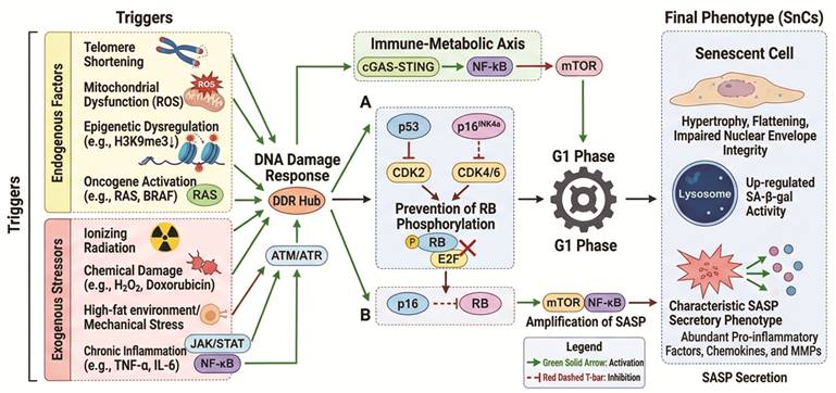
At the endogenous level, telomere shortening is the core cause of replicative senescence. As the number of divisions increases, telomere depletion triggers the ATM/ATR-p53-p21WAF1/CIP1 pathway, forcing the cells to exit the cell cycle [15, 18-21]. Additionally, mitochondrial dysfunction generates reactive oxygen species (ROS) that cause DNA double-strand breaks, which recruit γH2AX to trigger a persistent DNA damage response [22-25]. At the epigenetic level, disorders such as the reduction of H3K9me3 can promote the formation of senescence-associated heterochromatin foci, inhibiting the transcription of proliferation-related genes at the chromatin structure level [26-28]. Meanwhile, the activation of oncogenes (such as RAS, BRAF) can induce oncogene-induced senescence (OIS), constructing a crucial endogenous anti-tumor barrier [29, 30].
At the exogenous level, ionizing radiation or chemical damage (such as hydrogen peroxide, chemotherapeutic drugs like doxorubicin) can directly damage the genome and induce stress-induced premature senescence [31-35]. Metabolic disorders (such as a high-fat environment) and physical mechanical stress further induce aging by activating the cGAS-STING or TGF-β/Smad signaling pathways [36]. In addition, factors such as TNF-α and IL-6 in the chronic inflammatory microenvironment form a positive feedback loop through the JAK/STAT or NF-κB pathways, maintaining and strengthening the aging phenotype [37, 38].
The above stress signals are ultimately executed through two core pathways: one is the p53-p21-RB pathway, which inhibits the activity of CDK2 through p21; the other is the p16INK4a-RB pathway, which blocks RB phosphorylation by inhibiting CDK4/6, causing the cells to permanently remain in the G1 phase [39-42]. At the same time, the continuous activation of the mTOR/NF-κB axis amplifies the inflammatory effect of SASP, causing the complex pathological and physiological characteristics of SnCs [43].
2.2. Pathological characteristics of SnCs
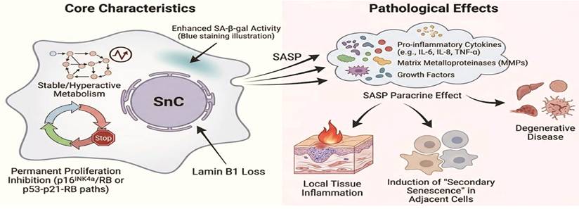
The most distinctive biological feature of SnCs is the restricted proliferation under active metabolism. Although permanent proliferation inhibition has been achieved through the p16INK4a/RB or p53-p21-RB pathways, the metabolic network of SnCs remains stable or even hyperactive [44, 45]. Besides the classic characteristics such as enhanced SA-β-gal activity and loss of nuclear lamina protein Lamin B1, the pathological driving force of SnCs mainly stems from the paracrine effect of SASP (Figure 2) [17, 46]. By continuously releasing pro-inflammatory cytokines (such as IL-6, IL-8, TNF-α), MMPs, and growth factors into the extracellular matrix, SASP not only directly promotes the inflammatory response and fibrotic remodeling in the local tissue, but also induces “secondary senescence” in adjacent healthy cells [47-51]. This spread of the pro-inflammatory microenvironment mediated by SnCs is a key mechanism driving the occurrence and development of various degenerative diseases.
The core characteristics of SnCs include: inactive/overactive metabolism, permanent cell cycle arrest, loss of lamin B1, and increased SA-β-gal activity. SnCs exhibit a senescence associated secretory phenotype, which involves the massive release of cytokines including pro-inflammatory cytokines, MMPs, etc., leading to a series of pathological changes, including local tissue inflammation, and secondary senescence of adjacent cells through paracrine effects, ultimately resulting in degenerative disease.
2.3. Association between SnCs and ARDs
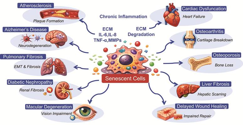
SnCs act as a key driver of various ARDs through their unique growth-inhibiting effect and imbalance in secretory activity [52, 53]. Specifically, the irreversible growth arrest of SnCs significantly weakens the regenerative potential of damaged tissues [54]. More importantly, the continuously secreted SASP (such as IL-6, IL-8, TNF-α, and MMPs) accumulates in the tissue microenvironment, inducing chronic low-grade inflammation, extracellular matrix degradation, and “secondary senescence” of adjacent healthy cells, thereby accelerating the functional deterioration of tissues [55-58]. Preclinical studies have confirmed the pathogenic role of SnCs in multiple organ system disorders. In the cardiovascular system, the inflammatory response mediated by senescent endothelial cells promotes lipid deposition and plaque instability [59]. In the central nervous system, senescent glial cells exacerbate neuronal damage and cognitive decline through pro-inflammatory responses [60, 61]. In the respiratory and movement systems, senescent fibroblasts and chondrocytes drive the evolution of pulmonary fibrosis and osteoarthritis through promoting abnormal collagen deposition and cartilage matrix degeneration [62, 63]. Furthermore, senescent renal tubular epithelial cells are the core pathological factor causing renal interstitial fibrosis in diabetic nephropathy [64].
Overall, the pathological aggregation of SnCs not only indicates the depletion of the body's functional reserve, but also amplifies the local cell damage into systemic organ dysfunction through the inflammatory cascade triggered by SASP. Thus, SnCs form a crucial link connecting basic biological aging and clinical pathological diseases (see Figure 3 and Table 1).
Mechanistic roles of SnCs in multisystem ARDs. SnCs disrupt tissue homeostasis through irreversible growth arrest and the pro-inflammatory effects of the SASP. SASP factors (including IL-6, TNF-α, and MMPs) drive chronic inflammation and extracellular matrix degradation, thereby promoting pathological degeneration across multiple organ systems, including the cardiovascular system (atherosclerosis and heart failure), nervous system (Alzheimer's disease), respiratory system (pulmonary fibrosis), skeletal system (osteoarthritis), and renal system (diabetic nephropathy).
Pathophysiological mechanisms of SnCs in ARDs
| Systems | Disease | Key SnCs Types | Pathophysiological Mechanisms | Ref., |
|---|---|---|---|---|
| Cardiovascular Systems | Atherosclerosis | Endothelial cells, smooth muscle cells, macrophages | Activating NF-κB signaling pathway. Promoting lipid deposition and ECM degradation. Reducing plaque stability. | [59] |
| Myocardial infarction or failure | Cardiomyocytes, cardiac fibroblasts, cardiac progenitor cells | Inducing a pro-inflammatory microenvironment and exacerbating myocardial fibrosis. Inhibit endogenous regeneration and impair contractile function. | [65, 66] | |
| Nervous system | Alzheimer's | Glial cells, neurons | 1.Slow down the clearance of amyloid beta protein. 2.Promote pathological aggregation of Tau protein. 3.Triggering neuroinflammation and cognitive impairment. | [67] |
| Parkinson's | Dopaminergic neurons, PC12 cells | 1.Toxic interaction between α-synuclein (α-syn-A53T) and iron overload (iron deposition) 2.SA-β-gal, p16, p21, H2A.X, γ-H2A.X, ROS accumulation, mitochondrial dysfunction 3.Dysregulation of iron-related proteins (L-ferritin, H-ferritin, DMT1, IRP1, IRP2), oxidative stress, preceding nigral dopaminergic neuron loss and motor dysfunction | [68] | |
| Respiratory system | Pulmonary fibrosis | Alveolar epithelial cells, fibroblasts | Drive epithelial mesenchymal transition (EMT). Abnormal deposition of collagen. Pulmonary parenchymal remodeling. | [69] |
| Chronic obstructive pulmonary disease | Small airway epithelial cells, fibroblasts, alveolar macrophages | Maintain chronic inflammation of the airway. | [70] | |
| Urinary system | Chronic and diabetes nephropathy | Renal tubular epithelium, mesangial cells, podocytes | Triggering renal tubular atrophy and interstitial fibrosis. Disrupting the glomerular filtration barrier. | [71, 72] |
| Bone system | Osteoarthritis | Chondrocytes, synovial cells, synovial macrophages | Release MMPs to degrade cartilage matrix and induce chronic synovitis. Reconstruct the subchondral bone microenvironment. | [62] |
| Osteoporosis | Osteoblasts, osteoclasts, and bone marrow myeloid cells | Inhibiting osteogenic differentiation and abnormally activating osteoclast activity. Disrupting bone metabolism homeostasis. | [73] | |
| Digestive system | Liver fibrosis | Hepatic stellate cells and liver cells | Continuously activating the pro fibrotic microenvironment and hindering tissue functional repair. | [74] |
| Visual system | Age-related macular degeneration | Retinal pigment epithelium cells | Disrupting the integrity of the blood retinal barrier and inducing pathological choroidal neovascularization. | [75] |
| Skin system | Skin aging and wound healing disorders | Keratinocytes, melanocytes, fibroblasts | Spread aging signals. Disrupt the skin barrier. Inhibit vascular regeneration, and delay healing. | [76, 77] |
3. Senotherapy: Intervention Strategies Targeting SnCs
Senotherapy represent a class of intervention paradigms designed to precisely modulate the fate and function of SnCs through pharmacological or bioengineering approaches [78]. The core logic of this field has evolved from a singular “cell clearance” strategy into an integrated pathway of “identification—clearance—suppression—homeostatic restoration”: first, SnCs are precisely identified by exploiting their specific SCAPs, surface markers, or metabolic features [79, 80]; second, apoptosis of SnCs is specifically induced through small-molecule senolytic drugs (senolytics) or immune-mediated senolytic strategies [81-83]; for the remaining SnCs, senomorphic drugs (senomorphics) are applied to intervene in key signaling axes, such as NF-κB and JAK/STAT, thereby suppressing the pro-inflammatory phenotype of the senescence-associated secretory phenotype (SASP) [84, 85]; finally, tissue microenvironmental homeostasis is restored through metabolic reprogramming [86]. This chapter will focus on reviewing the molecular targets, pharmacological characteristics, and translational prospects of senotherapy and related technical approaches.
3.1. Senolytics: Cleaning SnCs
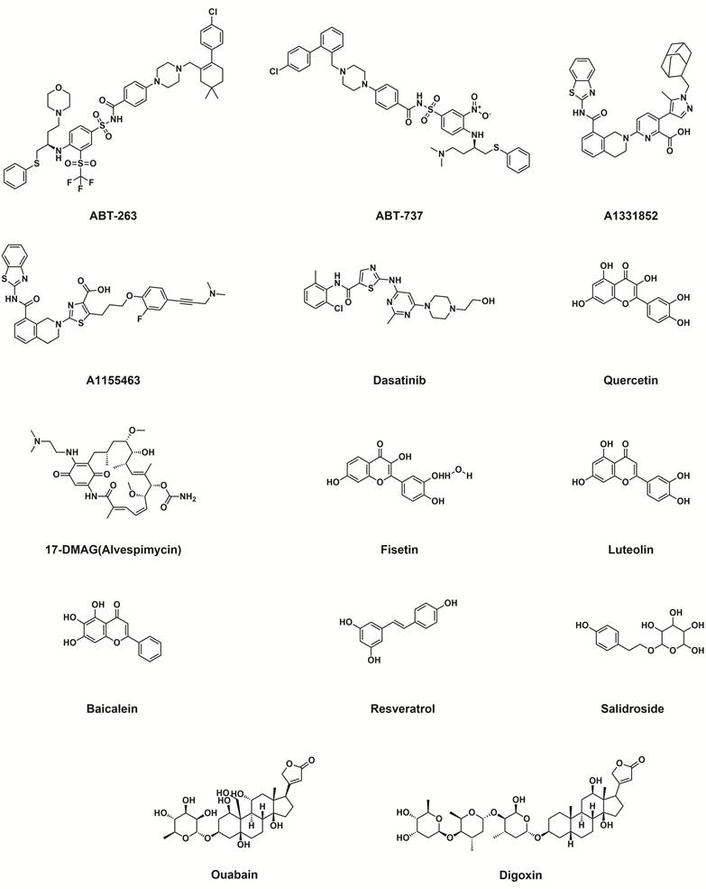
Senolytics aim to target SCAPs pathways, thereby overcoming the apoptotic resistance of SnCs and precisely inducing their programmed cell death [87]. The core molecular mechanisms center on antagonizing BCL-2 family proteins, inhibiting the PI3K/AKT/mTOR cascade, or blocking specific protein-protein interactions, which subsequently trigger mitochondrial outer membrane permeabilization and the apoptotic cascade [88]. This section will systematically discuss several major classes of representative senolytics, including small-molecule inhibitors and natural compounds (see Figure 4 and Table 2).
The chemical structures of representative small-molecule senolytics.
Classification, targets, and pharmacological characteristics of typical senolytics.
| Category | Representative drugs/compounds | Molecular targets/mechanisms | Pharmacological characteristics | Clinical evaluation | Ref. |
|---|---|---|---|---|---|
| Single-target inhibitors | ABT-263/ABT-737 | BCL-2/BCL-XL inhibitor | First generation broad-spectrum BH3 analogues | Dose-dependent thrombocytopenia | [94, 97-99] |
| A1331852/A1155463 | Highly selective BCL-XL inhibitor | Better hematological safety than ABT-263 | Efficiency exhibits heterogeneity in cell types | [100] | |
| Dasatinib + Quercetin | Src / SIRT1 | Synergy effect | Significantly downregulate cyclic SASP and repair multi system functionality | [101-105] | |
| FOXO4-DRI | Interference with p53-FOXO4 interaction axis | Competitive Binding of p53 | Heterogeneity of effects in different pathological conditions | [106-109] | |
| 17-DMAG | HSP90 inhibitor | Inducing key HSP degradation | Excellent potency (nM level) and broad-spectrum activity | [110, 113, 114] | |
| Multi-target inhibitors | Fisetin | p16INK4a / mTOR | Typical multi-target candidates | Exhibiting initial efficacy in reducing SASP factors and improving clinical outcomes across multiple human trials | [120, 124-127] |
| Luteolin | p16-CDK6 / SIRT1 | Antagonize stress-induced aging | Combining the potential to delay natural aging and resist fibrosis | [128-130] | |
| Baicalein | mTORC1 / Sirt3 | Dual mechanism | Inhibit tumor proliferation or alleviate pulmonary fibrosis | [131, 132] | |
| Resveratrol | SIRT1 / AMPK | Strengthen antioxidant defense, optimize mitochondrial function, and inhibit SASP production | Provides protective efficacy for various ARDs | [133-136] | |
| Salidroside | PI3K/Akt/TERT pathway activator | Neuroprotective senomorphic effects; improves neuronal viability, MAP2, cognition and alleviates hippocampal degeneration. | In preclinical studies, it demonstrated a good safety profile with no obvious adverse reactions observed. | [119] | |
| Homeostatic regulation | Ouabain/Digoxin | Na+/K+-ATPase (NKA) | Triggering Ca2+ overload mediated apoptosis | Narrow treatment window, requiring monitoring of cardiac toxicity | [140-144] |
| Fenofibrate/Pioglitazone | PPAR agonists | Regulating the metabolic inflammatory network; Regulating the expression of BCL-2 family proteins | Long term use requires vigilance against weight gain and edema | [146-153] |
3.1.1. Single-target inhibitors
(1) BCL-2 family inhibitors
SCAPs activation is the cornerstone of SnCs survival, with differential expression of BCL-2 family members playing a particularly critical role [89-94]. SnCs frequently construct a robust apoptotic defense mechanism through the specific upregulation of BCL-XL and BCL-W, thereby providing well-defined targets for the development of small-molecule inhibitors designed to disrupt this homeostasis [95, 96]. First-generation broad-spectrum BCL-2 inhibitors, represented by ABT-737 and its orally bioavailable formulation ABT-263, act as BH3-mimetics; they competitively bind to the hydrophobic pocket of anti-apoptotic proteins, blocking their interaction with pro-apoptotic counterparts [94, 97]. Although ABT-263 has demonstrated remarkable efficacy in clearing SnCs across multiple tissues, its clinical translation has been severely limited by dose-dependent thrombocytopenia resulting from systemic BCL-XL inhibition [98, 99].
To address this safety bottleneck, research has shifted toward highly selective BCL-XL inhibitors, such as A1331852 and A1155463. These compounds are designed to precisely eliminate BCL-XL-dependent senescent cells by triggering the caspase cascade [100]. However, such high selectivity also raises the issue of a “narrow therapeutic spectrum”: the apoptotic dependencies of SnCs are highly heterogeneous, and single-target inhibition often fails to cover all pathogenic subpopulations. Future studies should focus on identifying the specific apoptotic vulnerabilities of SnCs in different tissues and on exploring novel drug delivery systems to optimize the therapeutic index.
(2) Kinase inhibitors
The combination of dasatinib and quercetin (the D+Q regimen) represents one of the most rapidly translated therapeutic modalities in the field of senolytics. Dasatinib, a broad-spectrum tyrosine kinase inhibitor, primarily blocks pro-survival signaling by inhibiting Src family kinases, thereby overcoming the apoptotic resistance of specific SnCs, such as preadipocytes [101].
Quercetin in this combination exerts multi-target regulatory effects. Its biological actions are highly dependent on the allosteric activation of SIRT1, which subsequently remodels cellular homeostasis through the following pathways: (a) anti-inflammatory and immunomodulatory effects: suppressing NF-κB acetylation and blocking the pathological release of TNF-α and IL-1β; (b) redox homeostasis: scavenging excessive reactive oxygen species via the SIRT1/Nrf2/HO-1 axis; (c) metabolic and apoptotic control: coordinately modulating the SIRT1/FOXO pathway and optimizing the BCL-2/BAX ratio [102, 103].
Preclinical and early clinical evidence consistently demonstrates that the D+Q regimen significantly reduces the circulating SASP profile and restores multi-system tissue function [104, 105]; however, its relatively low bioavailability still needs to be optimized through novel delivery technologies.
(3) p53-FOXO4 axis regulator
Disrupting the physical interaction between p53 and FOXO4 is a highly efficient strategy for inducing apoptosis in SnCs. Interfering peptides represented by FOXO4-DRI can precisely overcome apoptotic resistance by releasing nuclear p53 into the cytoplasm to trigger cell death [106]. In cartilage aging models, FOXO4-DRI exhibits remarkable broad-spectrum senescent cell clearance efficacy and suppresses microenvironmental deterioration; however, it also reveals a kinetic disconnect between “clearance” and “functional regeneration”, as this peptide fails to effectively induce the redifferentiation of residual cells [107].
In complex pathological settings, p53-FOXO4 interfering agents display a pronounced “double-edged sword” characteristic. In fibrotic diseases such as radiation-induced pulmonary fibrosis, these agents confer significant protection by eliminating pathological fibroblasts; however, in models such as pulmonary arterial hypertension, non-specific induction of apoptosis in critical endothelial cells paradoxically exacerbates hemodynamic disturbances [108, 109].
These contradictory outcomes underscore the functional heterogeneity of SnCs across different tissues or pathological stages, and the development of inhibitors with higher cell-type selectivity is a critical prerequisite for clinical translation.
(4) HSP90 inhibitor
HSP90 inhibitors precisely disrupt the anti-apoptotic signaling network essential for SnCs survival by interfering with the interaction between molecular chaperones and client proteins. The core mechanism involves inducing ubiquitin-proteasomal degradation of downstream pro-survival factors, such as AKT and ERK, thereby dismantling the apoptotic barrier [110]. Recent studies have uncovered a novel role for HSP90 in regulating cell cycle checkpoints: for example, AT13387, through remodeling proteostasis, not only drives tumor cells into stable senescence but also suppresses their stemness features [111].
Moreover, the post-translational regulation of the tumor suppressor p14ARF by HSP90, mediated by CHIP-dependent lysosomal degradation, further underscores its multidimensionality as a therapeutic target [112]. In senolytic screening campaigns, derivatives such as 17-DMAG have demonstrated exceptional potency. In DNA damage repair-deficient mice, targeting HSP90 systemically reduces the SnCs burden across multiple organs with nanomolar efficacy, outperforming conventional chemotherapeutic agents [113, 114]. This broad-spectrum clearance capacity across diverse senescence models, including lung fibroblasts and endothelial cells, positions HSP90 inhibitors among the most promising clinical candidates.
3.1.2. Multi-target inhibitors
Naturally derived small-molecule compounds have garnered considerable attention for their exceptional senolytic potential. Representative active constituents, including fisetin, luteolin, baicalein, resveratrol, and salidroside, exhibit significant clearance efficacy by differentially modulating the anti-apoptotic pathways upon which SnCs depend [115-119]. The unique advantage of these phytochemicals lies in their multi-target effects: while specifically eliminating SnCs, they can synergistically remodel the immune microenvironment and initiate tissue regeneration programs, thereby offering new perspectives for the systemic intervention of ARDs.
(1) Fisetin
Fisetin is regarded as a senolytic agent with substantial clinical translational value. This profound clinical potential is currently being validated in several human clinical trials, highlighting its status as a premier candidate for age-related therapeutic interventions [120-123]. Studies have demonstrated that fisetin selectively eliminates p16INK4a-positive senescent endothelial cells, and by inhibiting the release of SASP components and restoring nitric oxide bioavailability, significantly ameliorates vascular endothelial dysfunction and arterial stiffness in aged individuals [124, 125]. In multiple animal models and experiments with human peripheral blood mononuclear cells, a “hit-and-run” intermittent dosing strategy not only effectively reduces the SnCs burden and circulating SASP levels, but its efficacy is even comparable to that achieved by genetic ablation approaches or synthetic senolytics [120, 126]. Furthermore, fisetin downregulates the expression of aging-related genes across multiple organs, such as the brain, liver, and lung, and exerts systemic anti-aging effects by lowering plasma S100B levels [127].
(2) Luteolin
Luteolin exerts protective effects through a dual mechanism. First, by inhibiting stress-induced p53 phosphorylation and upregulating SIRT1 expression, it antagonizes cellular senescence and collagen degradation triggered by oxidative stress [128] or ultraviolet A radiation [129]. Second, luteolin specifically disrupts the protein interaction between p16 and CDK6. In in vivo models, this mechanism mediates lifespan extension, amelioration of natural aging, and significant suppression of doxorubicin-induced inflammation and fibrosis [130].
(3) Baicalein
Baicalein exhibits distinct cell fate regulatory capabilities under different pathological contexts. For instance, in malignant tumor models such as melanoma, it induces compensatory senescence in tumor cells by inhibiting glucose uptake and blocking the mTORC1/HIF-1α pathway, thereby curbing their proliferation and migration [131]. In contrast, in models of idiopathic pulmonary fibrosis, baicalein significantly attenuates the senescent phenotype of lung fibroblasts, reduces aberrant collagen deposition, and delays pathological fibrotic progression by activating SIRT3 and antagonizing the TGF-β1/Smad signaling axis [132].
(4) Resveratrol
Resveratrol intervenes in the progression of aging through a multidimensional signaling network. Its core mechanism involves activation of the SIRT1/AMPK axis, which significantly suppresses the accumulation of SnCs and the production of the SASP by reinforcing antioxidant defenses and anti-inflammatory responses [133, 134]. Concurrently, resveratrol exhibits marked protective potential in neurodegenerative diseases, cardiac dysfunction, and metabolic disorders by optimizing mitochondrial biogenesis and modulating the apoptotic threshold [135, 136].
(5) Salidroside
Salidroside is regarded as a prominent nature-derived bioactive glucoside with substantial clinical translational value. This profound therapeutic potential in delaying aging and mitigating age-related pathologies highlights its status as a premier candidate for longevity interventions [137, 138]. Studies have demonstrated that salidroside effectively extends lifespan and improves healthspan across multiple species by inducing a characteristic hormetic biphasic dose response, which activates intracellular adaptive pathways at low concentrations [137]. At the molecular level, this longevity-promoting effect is directly driven by its capacity to bind HSP90 and inhibit its ATPase activity, thereby modulating downstream gene networks and reinforcing cellular stress resistance [138]. In multiple preclinical models of brain aging and neurodegeneration, such as naturally aged mice and Alzheimer's disease models, salidroside administration significantly ameliorates cognitive dysfunction, reduces amyloid-β (Aβ) deposition, and alleviates neuronal degeneration in the hippocampal CA1 region [119, 139]. These neuroprotective actions are tightly coupled with the selective downregulation of senescence-associated markers, including β-galactosidase, p21, and p16, an effect mediated by the upregulation of telomerase reverse transcriptase (TERT) expression via the PI3K/Akt signaling pathway [119]. Furthermore, salidroside exerts systemic anti-aging and antioxidative effects across diverse biological systems by augmenting superoxide dismutase (SOD) activity while consistently suppressing the accumulation of malondialdehyde (MDA) and pro-inflammatory cytokines, such as IL-1β, IL-6, and TNF-α [138, 139].
3.1.3. Senolytics based on homeostatic regulation
In addition to the aforementioned classical pathways, cardiac glycosides (CGs) and PPAR agonists also exhibit significant senolytic potential by modulating ion homeostasis and metabolic signaling networks.
(1) Cardiac glycosides (CGs)
CGs, such as digoxin and ouabain, exert their senolytic effects primarily by inhibiting the activity of the Na+/K+-ATPase (NKA) pump [140, 141]. The core mechanisms are as follows: first, NKA inhibition leads to intracellular Na+ and Ca2+ overload, which subsequently activates Ca2+-dependent signaling such as CaMKII, triggering mitochondrial membrane potential collapse and caspase-dependent apoptosis [142]; second, they suppress the secretion of SASP components by antagonizing the NF-κB signaling pathway, thereby ameliorating the chronic inflammatory microenvironment [143]. Furthermore, CGs can specifically induce autophagy inhibition and apoptosis in BRAFV600E-positive senescent cells by modulating the Src/Akt axis [144]. However, the efficacy of these agents exhibits pronounced cell-type dependence [145]: studies have revealed that human mesenchymal stem cells frequently acquire enhanced K+ compensatory capacity and apoptotic resistance during senescence, leading to resistance to CGs.
(2) PPAR agonists
PPAR agonists exert anti-aging effects by precisely modulating the “metabolism-inflammation” coupling network [146-148]. These agents exhibit dual molecular effects: PPAR-α activation mediates enhanced fatty acid oxidation, thereby alleviating lipotoxicity, whereas PPAR-γ activation simultaneously optimizes insulin sensitivity and synergistically suppresses pro-inflammatory responses, collectively disrupting the survival dependencies of SnCs [149, 150]. At the molecular level, these agonists bidirectionally regulate BCL-2 family proteins, triggering SnCs apoptosis by downregulating anti-apoptotic factors such as BCL-XL and upregulating pro-apoptotic proteins such as BAX [151, 152]. Notably, at the clinical level, PPAR-γ agonists, including rosiglitazone and pioglitazone, exhibit remarkable pleiotropic effects, encompassing the restoration of mitochondrial function and the blockade of SASP release [153]. Large-scale epidemiological data further corroborate that diabetic patients receiving long-term pioglitazone treatment demonstrate significantly reduced all-cause mortality, confirming a clear association between PPAR activation, human health lifespan extension, and a diminished risk of ARDs [148].
3.2. Immuno-senolytics: Chasing SnCs
Immune-mediated senescent cell clearance strategies (Immuno-senolytics) represent a novel intervention paradigm that achieves the precise elimination of SnCs by activating or enhancing endogenous immune system functions [154]. Distinct from conventional small-molecule Senolytics, this strategy aims to reinvigorate immune surveillance mechanisms and encompasses several innovative technological approaches. First, antibody-guided therapies target SnCs surface markers, such as urokinase-type plasminogen activator (uPAR), or block “don't eat me” signaling axes, such as CD47/SIRPα, thereby abrogating immune evasion and promoting macrophage-mediated phagocytosis [155, 156]. Second, adoptive cell therapies utilize engineered effector cells, such as CAR-T or NK cells, to achieve highly efficient killing by recognizing specific antigens, including NKG2D ligands [157, 158].
Furthermore, senolytic vaccines, for instance those targeting the GPNMB protein, are capable of inducing durable adaptive immune responses to suppress SnCs accumulation over the long term [159]. Concurrently, the application of immunomodulatory agents, such as cytokines (e.g., IL-15) and STING agonists, can further synergistically enhance the clearance efficiency of the immune system [160]. This combined strategy, characterized by high target specificity and immune memory effects, has emerged as a highly promising frontier in the field of aging intervention. Representative agents and technical approaches are summarized in Table 3.
Immune-mediated senescence cell clearance strategies (Immune-senolytics)
| Category | Representative drugs/technologies | Molecular targets/mechanisms | Pharmacological characteristics and clinical evaluation | Ref. |
|---|---|---|---|---|
| Antibody therapy | uPAR antibody | uPAR | Reshaping the fibrotic microenvironment through ADCC/ADC effect | [161, 162] |
| DcR2 antibody | Decoy receptor 2 | Reverse anti-apoptotic phenotype | [163] | |
| CD47 antibody | CD47-SIRPα axis | Block the “don't eat me” signal, enhance the phagocytic effect | [164-166] | |
| PD-L1 antibody | PD-L1 | Reverse immune escape and clear SnCs | [167] | |
| Cell therapy | HLA-E siRNA | HLA-E/NKG2A axis | Correction of NK cell subpopulations | [13, 168] |
| CAR-NK cells | Targeting uPAR | High targeting with strong infiltration of solid tissues | [169-171] | |
| Vaccines | GPNMB vaccine | GPNMB | Active CD8+ T cells and endogenous immune memory | [159, 172-174] |
| Regulating chemokines | CCL-2/CCR2 inhibitors | CCL-2-CCR2 axis | Inhibit the recruitment of Ly6C+ monocytes | [177-179] |
| Other immune regulation strategies | STING modulators | cGAS-STING-IRF3-RB axis | “pro-senescence/anti-tumor” and “pro-inflammation/immunosuppression” | [180-183] |
| TLR modulators | TLR receptor | Regulating immune homeostasis through multiple signaling | [186-189] |
3.2.1. Antibody therapy
(1) Cell-surface markers of SnCs
Studies have shown that uPAR is specifically and highly expressed on the surface of SnCs, where it is deeply involved in microenvironmental remodeling by regulating extracellular matrix degradation [161]. Based on this phenotype, antibody-based drugs targeting uPAR can precisely eliminate SnCs by synergistically harnessing antibody-dependent cellular cytotoxicity (ADCC) and complement-dependent cytotoxicity (CDC), and their efficacy has been validated in various pathological fibrosis models [162]. Furthermore, the decoy receptor 2 (DcR2), which is enriched on the surface of SnCs, is also a highly promising target: binding of a specific antibody to DcR2 can reverse the anti-apoptotic phenotype, reactivate the caspase-8-mediated extrinsic apoptotic pathway, and induce programmed cell death in SnCs [163].
(2) Immune checkpoints
SnCs can exploit immune checkpoint signaling to construct an immune-evasive microenvironment, and blockade of these inhibitory signaling axes can significantly enhance immune surveillance in aged individuals [164]. On the one hand, the overexpression of CD47 on the surface of SnCs effectively suppresses the phagocytic activity of macrophages through “don't eat me” signaling, i.e., the CD47-SIRPα interaction. Application of anti-CD47 antibodies specifically blocks this signaling axis, generating a marked pro-phagocytic effect that demonstrates significant therapeutic potential in aging-associated tumors and fibrotic diseases [165, 166]. On the other hand, reports have revealed that p16INK4a-positive cells evade immune recognition by upregulating PD-L1 expression, thereby accelerating the progression of pulmonary fibrosis and chronic obstructive pulmonary disease. Notably, activating PD-L1 antibodies can precisely ablate p16+/PD-L1+ SnCs via an Fcγ receptor-dependent mechanism, significantly alleviating the tissue inflammatory burden [167].
Antibody-based therapies, with their high target specificity and pharmacokinetic advantages, demonstrate potential superiority over small-molecule drugs in SnCs clearance. However, optimizing target specificity, enhancing penetration into solid tissues, and mitigating the potential risk of autoimmunity remain key challenges for translational research. Future studies should integrate single-cell sequencing technologies to identify candidate antigens and explore engineering approaches such as bispecific antibodies or antibody-drug conjugates (ADCs) to achieve more precise intervention.
3.2.2. Cell therapy
Natural killer (NK) cells, as the core effector components of the innate immune system, possess the endogenous capacity to recognize and eliminate SnCs, and functionally enhancing them through genetic engineering has emerged as a key approach to improving SnCs clearance efficiency [168].
Studies have shown that SnCs can construct an immune-evasive phenotype by upregulating the expression of non-classical human leukocyte antigen HLA-E, which binds to the inhibitory receptor NKG2A on the surface of NK cells. Targeted blockade of the NKG2A/HLA-E signaling axis effectively restores NK cell cytotoxicity, indicating that this pathway represents an important target for reversing the progression of aging-related pathologies [13]. Further reports have revealed that the aging process is accompanied by a characteristic remodeling of NK cell subsets, manifested by a decrease in the CD56bright subset and an increase in the CD56dim subset. This subset shift often leads to a decline in overall killing efficacy, thereby impairing immune surveillance and precipitating “immunosenescence” and associated degenerative diseases [168].
Currently, engineered CAR-NK cells constructed through synthetic biology exhibit more precise antigen recognition potential [169, 170]. Compared with CAR-T cells, CAR-NK cells demonstrate remarkable translational value in preclinical studies of age-related malignancies and chronic degenerative diseases, owing to their lower risk of autoimmune responses, enhanced capacity for solid tissue infiltration, and unique non-MHC-restricted killing mechanism [171].
3.2.3. Vaccines
Glycoprotein nonmetastatic melanoma protein B (GPNMB) is a transmembrane protein specifically upregulated on the surface of SnCs, widely distributed in tissues such as the liver, adipose tissue, and vascular endothelium, and has emerged as a highly promising molecular target in the field of aging intervention [172].
Studies have demonstrated that vaccines targeting GPNMB can effectively eliminate SnCs, markedly reverse pathological phenotypes in progeroid mice, and extend their lifespan [159]. Specifically, dendritic cell vaccines loaded with cationic protein-fused GPNMB have exhibited significant efficacy in various natural aging and stress-induced aging models: by downregulating SA-β-gal activity and the expression of classical senescence signaling axes such as p16INK4a, p21Cip1, and p53, this vaccine markedly improves glucose tolerance and insulin resistance, confirming its critical role in correcting metabolic dysfunction induced by SnCs accumulation [173].
Mechanistic analyses reveal that GPNMB, as a senescence-specific antigen, elicits active immunity that specifically activates CD8+ T cells to precisely eliminate GPNMB+ senescent vascular endothelial cells and adipocytes. This process effectively curbs the progression of atherosclerosis and metabolic disorders [174].
Beyond its therapeutic targeting potential, GPNMB has demonstrated translational value as a noninvasive biomarker for monitoring SnCs burden and senotherapy efficacy in vivo. Notably, plasma GPNMB levels are significantly elevated in Parkinson's disease (PD) patients compared with neurologically normal controls, positively correlating with disease severity as assessed by UPDRS Part III scores across two independent cohorts; furthermore, cerebrospinal fluid GPNMB levels exhibit a protein quantitative trait locus effect consistent with the PD risk-associated rs199347 haplotype, collectively establishing blood-based GPNMB quantification as a clinically accessible readout for tracking senescence-associated pathological progression and evaluating intervention outcomes [175].
The core advantage of senolytic vaccines lies in their ability to induce durable endogenous immune memory. Compared with antibody- or cell-based therapies, the vaccine strategy holds the promise of achieving a “single vaccination, long-term protection” intervention effect and features a more favorable safety profile [176]. However, this field still faces critical challenges, including the balance between antigen specificity and safety, immunogenicity optimization, and inter-individual variability.
3.2.4. Regulating chemokines
CCL-2, a core component of the SASP, is markedly upregulated in various SnCs models, and its secretion levels exhibit a stepwise increase with the progression of cellular replicative senescence [177]. Mechanistic studies have revealed that SnCs release substantial amounts of CCL-2 through a RAB27A-mediated lysosomal exocytosis mechanism; these pro-inflammatory factors are enriched within lysosomes and subsequently released into the circulatory system, overlapping considerably with known plasma senescence biomarkers and suggesting a pivotal role as systemic aging signals in the progression of ARDs [178].
It has been reported that in pathological models, DNA damage-induced type II alveolar epithelial cells enter senescence via a p21WAF1/CIP1-dependent pathway and activate the CCL-2-CCR2 signaling axis. This axis directionally recruits Ly6C+ inflammatory monocytes at the early stage of the lesion, and subsequently promotes excessive collagen deposition through paracrine crosstalk between activated macrophages and fibroblasts, thereby establishing a causal link between alveolar epithelial cells senescence and the initiation and progression of pulmonary fibrosis [179].
Chemokine-targeting strategies are designed to intervene in aging-associated pathologies through a dual mechanism: first, reversing the immunosuppressive microenvironment by inhibiting the recruitment of myeloid-derived suppressor cells; second, enhancing the immune surveillance capacity of NK cells and CD8+ T cells through the remodeling of chemotactic gradients. Although this field holds considerable promise, the construction of precise delivery systems and the optimization of multi-target combination regimens remain current bottlenecks in translational medicine.
3.2.5. Other immune regulation strategies
(1) STING modulators
STING (stimulator of interferon genes) agonists represent important tools for modulating senescence and the associated immune microenvironment, encompassing natural cyclic dinucleotides, such as 2'3'-cGAMP, and synthetic small molecules. Their core mechanism involves inducing STING protein oligomerization or the formation of biomolecular condensates, thereby activating type I interferon signaling [180]. In tumor biology, the cGAS-STING pathway has been demonstrated to induce DNA damage-associated senescence in tumor cells, suppress tumor proliferation by enhancing SASP secretion, and concurrently participate in the immune clearance of senescent cells [181, 182].
This pathway also plays a critically opposing role in certain diseases, including neurodegenerative disorders. Studies have revealed that excessive activation of cGAS within microglia triggers neurodegeneration, suggesting that this axis represents a core target for intervention in Alzheimer's disease, and the STING inhibitor H-151 can effectively alleviate neuroinflammation and cognitive impairment in the brain by suppressing microglial activation [56]. Furthermore, this pathway drives cell cycle arrest by assembling an IRF3-RB complex that inhibits CDK4/6-mediated RB phosphorylation. For instance, in hepatic stellate cells, the cGAS-STING-IRF3-RB axis exerts anti-fibrotic effects by inducing their senescence [40].
However, the effects of the cGAS-STING pathway are highly context-dependent. Chemotherapy-induced tumor cells can activate cGAS signaling in polymorphonuclear myeloid-derived suppressor cells (PMN-MDSCs) via the mtDNA-VDAC channel, paradoxically enhancing tumor immunosuppression [183]. This dichotomy reflects both the cellular context of cGAS-STING activation and the local immune composition. In the aged brain, cytosolic mtDNA from structurally compromised microglial mitochondria engages cGAS, triggering a type I IFN response and TNF-mediated neurotoxicity through microglial STING-TBK1 signaling, amplified by the paucity of immunosuppressive cells [56]. In contrast, senescent tumor cells release mtDNA via VDAC-dependent extracellular vesicles, selectively internalized by PMN-MDSCs, where STING signals predominantly through the PERK-NF-κB axis rather than IRF3-IFN due to low intrinsic IFN production and IFNAR1 expression, thereby upregulating immunosuppressive genes such as Arg1, Nos2, and PD-L1 [183]. Clinically, broad STING agonism may enhance anti-tumor senescence while reinforcing myeloid immunosuppression, whereas non-selective STING blockade, though neuroprotective, may impair anti-tumor immunity. These insights highlight the need for cell-type- or compartment-specific STING modulation, such as combining senolytics to remove mtDNA-releasing tumor cells with targeted VDAC inhibition, while preserving microglial STING functions for brain homeostasis. This dual effect of “pro-senescence/anti-tumor” versus “pro-inflammation/immunosuppression” necessitates the future development of more precise tissue-specific regulatory strategies to balance anti-aging efficacy with systemic immune homeostasis [184, 185].
(4) TLR modulators
The Toll-like receptor (TLR) family are core sensors of innate immunity, and their modulators exhibit multidimensional potential in reshaping aging-associated immune responses. Studies have revealed that TLR signaling serves multiple biological functions in the progression of aging. First, TLR3 plays a critical role in virus-induced senescence. SARS-CoV-2 RNA, upon recognition by TLR3, not only induces cellular senescence but also amplifies the expression of the pro-inflammatory SASP, whereas TLR3 blockade can suppress this pro-inflammatory cascade [186]. Second, TLR4 signaling exhibits synergistic effects in tumor senescence intervention. For example, utilizing lipid nanoparticles to co-deliver a TLR4 agonist and a STING agonist, in the context of tumor senescence induced by MEK/CDK4/6 inhibitors, effectively reverses the immunosuppressive microenvironment and activates the surveillance functions of NK cells and CD8+ T cells by enhancing type I interferon secretion [187]. Furthermore, in intervertebral disc degeneration, TLR2/6 activation has been demonstrated to be a key factor in inducing disc cell senescence and upregulating p16INK4a expression, and the antagonist o-vanillin can significantly reverse these senescent phenotypes [188].
Future TLR modulation strategies are evolving toward precision. In oncology, research is focused on enhancing tumor immunogenicity through TLR7/8 agonists and combining them with immune checkpoint inhibitors to reduce systemic toxicity. In inflammatory diseases, emphasis is placed on developing high-affinity antagonists targeting TLR7/9 to intervene in autoimmunity and COVID-19-associated cytokine storms [189-191].
3.3. Senomorphics: Calming SnCs
Senomorphics represent an aging intervention paradigm that is independent of eliminating SnCs, with a core mechanism centered on the precise modulation of SnCs secretory characteristics [192]. By targeting key regulatory nodes such as mTOR, NF-κB, and p38 MAPK, Senomorphics can significantly suppress the expression of pro-inflammatory SASP profiles. This strategy not only curbs the pathological deterioration of the senescent microenvironment but also circumvents the potential risk of impaired tissue regeneration associated with the direct clearance of SnCs, demonstrating exceptional therapeutic potential in multiple models of chronic degenerative diseases [193, 194].
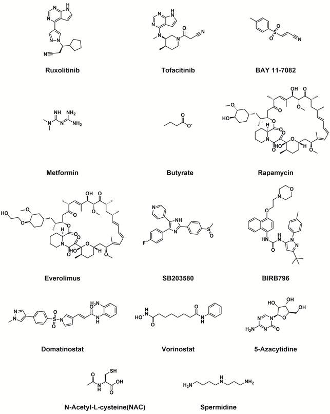
With the advancement of precision medicine, immunometabolism reprogramming based on natural polyphenolic compounds has become a research hotspot in the field of Senomorphics. By targeted modulation of cellular metabolic pathways, such agents hold promise in alleviating chronic inflammation while achieving systematic remodeling of immune functions [195]. This chapter will focus on discussing typical Senomorphics, including kinase inhibitors and epigenetic modifiers, and their molecular mechanisms (see Figure 5 and Table 4).
The chemical structure of typical senomorphics.
Summary of typical targets and mechanisms of senomorphics.
| Category | Molecular targets/ mechanisms | Representative drugs | Pharmacological characteristics and clinical evaluation | Ref. |
|---|---|---|---|---|
| SASP-related signaling pathway inhibitors | JAK2/STAT3 axis | Ruxolitinib | Inhibit SASP secretion in nucleus pulposus cells | [199] |
| JAK/STAT pathway | Tofacitinib | Regulating the aging phenotype and expression of γ-H2AX in memory T cells | [200] | |
| cGAS/STING/NF-κB | BAY 11-7082 | Inhibit stress-induced aging of lung epithelial cells and pro-inflammatory factors | [202] | |
| NF-κB/p65 activation | Metformin | 1.Regulating MMPs; 2.Inhibiting the abnormal proliferation and cancer tendency | [204, 205] | |
| NF-κB pathway | Butyrate (gut microbiota metabolite) | 1.Suppresses NF-κB, immune senescence and SASP 2.Microbiota-derived senomorphic for T-cell aging | [209] | |
| mTORC1 | Rapamycin | Enhance vaccine response; Improve cardiovascular function; Downregulate skin aging markers | [210, 211] | |
| mTORC1/p70/S6K pathway | Everolimus | Block the “Geroconversion” process; Protecting nucleus pulposus cells by enhancing autophagy | [213, 214] | |
| Inhibit p38 MAPK | SB203580 | Downregulate the expression of p53/p21 axis; Improve conjunctival laxity | [215, 216] | |
| Synergistic blockade of p38 MAPK/NF-κB | BIRB796 | Restore the localization of tight junction proteins in brain endothelial cells; Improve blood-brain barrier function. | [217] | |
| Epigenetic modulators | Dual inhibition of HDAC/LSD1 | Domatinostat | Induction clearance sequential mode; Synergistic USP7 inhibitor clearance of melanoma SnCs | [218] |
| Broad spectrum inhibition of HDAC | Vorinostat | Synergistic inhibition of NF-κB/mTOR; Relieve skin photoaging damage | [204, 219] | |
| Inhibition of DNMTs enzyme activity | 5-Azacytidine | Activate eIF2α-autophagy pathway; Eliminate senescent pancreatic β cells | [223] | |
| Other strategies | ROS/NF-κB/p38 axis | NAC | Eliminate ROS; Reverse lysosomal membrane permeability abnormality | [224, 226] |
| Activate AMPK while inhibiting mTOR | Spermidine | Enhance autophagy; Acetylation modification reshapes cartilage homeostasis | [227-229] |
3.3.1. SASP-related signaling pathway inhibitors
(1) JAK-STAT inhibitor
The JAK-STAT signaling pathway, an evolutionarily conserved intracellular signal transduction cascade, is composed of four Janus kinases (JAK1/2/3, TYK2) and seven signal transducers and activators of transcription (STATs), and is deeply involved in immune homeostasis regulation, inflammatory responses, and cell proliferation. JAK inhibitors specifically block cytokine-induced STAT phosphorylation and its nuclear translocation by competitively binding to the ATP-binding site of JAK proteins, thereby suppressing the transcriptional programs of downstream inflammatory genes [196, 197].
Studies have shown that STAT5 phosphorylation mediated by this pathway can upregulate anti-apoptotic factors such as BCL-2, sustaining the survival of SnCs; the application of JAK1/2 inhibitors can effectively intervene in this mechanism and promote the clearance of latently HIV-infected cells [198]. In intervertebral disc degeneration models, Ruxolitinib, by blocking the JAK2/STAT3 signaling axis, significantly downregulates the expression of p16, p21, and p53 in nucleus pulposus cells and suppresses the secretion of IL-6, TNF-α, and matrix-degrading enzymes (such as MMP-3), thereby delaying degenerative progression by maintaining extracellular matrix homeostasis [199]. Furthermore, Tofacitinib induces an immunosenescent phenotype in memory T cells and upregulates γH2AX expression, thereby exhibiting favorable efficacy in rheumatoid arthritis. This precise modulation of the activation and senescence status of immune effector cells provides a theoretical explanation for the balance between its clinical efficacy and the risk of infection [200].
(2) NF-κB inhibitor
The NF-κB signaling pathway encompasses both canonical and non-canonical activation pathways and, through the differential regulation of the IKKα/β complex, plays a pivotal role in pro-inflammatory gene expression and inflammatory homeostatic feedback [201].
Multiple lines of evidence support the value of targeting NF-κB in intervening in aging-associated pathologies. For instance, BAY 11-7082 significantly reduces the expression of pro-inflammatory SASP profiles induced by environmental stress and alleviates pulmonary epithelial cell senescence by suppressing the cGAS/STING/NF-κB cascade [202]. In osteoarthritis research, dendrobine delays the switch of chondrocytes to a SASP phenotype by reducing ROS generation and inhibiting IκBα degradation and p65 nuclear translocation [203]. Metformin, a clinically pleiotropic drug, has also been demonstrated to inhibit NF-κB/p65 activation induced by physical or metabolic stress, thereby suppressing the transformation propensity of SnCs by modulating the expression of MMPs and E-cadherin [204, 205].
An emerging research direction highlights the modulatory role of gut microbiota-derived metabolites in cellular senescence. Indeed, progressive alterations in the gut microbiota during chronological aging have been widely established to drive systemic frailty, orchestrating a complex, bidirectional crosstalk with host cellular senescence and immune homeostasis [206-208] Within this framework, accumulating evidence demonstrates that microbial short-chain fatty acids, particularly butyrate, exert potent senomorphic effects primarily by inhibiting the NF-κB pathway, thereby suppressing senescence in immune cells (especially T cells) and reducing SASP secretion. This gut microbiome-metabolite axis offers a novel, microecology-based therapeutic avenue for mitigating inflammaging and age-related immune dysfunction [209].
(3) mTOR inhibitor
mTOR inhibitors finely tune cellular metabolism and growth by differentially targeting the mTORC1 and mTORC2 complexes. Among them, rapamycin and its derivatives, as classical inhibitors, bind to FKBP12 and inhibit the FRB domain of mTORC1, thereby exerting anti-senescence effects across multiple organ systems [210].
Rapamycin has been demonstrated to improve immune responses, enhance vascular function, and downregulate skin aging markers in aged individuals; however, its benefits in the nervous and muscular systems remain controversial, and adverse effects such as hyperlipidemia and immunosuppression warrant caution [211]. Everolimus, a highly selective mTORC1 inhibitor, significantly reduces SASP production by blocking the “Geroconversion” process—the transition from functional proliferation to a senescent phenotype [212]. Further studies have confirmed that everolimus enhances autophagic flux by inhibiting the p70/S6K pathway, thereby protecting nucleus pulposus cells of the intervertebral disc from inflammation-induced apoptosis and offering new insights for the intervention of degenerative diseases [213].
(4) p38 MAPK inhibitor
The p38 MAPK inhibitors play a central role in modulating cellular stress responses and the maintenance of the SASP by blocking the mitogen-activated protein kinase pathway.
Studies have shown that SB203580 significantly suppresses the release of key SASP factors, such as IL-6, in SnCs, confirming the critical role of p38 signaling in sustaining the chronic inflammatory microenvironment [214]. In conjunctivochalasis models, targeting p38 MAPK effectively reduces SA-β-Gal activity and the expression of the p53/p21 axis, thereby restoring cellular viability [215]. Another potent inhibitor, BIRB 796, exhibits protective potential for the blood-brain barrier: by inhibiting oxidative stress-induced DNA damage responses and NF-κB activation, it improves the localization of tight junction proteins in endothelial cells, thus alleviating cerebrovascular dysfunction associated with neurological aging [216].
3.3.2. Epigenetic modulators
Epigenetic modulators target the dysregulated epigenetic landscape in SnCs, restoring cellular homeostasis at the source of gene transcriptional regulation and thereby suppressing the expression of pro-inflammatory SASP profiles. Compared with the aforementioned strategies that intervene in downstream signaling pathways, this class of agents exhibits broader-spectrum anti-aging potential and unique therapeutic advantages.
(1) HDACi
Histone deacetylase inhibitors (HDACi) remodel chromatin structure by modulating histone acetylation levels, thereby regulating the expression of aging-associated genes, and exhibit a complex “context-dependent” dual mechanism of action in intervening in the pathological processes of aging [217].
On the one hand, HDACi exhibit potential senolytic effects. In melanoma models, USP7 inhibition induces SnCs to display high HDAC activity and histone hypoacetylation features; applying domatinostat, a dual HDAC/LSD1 inhibitor, can specifically eliminate such SnCs by triggering the DNA damage-apoptosis pathway, producing synergistic anti-tumor effects. This “induce-then-clear” sequential therapy offers a new paradigm for precision intervention in aging-associated pathologies [218].
On the other hand, HDACi possess protective value as a senomorphic strategy in damage models such as photoaging. Although in young cells, the broad-spectrum inhibitor vorinostat induces typical senescence phenotypes, including proliferative arrest, SA-β-gal positivity, and upregulation of p16/p21, via NF-κB activation, in UVB-induced skin photoaging models, vorinostat significantly reduces the aberrant secretion of SASP factors and MMP-1/3/9 by synergistically inhibiting the NF-κB and mTOR signaling pathways, thereby effectively attenuating tissue damage [204, 219]. This functional divergence suggests that restoring the homeostatic levels of HDACs, such as HDAC7, rather than merely inhibiting them, may be key to delaying cell cycle arrest during natural aging [219].
(2) DNA methylation regulator
DNA methylation is a modification process mediated by DNA methyltransferases (DNMTs), involving the covalent addition of a methyl group to the 5' position of cytosine in CpG dinucleotides, and plays a central role in gene silencing and cellular identity remodeling by modulating transcription factor binding and genomic stability [220, 221].
Studies have demonstrated that the small-molecule drug TAC can upregulate telomerase reverse transcriptase (TERT) by activating the MEK/ERK/AP-1 signaling axis; TERT subsequently binds and recruits DNMT3B, inducing hypermethylation of the p16INK4a promoter and thereby silencing this gene, markedly reducing the systemic SnCs burden and downregulating inflammatory cytokine levels [222]. Furthermore, 5-azacytidine, a DNMT inhibitor, alleviates G2/M phase arrest by blocking DNA methyltransferase activity in models of oxidative stress-induced pancreatic β-cell senescence; moreover, it activates the eIF2α phosphorylation-autophagy pathway and nitric oxide-mediated necrotic death, thereby selectively eliminating SnCs and suppressing their pro-tumor paracrine effects [223].
In summary, epigenetic modulators target the aberrant epigenetic features of SnCs to correct cellular secretory dysregulation at the fundamental level of genetic information readout. These agents exhibit substantial clinical translational value in delaying tissue functional decline and preventing or treating ARDs.
3.3.3. Other senomorphic strategies
In addition to modulating core signaling pathways and epigenetic regulation, the SASP profile can also be effectively remodeled through interventions in cellular redox balance and proteostasis.
(1) Antioxidant
N-acetylcysteine (NAC) is a classical thiol-based antioxidant that promotes endogenous glutathione synthesis by providing cysteine precursors. NAC effectively scavenges excessively accumulated ROS within SnCs, thereby blocking ROS-dependent activation of the NF-κB and p38 MAPK signaling axis and significantly suppressing the transcriptional expression of key SASP factors such as IL-6 and IL-8.
Studies have demonstrated that the anti-aging effects of NAC involve multidimensional mechanisms. In a bleomycin-induced lung injury model, NAC reverses the lysosomal membrane permeabilization abnormalities and autophagic flux impairment commonly observed during senescence by eliminating intracellular ROS, thereby ameliorating the pathological phenotype of SnCs and alleviating pulmonary fibrosis [224]. At the clinical level, NAC significantly suppresses p16 expression in visceral adipose tissue, reduces SA-β-gal activity, and decreases the release of inflammatory markers by downregulating systemic oxidative stress, thereby improving insulin resistance while delaying obesity-associated systemic aging processes [225].
(2) Autophagy inducer
Autophagy is a core biological process essential for maintaining cellular proteostasis and organelle quality control. Autophagic function undergoes a progressive, stepwise decline with age, and this imbalance leads to the accumulation of damaged proteins and dysfunctional mitochondria, which in turn drives neurons and glial cells into a senescent state, thereby exacerbating the progression of Alzheimer's disease and Parkinson's disease through the release of pro-inflammatory SASP factors [226].
Spermidine, a natural polyamine that acts as an autophagy modulator, exhibits remarkable anti-aging potential. In the SAMP8 accelerated aging mouse model, spermidine enhances autophagic flux by activating the AMPK pathway, optimizes mitochondrial metabolic dynamics, and increases ATP production, thereby significantly delaying cognitive decline by attenuating oxidative stress and neuronal apoptosis [227]. Studies have further demonstrated that spermidine effectively suppresses the “senescent conversion” of alveolar epithelial cells and alleviates endoplasmic reticulum stress-mediated pro-inflammatory responses by upregulating core autophagy proteins such as LC3-II, Beclin-1, and ATG7, while concurrently inhibiting the mTOR pathway [228].
Notably, in pathological studies of osteoarthritis, spermidine has been found to correct autophagic defects in senescent chondrocytes by upregulating the acetyltransferase EP300-mediated acetylation of Beclin-1 and LC3, thereby restoring the chondrogenic potential of damaged tissues [229]. The above evidence indicates that targeting autophagy regulation—particularly the homeostatic remodeling achieved through spermidine—has emerged as an important strategy for intervening in multi-system aging-related diseases.
4. Conclusions and Prospects
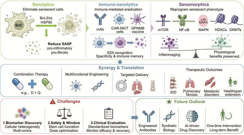
With the deepening understanding of aging biology, intervention strategies targeting SnCs have evolved from simplistic chemical screening into a multidimensional and precise pharmacological framework. As shown in Figure 6, this review has systematically summarized the latest advances in both basic research and clinical translation concerning senolytics, immuno-senolytics, and senomorphics [12, 192, 230].
Therapeutic strategies targeting cellular senescence: from elimination to reprogramming and future translation. The schematic outlines three core modalities: Senolytics, Immuno-senolytics and Senomorphics. Central goals include reducing the pro-inflammatory, pro-fibrotic SASP, achieving specificity through SSA recognition, establishing immune memory, and preserving physiological benefits while counteracting inflammaging. The synergy & translation panel highlights combination therapy (e.g., D + Q), multifunctional engineering, and targeted delivery. Key challenges—biomarker discovery via multi-omics, safety and therapeutic window (stem cell function, dose optimization), and standardized clinical evaluation—are listed alongside a future outlook embracing engineered antibodies, synthetic biology, AI-driven drug discovery, and one-time interventions for long-term benefit.
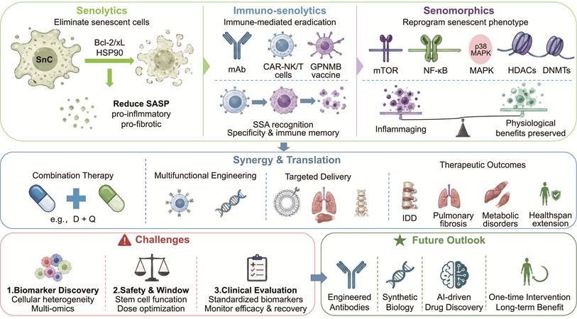
First, the evolution of intervention paradigms has enabled the systemic remodeling of the senescence microenvironment. By targeting pro-survival signaling pathways such as BCL-2/BCL-XL and HSP90, Senolytics selectively induce SnCs apoptosis, thereby reducing the reservoir of pro-inflammatory and pro-fibrotic SASP factors at the source [87, 88]. Immuno-senolytics, including monoclonal antibodies, engineered CAR-NK/T cells, and GPNMB vaccines, precisely recognize senescence-specific antigens, offering highly specific and durable protection with endogenous immune memory for age-related diseases. Senomorphics represent a “non-ablative” intervention paradigm; by inhibiting core signaling axes such as mTOR/NF-κB/p38 MAPK or correcting epigenetic dysregulation (e.g., modulation of HDACs and DNMTs), they markedly alleviate “inflammaging” while preserving the physiological benefits of senescent cells [194].
Second, multi-target synergy and tissue-specific delivery have become pivotal for enhancing therapeutic efficacy. Evidence demonstrates that the limitations of single-target approaches are being overcome through combination regimens (e.g., the D+Q protocol) or multifunctional engineering strategies. In various models of ARDs, these strategies not only exhibit remarkable capacity for pathological reversal but also reveal the feasibility of extending healthspan via “immunometabolic reprogramming”. Emerging evidence highlights the advantages of combined or sequential senotherapeutic strategies to address the inherent heterogeneity of senescent cells (SnCs), which limits the efficacy of single-modality approaches. Combination regimens integrating senolytics (e.g., D+Q) with senomorphics or immuno-senolytics have demonstrated synergistic effects by simultaneously clearing SnCs and suppressing residual SASP, leading to superior reductions in inflammation, improved tissue repair, and reduced toxicity compared to monotherapy [231]. Sequential protocols, such as pre-treatment with senomorphics followed by senolytic clearance, further minimize SASP rebound and enhance safety in models of ARDs [232].
Nevertheless, the translational leap to clinical application still faces formidable challenges [233]. Future research should concentrate on the following core scientific questions:
1. Discovery of specific markers: The heterogeneity of SnCs necessitates the integration of single-cell multi-omics technologies to identify tissue- and stage-specific senescence antigens and metabolic targets [234].
2. Balancing safety and therapeutic window: Further evaluation is required to assess the potential disruption of normal stem cell function by Senolytics and to optimize long-term dosing regimens of Senomorphics to circumvent systemic toxicity. Future studies should also focus on optimizing pharmacokinetic profiles through nano-delivery systems (such as exosome- or liposome-based encapsulation [235]) or chemical structural modifications (e.g., prodrug [236] and ADCs [237]) to achieve more precise senescent cell intervention.
3. Construction of a clinical evaluation system: There is an urgent need to establish consensus senescence biomarkers for accurately monitoring changes in SnCs burden and the recovery of tissue physiological function following clinical interventions.
Importantly, while senotherapeutic strategies hold great promise, long-term systemic clearance of senescent cells may carry risks due to the beneficial physiological roles of transient senescent cells. Accumulating evidence indicates that senescent cells contribute positively to tissue repair and wound healing, for instance by secreting PDGF-AA to promote myofibroblast differentiation and accelerate wound closure. A classic study demonstrated that eliminating senescent cells during the acute phase of injury significantly delays tissue repair, highlighting the need to carefully balance senescent cell removal with preservation of their reparative functions to avoid compromising the body's natural damage repair capacity [238].
In summary, anti-aging drug development is at a critical juncture of transition from “broad-spectrum intervention” to “personalized precision modulation”. With the interdisciplinary integration of engineered antibodies, synthetic biology, and artificial intelligence-driven drug screening technologies, a new era of aging intervention characterized by “single intervention, long-term benefit” is rapidly approaching. Furthermore, integrating metabolic reprogramming with microecological regulation, such as exploiting gut microbiota metabolites to alleviate systemic inflammaging and immune senescence, represents a highly promising frontier for next-generation senomorphic development.
Acknowledgements
Funding
This work was supported by National Natural Science Foundation of China [82404874], Natural Science Foundation of Sichuan [2025ZNSFSC1733, 2026NSFSC0626], Scientific and Technological Innovation Team for Qinghai-Tibetan Plateau Research in Southwest Minzu University [2024CXTD15], Sichuan Province Traditional Chinese Medicine Research Project [25MSZX428], Chengdu Medical Research Project [2024036], Dazhou Science and Technology Project [25YYJC0026], Dazhou Traditional Chinese Medicine Research Project [2025LHZRZD02], Sichuan Province Youth Medical Science Research Project [20250717-01], and Incentive Support Funding for the Personalized Drug Research and Therapy Key Laboratory of Sichuan Province [2025JDPT0081].
Author contributions
Qixiong Zhang and Shanshan Li were responsible for the design of conception and critically reviewing this article. Shasha Ran, Siyu Zhou and Haotian Ma contributed to the formation of the draft of the manuscript. Fang Zhang, Yanrui Yang, Xiujuan Yang and Maiheliya·Abulajiang are responsible for the design of figures in the manuscript. All authors agree to be responsible for all aspects of the work.
Competing Interests
The authors have declared that no competing interest exists.
References
1. Chang AY, Bolongaita S, Cao B, Castro MC, Karlsson O, Mao W. et al. Epidemiological and demographic trends and projections in global health from 1970 to 2050: a descriptive analysis from the third Lancet Commission on Investing in Health, Global Health 2050. Lancet. 2025;406:940-949
2. Childs BG, Durik M, Baker DJ, van Deursen JM. Cellular senescence in aging and age-related disease: from mechanisms to therapy. Nat Med. 2015;21:1424-1435
3. Tian Y, Shao S, Feng H, Zeng R, Li S, Zhang Q. Targeting senescent cells in atherosclerosis: Pathways to novel therapies. Ageing Res Rev. 2024;101:102502
4. Zhang K, Ma Y, Luo Y, Song Y, Xiong G, Ma Y. et al. Metabolic diseases and healthy aging: identifying environmental and behavioral risk factors and promoting public health. Front Public Health. 2023;11:1253506
5. GBD 2023 Demographics Collaborators. Global age-sex-specific all-cause mortality and life expectancy estimates for 204 countries and territories and 660 subnational locations, 1950-2023: a demographic analysis for the Global Burden of Disease Study 2023. Lancet. 2025;406:1731-1810
6. Herranz N, Gil J. Mechanisms and functions of cellular senescence. J Clin Invest. 2018;128:1238-1246
7. Di Micco R, Krizhanovsky V, Baker D, d'Adda di Fagagna F. Cellular senescence in ageing: from mechanisms to therapeutic opportunities. Nat Rev Mol Cell Biol. 2021;22:75-95
8. Li X, Chen M, Chen X, He X, Li X, Wei H. et al. TRAP1 drives smooth muscle cell senescence and promotes atherosclerosis via HDAC3-primed histone H4 lysine 12 lactylation. Eur Heart J. 2024;45:4219-4235
9. Björkegren JLM, Lusis AJ. Atherosclerosis: Recent developments. Cell. 2022;185:1630-1645
10. Feng X, Chen W, Ni X, Little PJ, Xu S, Tang L. et al. Metformin, Macrophage Dysfunction and Atherosclerosis. Front Immunol. 2021;12:682853
11. Gasek NS, Kuchel GA, Kirkland JL, Xu M. Strategies for Targeting Senescent Cells in Human Disease. Nat Aging. 2021;1:870-879
12. Zhang L, Pitcher LE, Prahalad V, Niedernhofer LJ, Robbins PD. Targeting cellular senescence with senotherapeutics: senolytics and senomorphics. FEBS J. 2023;290:1362-1383
13. Pereira BI, Devine OP, Vukmanovic-Stejic M, Chambers ES, Subramanian P, Patel N. et al. Senescent cells evade immune clearance via HLA-E-mediated NK and CD8(+) T cell inhibition. Nat Commun. 2019;10:2387
14. Birch J, Gil J. Senescence and the SASP: many therapeutic avenues. Genes Dev. 2020;34:1565-1576
15. Rossiello F, Jurk D, Passos JF, d'Adda di Fagagna F. Telomere dysfunction in ageing and age-related diseases. Nat Cell Biol. 2022;24:135-147
16. Ogrodnik M. Cellular aging beyond cellular senescence: Markers of senescence prior to cell cycle arrest in vitro and in vivo. Aging Cell. 2021;20:e13338
17. Fernandez C, Ormeno D, Villalobos V, Garrido M, Canelo J, Cerda O. et al. Characterization of senescence and nuclear reorganization in aging gingival cells. NPJ Aging. 2025;11:12
18. Turner KJ, Vasu V, Griffin DK. Telomere Biology and Human Phenotype. Cells. 2019 8
19. Slawinska N, Krupa R. Molecular Aspects of Senescence and Organismal Ageing-DNA Damage Response, Telomeres, Inflammation and Chromatin. Int J Mol Sci. 2021 22
20. Lascarez-Lagunas LI, Nadarajan S, Martinez-Garcia M, Quinn JN, Todisco E, Thakkar T. et al. ATM/ATR kinases link the synaptonemal complex and DNA double-strand break repair pathway choice. Curr Biol. 2022;32:4719-4726 e4714
21. Li C, Chen H, Chen X, Wang P, Shi Y, Xie X. et al. Identification of inflammatory response-related molecular mechanisms based on the ATM/ATR/p53 pathway in tumor cells. Comput Biol Med. 2024;180:108776
22. Yu B, Ma J, Li J, Wang D, Wang Z, Wang S. Mitochondrial phosphatase PGAM5 modulates cellular senescence by regulating mitochondrial dynamics. Nat Commun. 2020;11:2549
23. Georgoulis A, Vorgias CE, Chrousos GP, Rogakou EP. Genome Instability and gammaH2AX. Int J Mol Sci. 2017 18
24. Zhang Q, Shen Y, Zhang C, Zhang H, Li X, Yang S. et al. Immunoengineered mitochondria for efficient therapy of acute organ injuries via modulation of inflammation and cell repair. Sci Adv. 2025;11:eadj1896
25. Wang Z, Wang L, Ji M, Lou J, Yuan Y, Cui Y. et al. Mitochondrial dysfunction in immune cells during the perioperative period: mechanisms, emerging therapeutic strategies, and implications for multi-organ protection. Front Immunol. 2026;17:1734880
26. Liu Z, Liang Q, Ren Y, Guo C, Ge X, Wang L. et al. Immunosenescence: molecular mechanisms and diseases. Signal Transduct Target Ther. 2023;8:200
27. Klutstein M, Nejman D, Greenfield R, Cedar H. DNA Methylation in Cancer and Aging. Cancer Research. 2016:3446-3450
28. Deng P, Yuan Q, Cheng Y, Li J, Liu Z, Liu Y. et al. Loss of KDM4B exacerbates bone-fat imbalance and mesenchymal stromal cell exhaustion in skeletal aging. Cell Stem Cell. 2021;28:1057-1073 e1057
29. Dutchak K, Garnett S, Nicoll M, de Bruyns A, Dankort D. MOB3A Bypasses BRAF and RAS Oncogene-Induced Senescence by Engaging the Hippo Pathway. Mol Cancer Res. 2022;20:770-781
30. Garnett S, de Bruyns A, Provencher-Tom V, Dutchak K, Shu R, Dankort D. Metabolic Regulator IAPP (Amylin) Is Required for BRAF and RAS Oncogene-Induced Senescence. Mol Cancer Res. 2021;19:874-885
31. Barnes JL, Zubair M, John K, Poirier MC, Martin FL. Carcinogens and DNA damage. Biochemical Society Transactions. 2018 46
32. Deng Y, Chen Q, Yang X, Sun Y, Zhang B, Wei W. et al. Tumor cell senescence-induced macrophage CD73 expression is a critical metabolic immune checkpoint in the aging tumor microenvironment. Theranostics. 2024;14:1224-1240
33. Hari P, Millar FR, Tarrats N, Birch J, Quintanilla A, Rink CJ. et al. The innate immune sensor Toll-like receptor 2 controls the senescence-associated secretory phenotype. Sci Adv. 2019;5:eaaw0254
34. Jomova K, Raptova R, Alomar SY, Alwasel SH, Nepovimova E, Kuca K. et al. Reactive oxygen species, toxicity, oxidative stress, and antioxidants: chronic diseases and aging. Arch Toxicol. 2023;97:2499-2574
35. Sun T, Zhang L, Feng J, Bao L, Wang J, Song Z. et al. Characterization of cellular senescence in doxorubicin-induced aging mice. Exp Gerontol. 2022;163:111800
36. Mo X, Cheng R, Shen L, Sun Y, Wang P, Jiang G. et al. High-fat diet induces sarcopenic obesity in natural aging rats through the gut-trimethylamine N-oxide-muscle axis. J Adv Res. 2025;70:405-422
37. Ottone OK, Kim CJ, Collins JA, Risbud MV. The cGAS-STING Pathway Affects Vertebral Bone but Does Not Promote Intervertebral Disc Cell Senescence or Degeneration. Front Immunol. 2022;13:882407
38. Zhang J, Rong Y, Luo C, Cui W. Bone marrow mesenchymal stem cell-derived exosomes prevent osteoarthritis by regulating synovial macrophage polarization. Aging (Albany NY). 2020;12:25138-25152
39. Bateman G, Guo-Parke H, Rodgers AM, Linden D, Bailey M, Weldon S. et al. Airway Epithelium Senescence as a Driving Mechanism in COPD Pathogenesis. Biomedicines. 2023 11
40. Wu Q, Leng X, Zhang Q, Zhu YZ, Zhou R, Liu Y. et al. IRF3 activates RB to authorize cGAS-STING-induced senescence and mitigate liver fibrosis. Sci Adv. 2024;10:eadj2102
41. Zhang M, Bi X, Liu S, Liu Y, Wang Q. The novel polyfluoroalkyl benzenesulfonate OBS exposure induces cell cycle arrest and senescence of rat pituitary cell GH3 via the p53/p21/RB pathway. Toxicology. 2023;490:153511
42. Ke Y, Li D, Zhao M, Liu C, Liu J, Zeng A. et al. Gut flora-dependent metabolite Trimethylamine-N-oxide accelerates endothelial cell senescence and vascular aging through oxidative stress. Free Radic Biol Med. 2018;116:88-100
43. Xu T, Liu J, Li XR, Yu Y, Luo X, Zheng X. et al. The mTOR/NF-kappaB Pathway Mediates Neuroinflammation and Synaptic Plasticity in Diabetic Encephalopathy. Mol Neurobiol. 2021;58:3848-3862
44. Kirichenko TV, Markina YV, Markin AM, Vasilyev VS, Hua H, Li D. et al. Functional Features of Senescent Cells and Implications for Therapy. Int J Mol Sci. 2025 26
45. López-Otín C, Blasco MA, Partridge L, Serrano M, Kroemer G. Hallmarks of aging: An expanding universe. Cell. 2023;186:243-278
46. Guo S, Tang Q, Gao X, Hu L, Hu K, Zhang H. et al. EZH2 inhibition induces senescence via ERK1/2 signaling pathway in multiple myeloma. Acta Biochim Biophys Sin (Shanghai). 2024;56:1055-1064
47. Herbstein F, Sapochnik M, Attorresi A, Pollak C, Senin S, Gonilski-Pacin D. et al. The SASP factor IL-6 sustains cell-autonomous senescent cells via a cGAS-STING-NFkappaB intracrine senescent noncanonical pathway. Aging Cell. 2024;23:e14258
48. Horvath E, Solyom A, Szekely J, Nagy EE, Popoviciu H. Inflammatory and Metabolic Signaling Interfaces of the Hypertrophic and Senescent Chondrocyte Phenotypes Associated with Osteoarthritis. Int J Mol Sci. 2023 24
49. Kandhaya-Pillai R, Yang X, Tchkonia T, Martin GM, Kirkland JL, Oshima J. TNF-alpha/IFN-gamma synergy amplifies senescence-associated inflammation and SARS-CoV-2 receptor expression via hyper-activated JAK/STAT1. Aging Cell. 2022;21:e13646
50. Takasugi M, Yoshida Y, Hara E, Ohtani N. The role of cellular senescence and SASP in tumour microenvironment. Febs j. 2023;290:1348-1361
51. Wei L, Yang X, Wang J, Wang Z, Wang Q, Ding Y. et al. H3K18 lactylation of senescent microglia potentiates brain aging and Alzheimer's disease through the NFkappaB signaling pathway. J Neuroinflammation. 2023;20:208
52. Natarajan V, Chawla R, Mah T, Vivekanandan R, Tan SY, Sato PY. et al. Mitochondrial Dysfunction in Age-Related Metabolic Disorders. Proteomics. 2020;20:e1800404
53. Farr JN, Khosla S. Cellular senescence in bone. Bone. 2019;121:121-133
54. Gerosa L, Malvandi AM, Malavolta M, Provinciali M, Lombardi G. Exploring cellular senescence in the musculoskeletal system: Any insights for biomarkers discovery? Ageing Res Rev. 2023;88:101943
55. Faleeva M, Ahmad S, Theofilatos K, Lynham S, Watson G, Whitehead M. et al. Sox9 Accelerates Vascular Aging by Regulating Extracellular Matrix Composition and Stiffness. Circ Res. 2024;134:307-324
56. Gulen MF, Samson N, Keller A, Schwabenland M, Liu C, Glück S. et al. cGAS-STING drives ageing-related inflammation and neurodegeneration. Nature. 2023;620:374-380
57. He M, Zhou P, Chen H, Zhang J, Zhang Y, Zheng X. et al. (-)-α-Bisabolol inhibits D-Gal-induced HSF cellular senescence in vitro and prevents skin aging in vivo by reducing SASP. Iran J Basic Med Sci. 2024;27:1105-1114
58. Kirschner K, Rattanavirotkul N, Quince MF, Chandra T. Functional heterogeneity in senescence. Biochem Soc Trans. 2020;48:765-773
59. Fu Y, Zhang J, Liu Q, Yang L, Wu Q, Yang X. et al. Unveiling the role of ABI3 and hub senescence-related genes in macrophage senescence for atherosclerotic plaque progression. Inflamm Res. 2024;73:65-82
60. Baker DJ, Petersen RC. Cellular senescence in brain aging and neurodegenerative diseases: evidence and perspectives. J Clin Invest. 2018;128:1208-1216
61. Si Z, Sun L, Wang X. Evidence and perspectives of cell senescence in neurodegenerative diseases. Biomed Pharmacother. 2021;137:111327
62. Jeon OH, Kim C, Laberge RM, Demaria M, Rathod S, Vasserot AP. et al. Local clearance of senescent cells attenuates the development of post-traumatic osteoarthritis and creates a pro-regenerative environment. Nat Med. 2017;23:775-781
63. Yao C, Guan X, Carraro G, Parimon T, Liu X, Huang G. et al. Senescence of Alveolar Type 2 Cells Drives Progressive Pulmonary Fibrosis. Am J Respir Crit Care Med. 2021;203:707-717
64. Feng Q, Su C, Yang C, Wu M, Li X, Lin X. et al. RXRalpha/MR signaling promotes diabetic kidney disease by facilitating renal tubular epithelial cells senescence and metabolic reprogramming. Transl Res. 2024;274:101-117
65. Roh JD, Hobson R, Chaudhari V, Quintero P, Yeri A, Benson M. et al. Activin type II receptor signaling in cardiac aging and heart failure. Sci Transl Med. 2019 11
66. Salerno N, Marino F, Scalise M, Salerno L, Molinaro C, Filardo A. et al. Pharmacological clearance of senescent cells improves cardiac remodeling and function after myocardial infarction in female aged mice. Mech Ageing Dev. 2022;208:111740
67. Green GS, Fujita M, Yang HS, Taga M, Cain A, McCabe C. et al. Cellular communities reveal trajectories of brain ageing and Alzheimer's disease. Nature. 2024;633:634-645
68. Shen QQ, Jv XH, Ma XZ, Li C, Liu L, Jia WT. et al. Cell senescence induced by toxic interaction between α-synuclein and iron precedes nigral dopaminergic neuron loss in a mouse model of Parkinson's disease. Acta Pharmacol Sin. 2024;45:268-281
69. Schafer MJ, White TA, Iijima K, Haak AJ, Ligresti G, Atkinson EJ. et al. Cellular senescence mediates fibrotic pulmonary disease. Nat Commun. 2017;8:14532
70. Devulder JV, Baker JR, Fenwick PS, Odqvist L, Donnelly LE, Barnes PJ. Chronic Obstructive Pulmonary Disease Airway Epithelial Cell-derived Extracellular Vesicles Spread Cellular Senescence via MicroRNA-34a. Am J Respir Cell Mol Biol. 2025;73:210-220
71. Ha S, Kim HW, Kim KM, Kim BM, Kim J, Son M. et al. PAR2-mediated cellular senescence promotes inflammation and fibrosis in aging and chronic kidney disease. Aging Cell. 2024;23:e14184
72. Shen S, Ji C, Wei K. Cellular Senescence and Regulated Cell Death of Tubular Epithelial Cells in Diabetic Kidney Disease. Front Endocrinol (Lausanne). 2022;13:924299
73. Chandra A, Rajawat J. Skeletal Aging and Osteoporosis: Mechanisms and Therapeutics. Int J Mol Sci. 2021 22
74. Du K, Maeso-Diaz R, Oh SH, Wang E, Chen T, Pan C. et al. Targeting YAP-mediated HSC death susceptibility and senescence for treatment of liver fibrosis. Hepatology. 2023;77:1998-2015
75. Nashine S. Potential Therapeutic Candidates for Age-Related Macular Degeneration (AMD). Cells. 2021 10
76. Franco AC, Aveleira C, Cavadas C. Skin senescence: mechanisms and impact on whole-body aging. Trends Mol Med. 2022;28:97-109
77. Samarawickrama PN, Zhang G, Zhu E, Dong X, Nisar A, Zhu H. et al. Clearance of senescent cells enhances skin wound healing in type 2 diabetic mice. Theranostics. 2024;14:5429-5442
78. Zhang L, Pitcher LE, Yousefzadeh MJ, Niedernhofer LJ, Robbins PD, Zhu Y. Cellular senescence: a key therapeutic target in aging and diseases. J Clin Invest. 2022 132
79. Kay MM. Aging of cell membrane molecules leads to appearance of an aging antigen and removal of senescent cells. Gerontology. 1985;31:215-235
80. Kirkland JL, Tchkonia T. Cellular Senescence: A Translational Perspective. EBioMedicine. 2017;21:21-28
81. Mylonas KJ, O'Sullivan ED, Humphries D, Baird DP, Docherty MH, Neely SA. et al. Cellular senescence inhibits renal regeneration after injury in mice, with senolytic treatment promoting repair. Sci Transl Med. 2021 13
82. Palmer AK, Xu M, Zhu Y, Pirtskhalava T, Weivoda MM, Hachfeld CM. et al. Targeting senescent cells alleviates obesity-induced metabolic dysfunction. Aging Cell. 2019;18:e12950
83. Salminen A, Kaarniranta K, Kauppinen A. Immunosenescence: the potential role of myeloid-derived suppressor cells (MDSC) in age-related immune deficiency. Cell Mol Life Sci. 2019;76:1901-1918
84. Atasever E, Atayik MC, Cakatay U. Senotherapeutics: Milestones, innovations, and future prospects. Adv Pharmacol. 2025;104:1-35
85. Shao Z, Wang B, Shi Y, Xie C, Huang C, Chen B. et al. Senolytic agent Quercetin ameliorates intervertebral disc degeneration via the Nrf2/NF-kappaB axis. Osteoarthritis Cartilage. 2021;29:413-422
86. Liu L, Yue X, Sun Z, Hambright WS, Wei J, Li Y. et al. Reduction of senescent fibro-adipogenic progenitors in progeria-aged muscle by senolytics rescues the function of muscle stem cells. J Cachexia Sarcopenia Muscle. 2022;13:3137-3148
87. Chaib S, Tchkonia T, Kirkland JL. Cellular senescence and senolytics: the path to the clinic. Nat Med. 2022;28:1556-1568
88. Power H, Valtchev P, Dehghani F, Schindeler A. Strategies for senolytic drug discovery. Aging Cell. 2023;22:e13948
89. Wang E. Senescent human fibroblasts resist programmed cell death, and failure to suppress bcl2 is involved. Cancer Res. 1995;55:2284-2292
90. Yip KW, Reed JC. Bcl-2 family proteins and cancer. Oncogene. 2008;27:6398-6406
91. Cory S, Huang DC, Adams JM. The Bcl-2 family: roles in cell survival and oncogenesis. Oncogene. 2003;22:8590-8607
92. Azmi AS, Wang Z, Philip PA, Mohammad RM, Sarkar FH. Emerging Bcl-2 inhibitors for the treatment of cancer. Expert Opin Emerg Drugs. 2011;16:59-70
93. Zeitlin BD, Zeitlin IJ, Nor JE. Expanding circle of inhibition: small-molecule inhibitors of Bcl-2 as anticancer cell and antiangiogenic agents. J Clin Oncol. 2008;26:4180-4188
94. Tse C, Shoemaker AR, Adickes J, Anderson MG, Chen J, Jin S. et al. ABT-263: a potent and orally bioavailable Bcl-2 family inhibitor. Cancer Res. 2008;68:3421-3428
95. Yosef R, Pilpel N, Tokarsky-Amiel R, Biran A, Ovadya Y, Cohen S. et al. Directed elimination of senescent cells by inhibition of BCL-W and BCL-XL. Nat Commun. 2016;7:11190
96. Zhu Y, Tchkonia T, Fuhrmann-Stroissnigg H, Dai HM, Ling YY, Stout MB. et al. Identification of a novel senolytic agent, navitoclax, targeting the Bcl-2 family of anti-apoptotic factors. Aging Cell. 2016;15:428-435
97. Oltersdorf T, Elmore SW, Shoemaker AR, Armstrong RC, Augeri DJ, Belli BA. et al. An inhibitor of Bcl-2 family proteins induces regression of solid tumours. Nature. 2005;435:677-681
98. Ashkenazi A, Fairbrother WJ, Leverson JD, Souers AJ. From basic apoptosis discoveries to advanced selective BCL-2 family inhibitors. Nat Rev Drug Discov. 2017;16:273-284
99. Knight T, Luedtke D, Edwards H, Taub JW, Ge Y. A delicate balance - The BCL-2 family and its role in apoptosis, oncogenesis, and cancer therapeutics. Biochemical pharmacology. 2019;162:250-261
100. Zhu Y, Doornebal EJ, Pirtskhalava T, Giorgadze N, Wentworth M, Fuhrmann-Stroissnigg H. et al. New agents that target senescent cells: the flavone, fisetin, and the BCL-X(L) inhibitors, A1331852 and A1155463. Aging (Albany NY). 2017;9:955-963
101. Sun R, Feng J, Wang J. Underlying Mechanisms and Treatment of Cellular Senescence-Induced Biological Barrier Interruption and Related Diseases. Aging Dis. 2024;15:612-639
102. Erem S, Razzaque MS. Dietary phosphate toxicity: an emerging global health concern. Histochem Cell Biol. 2018;150:711-719
103. Cui Z, Zhao X, Amevor FK, Du X, Wang Y, Li D. et al. Therapeutic application of quercetin in aging-related diseases: SIRT1 as a potential mechanism. Front Immunol. 2022;13:943321
104. Hickson LJ, Langhi Prata LGP, Bobart SA, Evans TK, Giorgadze N, Hashmi SK. et al. Senolytics decrease senescent cells in humans: Preliminary report from a clinical trial of Dasatinib plus Quercetin in individuals with diabetic kidney disease. EBioMedicine. 2019;47:446-456
105. Zhang P, Kishimoto Y, Grammatikakis I, Gottimukkala K, Cutler RG, Zhang S. et al. Senolytic therapy alleviates Abeta-associated oligodendrocyte progenitor cell senescence and cognitive deficits in an Alzheimer's disease model. Nat Neurosci. 2019;22:719-728
106. Kong YX, Li ZS, Liu YB, Pan B, Fu X, Xiao R. et al. FOXO4-DRI induces keloid senescent fibroblast apoptosis by promoting nuclear exclusion of upregulated p53-serine 15 phosphorylation. Commun Biol. 2025;8:299
107. Huang Y, He Y, Makarcyzk MJ, Lin H. Senolytic Peptide FOXO4-DRI Selectively Removes Senescent Cells From in vitro Expanded Human Chondrocytes. Front Bioeng Biotechnol. 2021;9:677576
108. Meng J, Li Y, Wan C, Sun Y, Dai X, Huang J. et al. Targeting senescence-like fibroblasts radiosensitizes non-small cell lung cancer and reduces radiation-induced pulmonary fibrosis. JCI Insight. 2021 6
109. Born E, Lipskaia L, Breau M, Houssaini A, Beaulieu D, Marcos E. et al. Eliminating Senescent Cells Can Promote Pulmonary Hypertension Development and Progression. Circulation. 2023;147:650-666
110. Fuhrmann-Stroissnigg H, Niedernhofer LJ, Robbins PD. Hsp90 inhibitors as senolytic drugs to extend healthy aging. Cell Cycle. 2018;17:1048-1055
111. Chan KC, Ting CM, Chan PS, Lo MC, Lo KW, Curry JE. et al. A novel Hsp90 inhibitor AT13387 induces senescence in EBV-positive nasopharyngeal carcinoma cells and suppresses tumor formation. Mol Cancer. 2013;12:128
112. Han SY, Ko A, Kitano H, Choi CH, Lee MS, Seo J. et al. Molecular Chaperone HSP90 Is Necessary to Prevent Cellular Senescence via Lysosomal Degradation of p14ARF. Cancer Res. 2017;77:343-354
113. Fuhrmann-Stroissnigg H, Ling YY, Zhao J, McGowan SJ, Zhu Y, Brooks RW. et al. Identification of HSP90 inhibitors as a novel class of senolytics. Nat Commun. 2017;8:422
114. Dutta Gupta S, Pan CH. Recent update on discovery and development of Hsp90 inhibitors as senolytic agents. Int J Biol Macromol. 2020;161:1086-1098
115. Ji XM, Dong XX, Li JP, Tai GJ, Qiu S, Wei W. et al. Fisetin Clears Senescent Cells Through the Pi3k-Akt-Bcl-2/Bcl-xl Pathway to Alleviate Diabetic Aortic Aging. Phytother Res. 2025
116. Gendrisch F, Esser PR, Schempp CM, Wölfle U. Luteolin as a modulator of skin aging and inflammation. Biofactors. 2021;47:170-180
117. Wu CC, Chen YR, Lu DH, Hsu LH, Yang KC, Sumi S. Evaluation of the post-treatment anti-inflammatory capacity of osteoarthritic chondrocytes: An in vitro study using baicalein. Regen Ther. 2020;14:177-183
118. Gherardi G, Corbioli G, Ruzza F, Rizzuto R. CoQ(10) and Resveratrol Effects to Ameliorate Aged-Related Mitochondrial Dysfunctions. Nutrients. 2022 14
119. Zhu L, Liu Z, Ren Y, Wu X, Liu Y, Wang T. et al. Neuroprotective effects of salidroside on ageing hippocampal neurons and naturally ageing mice via the PI3K/Akt/TERT pathway. Phytother Res. 2021;35:5767-5780
120. Tavenier J, Nehlin JO, Houlind MB, Rasmussen LJ, Tchkonia T, Kirkland JL. et al. Fisetin as a senotherapeutic agent: Evidence and perspectives for age-related diseases. Mech Ageing Dev. 2024;222:111995
121. Senolytic Drugs Attenuate Osteoarthritis-Related Articular Cartilage Degeneration. A Clinical Trial. In: United States Department of D, Office of Naval R, editors. 2019
122. Targeting Cellular Senescence With Senolytics to Improve Skeletal Health in Older Humans. A Phase 2, Single-Center, 20-week, Open-Label, Randomized Controlled Trial. 2020.
123. Phase 2 Clinical Trial of FIsetin to Treat CArpal Tunnel Syndrome (FITCATS). 2022
124. Mahoney SA, Venkatasubramanian R, Darrah MA, Ludwig KR, VanDongen NS, Greenberg NT. et al. Intermittent supplementation with fisetin improves arterial function in old mice by decreasing cellular senescence. Aging Cell. 2024;23:e14060
125. Yousefzadeh MJ, Zhu Y, McGowan SJ, Angelini L, Fuhrmann-Stroissnigg H, Xu M. et al. Fisetin is a senotherapeutic that extends health and lifespan. EBioMedicine. 2018;36:18-28
126. Murray KO, Mahoney SA, Ludwig KR, Miyamoto-Ditmon JH, VanDongen NS, Banskota N. et al. Intermittent Supplementation With Fisetin Improves Physical Function and Decreases Cellular Senescence in Skeletal Muscle With Aging: A Comparison to Genetic Clearance of Senescent Cells and Synthetic Senolytic Approaches. Aging Cell. 2025;24:e70114
127. Huard CA, Gao X, Dey Hazra ME, Dey Hazra RO, Lebsock K, Easley JT. et al. Effects of Fisetin Treatment on Cellular Senescence of Various Tissues and Organs of Old Sheep. Antioxidants (Basel). 2023 12
128. Zhu RZ, Li BS, Gao SS, Seo JH, Choi BM. Luteolin inhibits H(2)O(2)-induced cellular senescence via modulation of SIRT1 and p53. Korean J Physiol Pharmacol. 2021;25:297-305
129. Yan Y, Huang H, Su T, Huang W, Wu X, Chen X. et al. Luteolin Mitigates Photoaging Caused by UVA-Induced Fibroblast Senescence by Modulating Oxidative Stress Pathways. Int J Mol Sci. 2025 26
130. Zumerle S, Sarill M, Saponaro M, Colucci M, Contu L, Lazzarini E. et al. Targeting senescence induced by age or chemotherapy with a polyphenol-rich natural extract improves longevity and healthspan in mice. Nat Aging. 2024;4:1231-1248
131. Huang L, Peng B, Nayak Y, Wang C, Si F, Liu X. et al. Baicalein and Baicalin Promote Melanoma Apoptosis and Senescence via Metabolic Inhibition. Front Cell Dev Biol. 2020;8:836
132. Ji-Hong Y, Yu M, Ling-Hong Y, Jing-Jing G, Ling-Li X, Lv W. et al. Baicalein attenuates bleomycin-induced lung fibroblast senescence and lung fibrosis through restoration of Sirt3 expression. Pharm Biol. 2023;61:288-297
133. Pyo IS, Yun S, Yoon YE, Choi JW, Lee SJ. Mechanisms of Aging and the Preventive Effects of Resveratrol on Age-Related Diseases. Molecules. 2020 25
134. Zhou DD, Luo M, Huang SY, Saimaiti A, Shang A, Gan RY. et al. Effects and Mechanisms of Resveratrol on Aging and Age-Related Diseases. Oxid Med Cell Longev. 2021;2021:9932218
135. Novelle MG, Wahl D, Diéguez C, Bernier M, de Cabo R. Resveratrol supplementation: Where are we now and where should we go? Ageing Res Rev. 2015;21:1-15
136. Zhang LX, Li CX, Kakar MU, Khan MS, Wu PF, Amir RM. et al. Resveratrol (RV): A pharmacological review and call for further research. Biomed Pharmacother. 2021;143:112164
137. Calabrese EJ, Dhawan G, Kapoor R, Agathokleous E, Calabrese V. Rhodiola rosea and salidroside commonly induce hormesis, with particular focus on longevity and neuroprotection. Chem Biol Interact. 2023;380:110540
138. Zhang J, Li Z, Song J, Zhou L, Chen X, Ge W. et al. Salidroside promotes healthy longevity by interfering with HSP90 activity. Geroscience. 2024;46:1641-1655
139. Zhang N, Nao J, Dong X. Neuroprotective Mechanisms of Salidroside in Alzheimer's Disease: A Systematic Review and Meta-analysis of Preclinical Studies. J Agric Food Chem. 2023;71:17597-17614
140. Lee H, Wilson D, Bunting KV, Kotecha D, Jackson T. Repurposing digoxin for geroprotection in patients with frailty and multimorbidity. Ageing Res Rev. 2023;86:101860
141. Martin N, Soriani O, Bernard D. Cardiac Glycosides as Senolytic Compounds. Trends Mol Med. 2020;26:243-245
142. Guerrero A, Herranz N, Sun B, Wagner V, Gallage S, Guiho R. et al. Cardiac glycosides are broad-spectrum senolytics. Nat Metab. 2019;1:1074-1088
143. Triana-Martinez F, Picallos-Rabina P, Da Silva-Alvarez S, Pietrocola F, Llanos S, Rodilla V. et al. Identification and characterization of Cardiac Glycosides as senolytic compounds. Nat Commun. 2019;10:4731
144. L'Hote V, Courbeyrette R, Pinna G, Cintrat JC, Le Pavec G, Delaunay-Moisan A. et al. Ouabain and chloroquine trigger senolysis of BRAF-V600E-induced senescent cells by targeting autophagy. Aging Cell. 2021;20:e13447
145. Deryabin PI, Shatrova AN, Borodkina AV. Apoptosis resistance of senescent cells is an intrinsic barrier for senolysis induced by cardiac glycosides. Cell Mol Life Sci. 2021;78:7757-7776
146. Imanishi T, Kobayashi K, Kuroi A, Ikejima H, Akasaka T. Pioglitazone inhibits angiotensin II-induced senescence of endothelial progenitor cell. Hypertens Res. 2008;31:757-765
147. Nogueira-Recalde U, Lorenzo-Gomez I, Blanco FJ, Loza MI, Grassi D, Shirinsky V. et al. Fibrates as drugs with senolytic and autophagic activity for osteoarthritis therapy. EBioMedicine. 2019;45:588-605
148. Xu L, Ma X, Verma N, Perie L, Pendse J, Shamloo S. et al. PPARgamma agonists delay age-associated metabolic disease and extend longevity. Aging Cell. 2020;19:e13267
149. Cai Y, Liu H, Song E, Wang L, Xu J, He Y. et al. Deficiency of telomere-associated repressor activator protein 1 precipitates cardiac aging in mice via p53/PPARalpha signaling. Theranostics. 2021;11:4710-4727
150. Liu X, Gu Y, Kumar S, Amin S, Guo Q, Wang J. et al. Oxylipin-PPARgamma-initiated adipocyte senescence propagates secondary senescence in the bone marrow. Cell Metab. 2023;35:667-684 e666
151. You BJ, Hour MJ, Chen LY, Luo SC, Hsu PH, Lee HZ. Fenofibrate induces human hepatoma Hep3B cells apoptosis and necroptosis through inhibition of thioesterase domain of fatty acid synthase. Sci Rep. 2019;9:3306
152. Jang JH, Lee TJ, Sung EG, Song IH, Kim JY. Pioglitazone mediates apoptosis in Caki cells via downregulating c-FLIP((L)) expression and reducing Bcl-2 protein stability. Oncol Lett. 2021;22:743
153. Pompili S, Vetuschi A, Latella G, Smakaj A, Sferra R, Cappariello A. PPAR-Gamma Orchestrates EMT, AGE, and Cellular Senescence Pathways in Colonic Epithelium and Restrains the Progression of IBDs. Int J Mol Sci. 2023 24
154. Prata L, Ovsyannikova IG, Tchkonia T, Kirkland JL. Senescent cell clearance by the immune system: Emerging therapeutic opportunities. Semin Immunol. 2018;40:101275
155. Amor C, Feucht J, Leibold J, Ho YJ, Zhu C, Alonso-Curbelo D. et al. Senolytic CAR T cells reverse senescence-associated pathologies. Nature. 2020;583:127-132
156. Hu LY, Zhuang WT, Chen MJ, Liao J, Wu DF, Zhang YX. et al. EGFR Oncogenic Mutations in NSCLC Impair Macrophage Phagocytosis and Mediate Innate Immune Evasion Through Up-Regulation of CD47. J Thorac Oncol. 2024;19:1186-1200
157. Gergues M, Bari R, Koppisetti S, Gosiewska A, Kang L, Hariri RJ. Senescence, NK cells, and cancer: navigating the crossroads of aging and disease. Front Immunol. 2025;16:1565278
158. Yang D, Sun B, Li S, Wei W, Liu X, Cui X. et al. NKG2D-CAR T cells eliminate senescent cells in aged mice and nonhuman primates. Sci Transl Med. 2023;15:eadd1951
159. Suda M, Shimizu I, Katsuumi G, Yoshida Y, Hayashi Y, Ikegami R. et al. Senolytic vaccination improves normal and pathological age-related phenotypes and increases lifespan in progeroid mice. Nat Aging. 2021;1:1117-1126
160. Nicolai CJ, Wolf N, Chang IC, Kirn G, Marcus A, Ndubaku CO. et al. NK cells mediate clearance of CD8(+) T cell-resistant tumors in response to STING agonists. Sci Immunol. 2020 5
161. Amor C, Fernández-Maestre I, Chowdhury S, Ho YJ, Nadella S, Graham C. et al. Prophylactic and long-lasting efficacy of senolytic CAR T cells against age-related metabolic dysfunction. Res Sq. 2023
162. Zhang Z, Ma B, Li B, Li Z, Gao M, Zhao H. et al. Cardiolipin-mimic lipid nanoparticles without antibody modification delivered senolytic in vivo CAR-T therapy for inflamm-aging. Cell Rep Med. 2025;6:102209
163. Zhang X, Zhao J, Zhu W, Gou H, Cao D, Yang Y. et al. Synergistic Effect of Subtoxic-dose Cisplatin and TRAIL to Mediate Apoptosis by Down-regulating Decoy Receptor 2 and Up-regulating Caspase-8, Caspase-9 and Bax Expression on NCI-H460 and A549 Cells. Iran J Basic Med Sci. 2013;16:710-718
164. Singh P, Kapahi P, van Deursen JM. Immune checkpoint inhibitors as senolytic agents. Cell Res. 2023;33:197-198
165. Shi H, Wang X, Li F, Gerlach BD, Yurdagul A Jr, Moore MP. et al. CD47-SIRPalpha axis blockade in NASH promotes necroptotic hepatocyte clearance by liver macrophages and decreases hepatic fibrosis. Sci Transl Med. 2022;14:eabp8309
166. Zhang W, Zeng Y, Xiao Q, Wu Y, Liu J, Wang H. et al. An in-situ peptide-antibody self-assembly to block CD47 and CD24 signaling enhances macrophage-mediated phagocytosis and anti-tumor immune responses. Nat Commun. 2024;15:5670
167. Majewska J, Agrawal A, Mayo A, Roitman L, Chatterjee R, Sekeresova Kralova J. et al. p16-dependent increase of PD-L1 stability regulates immunosurveillance of senescent cells. Nat Cell Biol. 2024;26:1336-1345
168. Brauning A, Rae M, Zhu G, Fulton E, Admasu TD, Stolzing A. et al. Aging of the Immune System: Focus on Natural Killer Cells Phenotype and Functions. Cells. 2022 11
169. Maalej KM, Merhi M, Inchakalody VP, Mestiri S, Alam M, Maccalli C. et al. CAR-cell therapy in the era of solid tumor treatment: current challenges and emerging therapeutic advances. Mol Cancer. 2023;22:20
170. Silvestre RN, Eitler J, de Azevedo JTC, Tirapelle MC, Fantacini DMC, de Souza LEB. et al. Engineering NK-CAR.19 cells with the IL-15/IL-15Ralpha complex improved proliferation and anti-tumor effect in vivo. Front Immunol. 2023;14:1226518
171. Xie G, Dong H, Liang Y, Ham JD, Rizwan R, Chen J. CAR-NK cells: A promising cellular immunotherapy for cancer. EBioMedicine. 2020;59:102975
172. Ma Y, Zhao C, Feng J, Gou J, Kang E, Guan F. et al. MSC-sEVs exacerbate senescence by transferring bisecting GlcNAcylated GPNMB. Stem Cell Res Ther. 2025;16:23
173. Cao Y, Du X, Yu J, Wang Y, Jin X, Gu B. et al. Seno-antigen-pulsed dendritic cell vaccine induce anti-aging immunity to improve adipose tissue senescence and metabolic abnormalities. Biomed Pharmacother. 2024;179:117433
174. Suda M, Shimizu I, Katsuumi G, Hsiao CL, Yoshida Y, Matsumoto N. et al. Glycoprotein nonmetastatic melanoma protein B regulates lysosomal integrity and lifespan of senescent cells. Sci Rep. 2022;12:6522
175. Diaz-Ortiz ME, Seo Y, Posavi M, Carceles Cordon M, Clark E, Jain N. et al. GPNMB confers risk for Parkinson's disease through interaction with α-synuclein. Science. 2022;377:eabk0637
176. Iordache F, Petcu ACI, Alexandru DM. Genetic and Epigenetic Interactions Involved in Senescence of Stem Cells. Int J Mol Sci. 2024 25
177. Taylor RN, Berga SL, Zou E, Washington J, Song S, Marzullo BJ. et al. Interleukin-1beta induces and accelerates human endometrial stromal cell senescence and impairs decidualization via the c-Jun N-terminal kinase pathway. Cell Death Discov. 2024;10:288
178. Rovira M, Sereda R, Pladevall-Morera D, Ramponi V, Marin I, Maus M. et al. The lysosomal proteome of senescent cells contributes to the senescence secretome. Aging Cell. 2022;21:e13707
179. Yamada Z, Nishio J, Motomura K, Mizutani S, Yamada S, Mikami T. et al. Senescence of alveolar epithelial cells impacts initiation and chronic phases of murine fibrosing interstitial lung disease. Front Immunol. 2022;13:935114
180. Bennett ZT, Li S, Sumer BD, Gao J. Polyvalent design in the cGAS-STING pathway. Semin Immunol. 2021;56:101580
181. Du H, Xu T, Cui M. cGAS-STING signaling in cancer immunity and immunotherapy. Biomed Pharmacother. 2021;133:110972
182. He X, Wedn A, Wang J, Gu Y, Liu H, Zhang J. et al. IUPHAR ECR review: The cGAS-STING pathway: Novel functions beyond innate immune and emerging therapeutic opportunities. Pharmacol Res. 2024;201:107063
183. Lai P, Liu L, Bancaro N, Troiani M, Calì B, Li Y. et al. Mitochondrial DNA released by senescent tumor cells enhances PMN-MDSC-driven immunosuppression through the cGAS-STING pathway. Immunity. 2025;58:811-825.e817
184. Yang H, Wang H, Ren J, Chen Q, Chen ZJ. cGAS is essential for cellular senescence. Proc Natl Acad Sci U S A. 2017;114:E4612-E4620
185. Li T, Chen ZJ. The cGAS-cGAMP-STING pathway connects DNA damage to inflammation, senescence, and cancer. J Exp Med. 2018;215:1287-1299
186. Tripathi U, Nchioua R, Prata L, Zhu Y, Gerdes EOW, Giorgadze N. et al. SARS-CoV-2 causes senescence in human cells and exacerbates the senescence-associated secretory phenotype through TLR-3. Aging (Albany NY). 2021;13:21838-21854
187. Chibaya L, DeMarco KD, Lusi CF, Kane GI, Brassil ML, Parikh CN. et al. Nanoparticle delivery of innate immune agonists combined with senescence-inducing agents promotes T cell control of pancreatic cancer. Sci Transl Med. 2024;16:eadj9366
188. Mannarino M, Cherif H, Li L, Sheng K, Rabau O, Jarzem P. et al. Toll-like receptor 2 induced senescence in intervertebral disc cells of patients with back pain can be attenuated by o-vanillin. Arthritis Res Ther. 2021;23:117
189. Patel AM, Willingham A, Cheng AC, Tomazela D, Bowman E, Kofman E. et al. Design and Optimization of Selectivity-Tunable Toll-like Receptor 7/8 Agonists as Novel Antibody-Drug Conjugate Payloads. J Med Chem. 2024;67:15756-15779
190. Talukdar A, Ganguly D, Roy S, Das N, Sarkar D. Structural Evolution and Translational Potential for Agonists and Antagonists of Endosomal Toll-like Receptors. J Med Chem. 2021;64:8010-8041
191. Wang Y, Zhang S, Li H, Wang H, Zhang T, Hutchinson MR. et al. Small-Molecule Modulators of Toll-like Receptors. Acc Chem Res. 2020;53:1046-1055
192. Lagoumtzi SM, Chondrogianni N. Senolytics and senomorphics: Natural and synthetic therapeutics in the treatment of aging and chronic diseases. Free Radic Biol Med. 2021;171:169-190
193. Czajkowski K, Herbet M, Murias M, Piatkowska-Chmiel I. Senolytics: charting a new course or enhancing existing anti-tumor therapies? Cell Oncol (Dordr). 2025;48:351-371
194. Martel J, Ojcius DM, Wu CY, Peng HH, Voisin L, Perfettini JL. et al. Emerging use of senolytics and senomorphics against aging and chronic diseases. Med Res Rev. 2020;40:2114-2131
195. Luís C, Maduro AT, Pereira P, Mendes JJ, Soares R, Ramalho R. Nutritional senolytics and senomorphics: Implications to immune cells metabolism and aging - from theory to practice. Frontiers in Nutrition. 2022;9:958563
196. Banerjee S, Biehl A, Gadina M, Hasni S, Schwartz DM. JAK-STAT Signaling as a Target for Inflammatory and Autoimmune Diseases: Current and Future Prospects. Drugs. 2017;77:521-546
197. Traves PG, Murray B, Campigotto F, Galien R, Meng A, Di Paolo JA. JAK selectivity and the implications for clinical inhibition of pharmacodynamic cytokine signalling by filgotinib, upadacitinib, tofacitinib and baricitinib. Ann Rheum Dis. 2021;80:865-875
198. Reece MD, Song C, Hancock SC, Pereira Ribeiro S, Kulpa DA, Gavegnano C. Repurposing BCL-2 and Jak 1/2 inhibitors: Cure and treatment of HIV-1 and other viral infections. Front Immunol. 2022;13:1033672
199. Hao H, Liang W, Zhang S, Cai X, Yakefu A, Gao S. et al. Ruxolitinib Delays Nucleus Pulposus Cell Senescence in Rat Intervertebral Discs. JOR Spine. 2025;8:e70044
200. Alamino VA, Onofrio LI, Acosta CDV, Ferrero PV, Zacca ER, Cadile II. et al. Tofacitinib treatment of rheumatoid arthritis increases senescent T cell frequency in patients and limits T cell function in vitro. Eur J Immunol. 2023;53:e2250353
201. Lawrence T. The Nuclear Factor NF-κB Pathway in Inflammation. Cold Spring Harbor Perspectives in Biology. 2009;1:a001651
202. Wang X, Lu W, Xia X, Zhu Y, Ge C, Guo X. et al. Selenomethionine mitigate PM2.5-induced cellular senescence in the lung via attenuating inflammatory response mediated by cGAS/STING/NF-kappaB pathway. Ecotoxicol Environ Saf. 2022;247:114266
203. Chen H, Tu M, Liu S, Wen Y, Chen L. Dendrobine Alleviates Cellular Senescence and Osteoarthritis via the ROS/NF-kappaB Axis. Int J Mol Sci. 2023 24
204. Dai Q, Wang Z, Wang X, Lian W, Ge Y, Song S. et al. Vorinostat attenuates UVB-induced skin senescence by modulating NF-kappaB and mTOR signaling pathways. Sci Rep. 2025;15:10905
205. Kanigur Sultuybek G, Soydas T, Yenmis G. NF-kappaB as the mediator of metformin's effect on ageing and ageing-related diseases. Clin Exp Pharmacol Physiol. 2019;46:413-422
206. Kawamoto S, Hara E. Crosstalk between gut microbiota and cellular senescence: a vicious cycle leading to aging gut. Trends Cell Biol. 2024;34:626-635
207. Xiao Y, Feng Y, Zhao J, Chen W, Lu W. Achieving healthy aging through gut microbiota-directed dietary intervention: Focusing on microbial biomarkers and host mechanisms. J Adv Res. 2025;68:179-200
208. Ling Z, Liu X, Cheng Y, Yan X, Wu S. Gut microbiota and aging. Crit Rev Food Sci Nutr. 2022;62:3509-3534
209. Rees NP, Conway J, Dugan B, Amir SS, Parker A, Carding SR. et al. Defining Microbiota-Derived Metabolite Butyrate as a Senomorphic: Therapeutic Potential in the Age-Related T Cell Senescence. Aging Cell. 2025;24:e70257
210. Mao B, Zhang Q, Ma L, Zhao DS, Zhao P, Yan P. Overview of Research into mTOR Inhibitors. Molecules. 2022 27
211. Lee DJW, Hodzic Kuerec A, Maier AB. Targeting ageing with rapamycin and its derivatives in humans: a systematic review. Lancet Healthy Longev. 2024;5:e152-e162
212. Blagosklonny MV. From rapalogs to anti-aging formula. Oncotarget. 2017;8:35492-35507
213. Kakiuchi Y, Yurube T, Kakutani K, Takada T, Ito M, Takeoka Y. et al. Pharmacological inhibition of mTORC1 but not mTORC2 protects against human disc cellular apoptosis, senescence, and extracellular matrix catabolism through Akt and autophagy induction. Osteoarthritis Cartilage. 2019;27:965-976
214. Alimbetov D, Davis T, Brook AJ, Cox LS, Faragher RG, Nurgozhin T. et al. Suppression of the senescence-associated secretory phenotype (SASP) in human fibroblasts using small molecule inhibitors of p38 MAP kinase and MK2. Biogerontology. 2016;17:305-315
215. Xiang M, Mo L, Zhan Y, Wen H, Zhou H, Miao W. P38-Mediated Cellular Senescence in Conjunctivochalasis Fibroblasts. Invest Ophthalmol Vis Sci. 2019;60:4643-4651
216. Ya J, Bayraktutan U. Senolytics and Senomorphics Targeting p38MAPK/NF-kappaB Pathway Protect Endothelial Cells from Oxidative Stress-Mediated Premature Senescence. Cells. 2024 13
217. Peng X, Liao G, Sun P, Yu Z, Chen J. An Overview of HDAC Inhibitors and their Synthetic Routes. Curr Top Med Chem. 2019;19:1005-1040
218. Granieri L, Marocchi F, Melixetian M, Mohammadi N, Nicoli P, Cuomo A. et al. Targeting the USP7/RRM2 axis drives senescence and sensitizes melanoma cells to HDAC/LSD1 inhibitors. Cell Rep. 2022;40:111396
219. Warnon C, Bouhjar K, Ninane N, Verhoyen M, Fattaccioli A, Fransolet M. et al. HDAC2 and 7 down-regulation induces senescence in dermal fibroblasts. Aging (Albany NY). 2021;13:17978-18005
220. Maimaiti A, Turhon M, Abulaiti A, Dilixiati Y, Zhang F, Axieer A. et al. DNA methylation regulator-mediated modification patterns and risk of intracranial aneurysm: a multi-omics and epigenome-wide association study integrating machine learning, Mendelian randomization, eQTL and mQTL data. J Transl Med. 2023;21:660
221. Mattei AL, Bailly N, Meissner A. DNA methylation: a historical perspective. Trends Genet. 2022;38:676-707
222. Shim HS, Iaconelli J, Shang X, Li J, Lan ZD, Jiang S. et al. TERT activation targets DNA methylation and multiple aging hallmarks. Cell. 2024;187:4030-4042 e4013
223. Filip K, Lewinska A, Adamczyk-Grochala J, Marino Gammazza A, Cappello F, Lauricella M. et al. 5-Azacytidine Inhibits the Activation of Senescence Program and Promotes Cytotoxic Autophagy during Trdmt1-Mediated Oxidative Stress Response in Insulinoma beta-TC-6 Cells. Cells. 2022 11
224. Qi Z, Yang W, Xue B, Chen T, Lu X, Zhang R. et al. ROS-mediated lysosomal membrane permeabilization and autophagy inhibition regulate bleomycin-induced cellular senescence. Autophagy. 2024;20:2000-2016
225. Sohouli MH, Eslamian G, Malekpour Alamdari N, Abbasi M, Fazeli Taherian S, Behtaj D. et al. Effects of N-acetylcysteine on aging cell and obesity complications in obese adults: a randomized, double-blind clinical trial. Front Nutr. 2023;10:1237869
226. Palmer JE, Wilson N, Son SM, Obrocki P, Wrobel L, Rob M. et al. Autophagy, aging, and age-related neurodegeneration. Neuron. 2025;113:29-48
227. Xu TT, Li H, Dai Z, Lau GK, Li BY, Zhu WL. et al. Spermidine and spermine delay brain aging by inducing autophagy in SAMP8 mice. Aging (Albany NY). 2020;12:6401-6414
228. Baek AR, Hong J, Song KS, Jang AS, Kim DJ, Chin SS. et al. Spermidine attenuates bleomycin-induced lung fibrosis by inducing autophagy and inhibiting endoplasmic reticulum stress (ERS)-induced cell death in mice. Exp Mol Med. 2020;52:2034-2045
229. Sacitharan PK, Lwin S, Gharios GB, Edwards JR. Spermidine restores dysregulated autophagy and polyamine synthesis in aged and osteoarthritic chondrocytes via EP300. Exp Mol Med. 2018;50:1-10
230. Lee YI, Choi S, Roh WS, Lee JH, Kim TG. Cellular Senescence and Inflammaging in the Skin Microenvironment. Int J Mol Sci. 2021 22
231. Saliev T, Singh PB. Targeting Senescence: A Review of Senolytics and Senomorphics in Anti-Aging Interventions. Biomolecules. 2025 15
232. Alum EU, Izah SC, Uti DE, Ugwu OP, Betiang PA, Basajja M. et al. Targeting Cellular Senescence for Healthy Aging: Advances in Senolytics and Senomorphics. Drug Des Devel Ther. 2025;19:8489-8522
233. Raffaele M, Vinciguerra M. The costs and benefits of senotherapeutics for human health. Lancet Healthy Longev. 2022;3:e67-e77
234. Konopka AR, Lamming DW. Blazing a trail for the clinical use of rapamycin as a geroprotecTOR. Geroscience. 2023;45:2769-2783
235. Zhang K, Yin Q, Ma Y, Cao M, Li L, Jin X. et al. Nanovaccine loaded with seno-antigen target senescent cells to improve metabolic disorders of adipose tissue and cardiac dysfunction. Hum Vaccin Immunother. 2025;21:2479229
236. Zhang C, Yuan Y, Xia Q, Wang J, Xu K, Gong Z. et al. Machine Learning-Driven Prediction, Preparation, and Evaluation of Functional Nanomedicines Via Drug-Drug Self-Assembly. Adv Sci (Weinh). 2025;12:e2415902
237. Duro-Sanchez S, Nadal-Serrano M, Lalinde-Gutierrez M, Arenas EJ, Bernado Morales C, Morancho B. et al. Therapy-Induced Senescence Enhances the Efficacy of HER2-Targeted Antibody-Drug Conjugates in Breast Cancer. Cancer Res. 2022;82:4670-4679
238. Demaria M, Ohtani N, Youssef SA, Rodier F, Toussaint W, Mitchell JR. et al. An essential role for senescent cells in optimal wound healing through secretion of PDGF-AA. Dev Cell. 2014;31:722-733
Author contact
Corresponding authors: Qixiong Zhang, PhD, Prof., Department of Pharmacy, Personalized Drug Research and Therapy Key Laboratory of Sichuan Province, Sichuan Academy of Medical Sciences & Sichuan Provincial People's Hospital, School of Medicine, University of Electronic Science and Technology of China, Chengdu 610000, China, E-mail: qixiongzhangcom, zhangqixiongedu.cn. Shanshan Li, PhD, Prof., College of Pharmacy and Food, Key Laboratory of Research and Application of Ethnic Medicine Processing and Preparation on the Qinghai-Tibet Plateau, Southwest Minzu University, Chengdu 610000, China, E-mail: sslscu2009com.