Impact Factor ISSN: 1449-2288




Int J Biol Sci 2026; 22(13):7018-7052. doi:10.7150/ijbs.139733 This issue Cite
Review
Targeting Folate Receptors for the Detection and Selective Treatment of Cancer: Advanced Precision Oncology in Practice
Michal Stark, Yehuda G. Assaraf 
The Fred Wyszkowski Cancer Research Laboratory, Faculty of Biology, The Technion-Israel Institute of Technology, Haifa 3200003, Israel.
Received 2026-6-23; Accepted 2026-7-23; Published 2026-7-30
Abstract
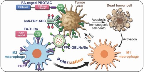
The modern era of precision oncology strives to identify novel and selective druggable targets that, on the one hand, achieve efficacious anti-tumor activity, and on the other hand inflict minimal adverse effects to healthy tissues. The realization that both the tumor microenvironment and the tumor immune microenvironment (TIME) modulate tumor progression and response to chemotherapeutics, has pushed tumor-associated immune cells to the forefront as bona fide druggable targets. Based on their unique overexpression pattern on the cell surface of specific human malignancies and suppressor immune cells, folate receptors FRα and FRβ constitute optimal facilitators of noninvasive cancer detection and localization, as well as efficacious selective delivery of potent therapeutic payloads. Along this vein, this review highlights recent advanced strategies utilizing FR-targeting for precise tumor and metastatic lesion localization for guided surgical precision resection, potent anti-cancer efficacy and reprogramming of the suppressive TIME. Notably, these activities were accomplished with minimal side effects to healthy tissues. AZD5335, a recent clinically tested FRα-targeted antibody-drug conjugate carrying a topoisomerase I inhibitor payload, exceeded its predecessors by demonstrating remarkable efficacy against low FRα-expressing tumors as well. Thus, FRs emerge as selective and promising targets for advanced precision oncology of various malignancies including those displaying chemoresistance.
Keywords: folate receptor, cancer detection, selective targeting, tumor associated macrophages
1. Introduction
One carbon metabolism, and specifically the folate cycle, provides one carbon units for the vital de novo biosynthesis of DNA (purine nucleotides and thymidylate) and amino acids (glycine and methionine), as well as for various cellular methylation reactions (DNA, RNA, histones, phospholipids and neurotransmitters) [1,2]. As such, folates (vitamin B9) are imperative for normal fetal development [3,4] and cellular health [5], however, they are also essential for carcinogenesis and cancer progression [6,7]. While folate cofactors are crucial for cellular functions, most eukaryotic cells lack the capability for de novo folate biosynthesis [1]. Hence, several transport routes mediate the uptake of folates into cells [8]. These include: 1) The predominant and ubiquitously expressed reduced folate carrier (RFC, SLC19A1) [9], 2) The principal intestinal folate absorption transporter, known as the proton-coupled folate transporter (PCFT, SLC46A1) [10], and 3) The glycosyl-phosphatidylinositol (GPI)-anchored membrane and soluble α, β and γ folate receptors (FRs) [11]. Folic acid (FA) is a poor substrate of RFC (Km ~ 200 µM) which efficiently takes up the predominant blood circulating folate 5-methyltetrahydrofolate (5-MTHF) and other reduced folates with much higher affinity (Km ~ 1 µM) [12,13]. PCFT functions as a proton-driven folate influx transporter. As such, PCFT displays comparable Km values (~ 1.5 µM) for FA and 5-MTHF at an acidic pH of 5.5, [14,15], whereas its affinity for FA markedly drops to 56 µM at pH 7.5 [15]. In comparison, at physiological pH, the acceptable dissociation constant (Kd) values for FA binding to FRα and FRβ reveal a high-affinity interaction in the range of 0.1-0.4 nM and 1-3 nM, respectively [16-19]. These receptors exhibit slightly lower affinity towards 5-MTHF, i.e., Kd = 2-3 nM [16]. Their soluble counterpart FRγ displayed Kd values of 5 and 2.5 nM for FA and 5-MTHF, respectively [20]. It should be emphasized that these measured Kd values may vary in different reports based on both the distinct assays employed, like the gold standard isothermal titration calorimetry (ITC) [19] or competition assays [21], as well as the variable conditions used like pH, temperature, ionic strength and cell types. Notably, in studies using ITC measurements in which Kd values as low as 10 pM were reported [19], one has to take caution as the lower end of sensitivity of ITC is ~ 1 nM [22]. Under acidic pH, the Kd of FRα for FA increases to ~ 20 nM [19], thereby allowing the release of FA within lysosomes.
Apart from their high affinity for FA, membrane bound FRs have unique tissue expression patterns. While FRα is often overexpressed in various malignancies [23], predicting dismal prognosis [24,25], FRβ is highly expressed by suppressive tumor-associated immune cells [26-29]. For these reasons, FRs have been exploited for decades for targeted delivery of FA-conjugated anticancer drugs [30,31]. The normal plasma concentration of circulating folates is at a low range of 10-40 nM [32,33]. However, the specialized membrane folate transport routes and, most importantly, folate-polyglutamylation catalyzed by folylpoly-γ-glutamate synthetase (FPGS) [1], achieve an intracellular folate pool of 0.5-1 µM [34-36]. Conversely, the serum folate concentration in mice used for in vivo experiments, is much higher than in humans, attaining up to 800 nM [37-39]. Hence, the large difference between the serum folate concentration in men and xenograft murine models can markedly diminish the efficacy of folate-targeted experimental therapeutics in the preclinical setting [39]. Specifically, the high concentrations of folates found in mouse serum readily compete with the tested agent on binding to FRs, thereby markedly compromising its cytotoxic efficacy [39]. It has been established during the 1990's that mice should be fed with a folate-deficient diet before testing FR-targeted agents [37]. Indeed, following feeding of mice with a folate-deficient chow, their serum folate concentration was reduced to 20-70 nM [37-39], thus being clinically representative of the human plasma folate concentration range.
In the present review, we summarize recent novel strategies exploiting FRs for the detection and targeted treatment of various malignancies, as well as the reprograming of the tumor immune microenvironment (TIME) [40]. However, one should emphasize that most of the discussed results that were obtained in xenograft murine models, are apparently an underestimation, since almost all of the studies did not use a folate-deficient diet.
2. FRα in cancer detection
2.1. Fluorescence guided surgery with OTL38
The successful eradication of solid tumors primarily relies on the complete resection of the cancerous tissue. Various publications have validated the correlation between gross-total resection and prolonged survival [41-45]. However, the “complete” resection based on a surgeon's assessment can be underachieved. This was demonstrated in a post-trial ad hoc analysis of a phase III randomized clinical trial, where 40% of advanced stage ovarian cancer (OC) patients (n = 627) displayed a residual disease (RD) >1 cm post optimal surgical cytoreduction [46]. Consistently, an independent study also reported that 49% of advanced OC patients (n = 117) presented with RD >1 cm following optimal primary cytoreduction [47]. In this respect, the gold standard for evaluating surgical margins is histopathological examination, which is restricted to examining a small portion of the resected surface, and requires a relatively long processing time of 2-5 days [48,49], frequently resulting in the need for re-excision [50].
Human FRα displays low baseline expression levels in normal tissues (0.3-3 pmol FR/mg solubilized membrane protein) except for the lungs and kidneys, which exhibit a mean of 7.8 and 14.4 pmol/mg, respectively [51]. In contrast, OC is characterized by FRα overexpression with levels attaining a mean of ~ 35 and ~ 45 pmol/mg in 100% of papillary serous cystadenocarcinoma and metastatic ovarian adenocarcinoma, respectively [51]. This led to the development of an FRα-targeting tumor-specific fluorescence imaging agent named OTL38 (Pafolacianine, Cytalux®) for real-time fluorescence-guided surgery of cancer. OTL38 is a folate analog ligand (pteroyl moiety) conjugated to a S0456 dye which absorbs at 774-775 nm and emits at 794-796 nm [52,53]. These wavelengths are within the near infra-red (NIR) region, which is ideal for in vivo imaging, as it can penetrate several cm of tissue with minimal light absorption by hemoglobin (<650 nm) and water (>900 nm) [54,55]. Following intravenous administration, the OTL38 folate analog targets FRα overexpressing cells with ~ 1 nM binding affinity, cells then internalize this probe and emit fluorescence upon excitation with a NIR laser [52].
In a randomized controlled trial, Charlotte E.S. Hoogstins et al., examined the tolerability and pharmacokinetics of OTL38 in healthy volunteers, determining the optimal dosage and imaging time window [56]. The low dose of 0.025 mg/kg OTL38 was selected as optimal since it elicited a few mild treatment emergent adverse events (TEAEs) and achieved the highest tumor-to-background ratio (TBR). OTL38 was then administered to 12 OC patients, allowing the intraoperative in situ fluorescent detection of FRα-positive tumors and metastases. Remarkably, 77% (n = 62) of fluorescent-guided resected lesions were confirmed as malignancies by histopathology, with 29% of them being undetected by standard methods [56]. Around fifty percent of the false positive resected lesions were lymph nodes containing activated macrophages that, as will be further detailed below, express FRβ. The latter was found to be bound by OTL38 with equal affinity to FRα [52]. The rest of the false positive lesions included FRα-positive epithelial cells that are routinely resected during cytoreductive surgery. Two false negative lesions were found, representing ~ 3% of malignant lesions. This study represented the first successful use of tumor-specific NIR fluorescence-guided OC surgery [56].
The overwhelming promising results obtained with OTL38 in OC led to a phase II, multicenter, open-label trial of intraoperative molecular imaging (IMI) (NCT02317705) [57]. A sensitivity of 98% was estimated for the correlation of detection among multiple lesions within a single patient, and at least one undetected lesion was identified by OTL38 in 48% of the patients (n = 14) [57]. A phase III clinical trial soon followed, encompassing 11 centers and 109 FR-positive OC patients (NCT03180307) [58]. OTL38 identified undetected cancer tissues in ~ 35% of patients, its OC detection sensitivity was 83%, complete R0 resection was achieved in 62.4% of patients, and its false-positive rate was 24.8% [58]. Systematic reviews of clinical and preclinical studies assessing the use of OTL38 IMI in OC have been reported [59,60]. OTL38 was approved by the US Food and Drug Administration (FDA) in 2021 for the IMI and detection of OC [61].
Since several studies demonstrated overexpression of FRα in lung adenocarcinoma [62-65], OTL38 was tested during surgery of dogs with primary lung tumors and compared to video-assisted thoracoscopic surgery (VATS) [53]. Tumor margins were evaluated by immunohistochemistry (IHC) of formalin-fixed paraffin-embedded (FFPE) tissue sections and additionally tested for FRα expression. OTL38 fluorescence was not only consistent with the pathological results but also facilitated the identification of undiscovered positive lymph nodes harboring cancer cells as well as retained tumor margin from the VATS procedure. Following these promising results, Jane Keating et al., conducted a proof-of-principle study on three patients with diagnosed pulmonary adenocarcinoma. The test subjects were administered 3 hours prior to imaging with an injection of 0.025 mg/kg OTL38 via the median cubital vein, with no TEAEs. Post-operative pathology confirmed negative margins [53].
Following a small-scale phase I trial for the IMI of pulmonary adenocarcinomas [66], a multi-institutional phase II clinical trial was conducted, evaluating the use of OTL38 for IMI in non-small cell lung cancer (NSCLC) patients (n = 92) (NCT02872701) [67]. OTL38-based IMI allowed the discovery of wrongly classified positive margins (9%) and undetected malignant lesions (12%). The IMI also detected synchronous cancer lesions that included primary adenocarcinomas, metastatic papillary thyroid cancer and adenoid cystic carcinoma. False negative detection by IMI included pre-invasive lung lesions and inflamed tissues containing activated macrophages [67]. In a phase III, 12-center trial (NCT04241315) [68], among 100 lung cancer patients, OTL38 IMI identified undetected primary nodules in 19% of patients, as well as undetectable synchronous cancers (adenocarcinomas and metastatic chordomas) in 8% of them. The cancer detection sensitivity was 76% with a 17% false-positive detection. IMI-guided detection in 29% of the patients resulted in a change of the scope of the surgical procedure [68]. OTL38 was also shown to be useful and safe for IMI of pulmonary metastases in young adults [69].
Clinical trials assessing the use of OTL38 for the IMI of other cancers are ongoing. In this respect, NCT06235125 and NCT06915727 are evaluating the safety of OTL38 in children with confirmed diagnosis of osteosarcoma, synovial sarcoma, hepatoblastoma, rhabdomyosarcoma, Ewing sarcoma, Wilms tumor and other solid tumors. NCT07124351 for gastrointestinal malignancies, NCT07278986 for endometrial cancer, NCT06434909 for pancreatic cancer, NCT06511037 for peritoneal carcinomatosis as well as NCT07039526 for digestive tract and gynecological malignancies.
Since OTL38 is restricted to FRα-positive tumors and FRβ-positive macrophages, the group of the pioneer PS Low set out to design a tumor fluorescence imaging cocktail with a broader scope [70]. Towards this end, they generated a fibroblast activation protein (FAP)-targeted NIR dye conjugate (i.e., FAP9-S0456) with a FAP-specificity IC50 value of 2 nM. Since this conjugate consists of the same fluorophore as OTL38, both probes exhibited comparable excitation and emission spectra, allowing their co-excitation with a single laser [70]. FAP, a serine protease, is a cell surface, single pass transmembrane protein [71]. It is expressed on various cells of the tumor microenvironment (TME), primarily on cancer-associated fibroblasts (CAFs) [71,72]. CAFs constitute a major component of the TME [73], where they support carcinogenesis, tumor growth and metastasis [74-76]. As such, they are good indicators for tumor localization, and their counterparts metastasis-associated fibroblasts (MAFs, mCAFs) are indicators of the localization of metastases [77,78]. The combination of FAP9-S0456 and OTL38 was predicted to detect the vast majority of tumor types. The actual efficiency of this cocktail was assessed using murine models harboring tumor xenografts from multiple non FRα/FAP overexpressing malignant origins: murine breast cancer 4T1 cells, colon carcinoma CT26 cells and renal cortical adenocarcinoma RENCA cells, as well as human NSCLC A549 cells and pancreatic cancer MIA PaCa-2 cells [70]. While the tumor fluorescence intensity was higher (2-3-fold) with the cocktail than with each probe alone, the TBR was hampered by the presence of FRα in healthy tissues. Thus, FAP9-S0456 had the highest TBR. When FRα-overexpressing human breast cancer MDA-MB-231 cells or FAP-overexpressing human glioblastoma U87MG cell xenografts were used, the cocktail-based tumor fluorescence did not exceed that of OTL38 or FAP9-S0456 alone, respectively. Taken together, the use of the cocktail may provide an advantage in ~ 50% of human cancers which neither overexpress FAP nor FRα [70].
2.2. The companion diagnostic tool for immunohistochemical detection of FRα
FRα-targeted therapy is based upon FRα overexpression by malignant cancers. In this respect, patients should be first scanned to validate their qualification for FRα-targeted therapy. Moreover, the use of a single detection system with a global FRα scoring method will help to minimize variations in interpreting FRα expression and allow to achieve diagnostic consistency. Along this vein, a test was developed to measure the expression of FRα in patient-derived FFPE blocks of epithelial OC. The VENTANA FOLR1 (FOLR1-2.1) RxDx Assay, developed by Ventana Medical Systems Inc and Roche Tissue Diagnostics, is a fully automated IHC assay, based on a mouse monoclonal anti-FOLR1 primary antibody (FOLR1-2.1) and an OptiView DAB IHC Detection Kit [79]. Following the validation of the repeatability and reproducibility of the VENTANA FOLR1 assay, it was approved by the FDA as a companion diagnostic tool for the treatment with mirvetuximab soravtansine-gynx (MIRV, IMGN853, ELAHERE) [80]. The latter is an FRα-targeted antibody-drug conjugate (ADC) on which we elaborate below. The VENTANA FOLR1 assay was utilized to screen and select patients as a participation criterion for a single-arm phase II study, named SORAYA (NCT04296890) [81,82] and a phase III randomized trial named MIRASOL (NCT04209855) [83]. The trials evaluated the efficacy and safety of MIRV in advanced high-grade OC patients with high FRα expression, and compared MIRV to standard chemotherapy, respectively. Following these two pivotal studies, MIRV was approved by the FDA for the treatment of platinum-resistant epithelial ovarian cancer (PROC) [84,85]. A retrospective study encompassing 425 OC patient specimens, primarily high-grade serous carcinoma, tested for FRα expression using the VENTANA FOLR1 assay [86]. High FRα expression was found in 36.3% of the cases and was significantly associated with high-grade serous ovarian histology. Primary masses exhibited higher FRα levels than metastases (positive rates of 44.4% vs 32.5%, p = 0.02) [86]. A second study was recently published with comparable results from a cohort of Chinese OC patients, where the VENTANA FOLR1 assay was used to analyze 313 samples for FRα expression [87]. Three independent pathologists demonstrated a strong concordance (>97%) in the interpretation of the assay's results. A total of 40.9% of cases presented high FRα levels, and primary masses exhibited higher FRα levels than metastases (positive rates of 44.2% vs 32.2%, p = 0.04) [87]. These results emphasize the requirement for standardized FRα testing before FRα-targeted treatment is administered and the advantage of a uniform FRα scoring method.
2.3. Serum FRα
While the VENTANA FOLR1 assay standardized patient specimen testing for FRα positivity, it is based on an invasive biopsy procedure. In search of a non-invasive, rapid and inexpensive procedure for assessing FRα positivity in tumors, researchers turned to the rapidly growing field of cancer serum biomarkers [88-91]. The elevated expression of FRα in OC along with its ability to be secreted or shed from cells, despite its GPI-membrane-anchoring [92], has brought forth soluble FRα (sFRα) as a serum biomarker for the detection, diagnosis and monitoring of OC [89,93-95]. Beyond OC detection, sFRα protein levels were shown to be associated with tumor burden, allowing the indirect monitoring of tumor shrinkage following treatment [94], and disease recurrence [95]. One clinical trial, discussed in section 3.1.2.2, quantified the levels of circulating sFRα in patients during treatment, and demonstrated a transient peak in serum sFRα levels due to tumor cell death [96]. Serum sFRα was also demonstrated to predict histological subgroups of medulloblastoma with sensitivity and specificity [97]. FRα can be readily detected in serum samples using an ELISA and the variety of commercially available anti-FRα antibodies detailed below, as well as others which are being continuously developed [98].
3. FRα-targeted cancer therapeutics
3.1. Antibody-based cancer therapeutics
Antibodies have been utilized in biomedical research and diagnostics since the early 1940s, a use that has expanded with the introduction of monoclonal antibodies (mAbs) half a century ago by Kohler and Milstein [99]. The increase in clinical demand along with technological advancements and the high specificity of mAbs, established them as a cornerstone in contemporary diagnostics and therapeutics. The FDA and the European Medicines Agency (EMA) have approved over 50 antibody-based therapeutics for oncology applications [100], in addition to chimeric antigen receptors (CARs) for T cell engineering [101,102]. Antibody-based therapeutics include three main modalities, monospecific and bispecific antibodies [103], as well as ADCs [104,105]. Monospecific antibodies are full-length immunoglobulins, typically IgG since it is the only isotype that is bound and protected by the neonatal Fc receptor (FcRn), leading to its long circulation half-life [106]. Monospecific antibodies are designed to target cell surface antigens such as receptors for growth factors and differentiation marker glycoproteins that are overexpressed in solid tumors and immune cells, respectively [100]. By targeting these ligands, monospecific antibodies elicit cancer cell death via several mechanisms [107,108]: 1) Disruption of signaling; by binding cell surface receptors, monospecific antibodies directly block survival and proliferation signals by growth factors such as epidermal growth factor (EGF) and transforming growth factor alpha (TGF-α) [109]. Furthermore, blocking the signaling of vascular endothelial growth factor (VEGF) abolishes angiogenesis and disrupts tumor blood supply. In this respect, bevacizumab (Avastin), used in the treatment of PROC, is a humanized mAb that targets VEGF itself and blocks its pro-tumoral signaling pathways [110,111]. 2) Immunologic tumor destruction, including antibody-dependent cell-mediated cytotoxicity (ADCC) and antibody-dependent cell-mediated phagocytosis (ADCP). In these mechanisms, the antibody binds a cancer cell surface antigen via its variable regions, while its Fc region binds an Fcγ receptor (FcγR) on the surface of a leukocyte cell, such as a natural killer (NK) cell [112] or macrophage [113]. The antibody recruits and activates effector immune cells which can either secrete proteolytic granules that induce cancer cell apoptosis [114], secrete cytokines that activate complement-dependent cell death (CDC) [115,116], or phagocytose the cancer cell [117]. 3) Immune checkpoint blockade [118]. Cancer cells often increase the expression of immune checkpoint surface proteins which allow them to evade host immune surveillance by suppressing the activation of immune cells, primarily cytotoxic T-lymphocytes (CTLs). Hence, targeted blocking of these checkpoint molecules by antibodies eliminate the immune suppression, thereby restoring the activation of the immune system against cancer cells [119].
3.1.1. Anti-FRα antibodies
During the 1980s three cornerstone anti-FRα mAbs were raised against human ovarian carcinoma and choriocarcinoma using hybridoma technology, MOv18 and MOv19 [120,121] and LK26 [122].
3.1.1.1. Farletuzumab (MORAb-003)
The murine LK26 mAb exhibited a Kd of 7 nM for native FRα and reduced the growth of SKOV3 cell xenografts in nude mice by ~ 62% compared to control IgG (p < 0.05) [123]. For clinical application purposes, this antibody had to be humanized and preclinically evaluated. To this end, optimization of humanized LK26 mAb was performed using a whole cell genetic evolution platform, i.e., morphogenics technology [124]. The latter platform has proven highly successful in generating antibody-producing cell lines which secrete antibodies with superb binding affinities as well as enhanced titers that are suitable for scalable manufacturing. Thus, this morphogenics technology platform yielded farletuzumab (MORAb-003), a high affinity (Kd = 2.2 nM) anti-FRα IgG mAb [123]. With the use of FRα-expressing cell lines and human serum, Wolfgang Ebel et al., demonstrated the ability of farletuzumab to induce CDC resulting in up to 98% cytotoxicity in vitro, compared to negligible toxicity by farletuzumab or serum alone. Moreover, by using a combination of farletuzumab and peripheral blood mononuclear cells (PBMCs) from healthy donors, the authors demonstrated the in vitro activation of ADCC, resulting in >70% cytotoxicity in FRα-positive cells vs. no toxicity in FRα-negative cells. Toxicology studies in female cynomolgus macaque monkeys receiving intravenous infusions of farletuzumab, revealed a very long antibody half-life of ~ 250 hours, with measurable serum levels up to 4 weeks following last infusion. Importantly, almost no anti-human antibodies were developed in response to the administration of farletuzumab [123].
A phase I study of farletuzumab was conducted with 25 PROC patients who previously underwent ≥3 lines of treatment (NCT00428766) [125]. Dose escalation experiments attained 400 mg/m2 farletuzumab with no grade ≥3 TEAEs. Since no maximum tolerated dose (MTD) of farletuzumab was reached, a recommended phase II dose (RP2D) could not be determined. Tumor targeting was monitored via a radiolabeled farletuzumab in SW620 colorectal cancer xenograft mouse models and PROC patients via planar whole body gamma camera images [126]. Following 4 weeks of farletuzumab treatment, 36% of patients had radiologically stable disease (SD) and 4 patients exhibited a ≤43% decrease in carcinoma-associated antigen CA-125 levels [125]. Further clinical trials evaluating farletuzumab in combination therapy in OC patients did not show any improvement in treatment outcome [127-129]. This suggests that the farletuzumab-dependent ADCC and CDC effects were not recapitulated in vivo, possibly due to the immune-suppressive TME generated following platinum treatment of OC [130,131]. Thus, the high specificity and affinity of farletuzumab along with its safety were exploited for the design of the FRα-targeted ADC MORAb-202 [132], which is discussed below.
3.1.1.2. MOv18
Although FDA approved mAbs for cancer therapy have all been of the IgG isotype [108], the chimeric IgE MOv18 antibody (i.e., consisting of murine variable and human constant regions) was found to exhibit greater efficacy for a longer time against a murine xenograft model of OC compared to the IgG isotype [133]. While IgE antibodies are established mediators of histamine degranulation-based allergic reactions, their ability to facilitate tumor destruction has gained much interest in the past two decades [134], leading to AllergoOncology, an evolving interdisciplinary new field encompassing allergy, immunology and oncology [135,136]. IgE has the highest affinity to its cognate receptors of all immunoglobulins [134,137] and it has no known inhibitory Fc receptors such as the FcγRIIb, which potentiates immunosuppression in the TME [138]. The development of IgE-mediated cancer therapy has not reached its full potential since the expression of FcεRI, the high-affinity receptor of IgE, is restricted to mast cells and basophils in mice, hindering preclinical evaluation. In humans, FcεRI is also expressed by dendritic cells (DCs) and monocytes, thus preclinical testing of IgE has been limited to in vitro experiments using donor-derived DCs and monocytes [139]. Unlike IgG, IgE does not elicit ADCC in vitro [133,140]. However, when accompanied by human-derived PBMCs, the chimeric IgE MOv18 antibody exhibited a more potent anti-tumor effect in a severe combined immunodeficient (SCID) xenograft mouse model of OC compared to its IgG counterpart [133]. A subsequent study in a nude-mouse model of OC injected with PBMCs revealed, for the first time, that the IgE MOv18 chimeric antibody promoted ADCP through tumor infiltrating monocytes [141]. This resulted in longer survival of the OC xenograft bearing mice, i.e., 40 ± 5.8 days vs. 22 ± 2.4 days, p = 0.012, for IgE and IgG MOv18, respectively. The chimeric IgE MOv18 antibody was able to elicit both ADCC and ADCP in a nude mouse OC model when injected in combination with U937 monocytes before and after interleukin-4 (IL-4) stimulation [142]. U937 monocytes express both the IgE high affinity FcεRI and the low affinity FcεRII (CD23) receptors [143]; with the expression of the latter being upregulated upon IL-4 stimulation. Hence, the authors demonstrated that ADCC was mediated via FcεRI, whereas FcεRII facilitated ADCP. This resulted in an increase in mean survival from 16 days to 27 days (p < 0.005), and from 17.5 days to 35 days (p < 0.0005), for MOv18 + untreated U937 and MOv18 + IL-4-treated U937 cells, respectively [142]. Consistently, these findings were recapitulated in a nude mouse OC model using human-derived monocytes [144].
Further mechanistic insights were obtained by utilizing an immunocompetent syngeneic rat model harboring FRα-expressing lung adenocarcinoma metastases [145]. The rat model better recapitulates the human IgE-FcεR system, since rat effector cells (e.g., monocytes and macrophages) express trimeric FcεRI [146]. Using chimeric IgE and IgG MOv18 antibodies, consisting of rat Fc sequences (i.e., rMOv18 IgE/IgG2b), the authors uncovered a role for IgE-mediated tumor-infiltrating macrophages in combating cancer in vivo [145].
As detailed in our recent review [147], M0 monocyte-differentiated quiescent cells undergo polarization to two distinct macrophage phenotypes, the pro-inflammatory/anti-tumoral M1 or the anti-inflammatory/pro-tumoral M2 phenotype. M1 macrophages directly eliminate malignant cells by phagocytosis and consequent autophagy, elicit cancer cell death by secreting cytokines such as tumor necrosis factor (TNF), as well as promote anti-tumor activities of other leukocytes. In stark contrast, M2 macrophages both abolish anti-tumor immune response and secrete pro-tumoral growth factors, thus representing a major component of the immune-suppressive TME [147]. In the above syngeneic rat model, superior infiltration of rat macrophages into tumors was observed upon IgE rMOv18 treatment, compared to PBS (p = 0.003) and IgG rMOv18 (p = 0.03) [145]. More importantly, IgE rMOv18 induced the highest percentage of tumor infiltrating CD80+ macrophages (p = 0.01), indicating they were of the M1-phenotype [148]. Indeed, enhanced intracellular macrophage expression (p = 0.04) and secretion (p = 0.017) of TNF was measured upon IgE rMOv18 treatment [145]. The M1 macrophage activation culminated in significantly lower lung metastases and tumor occupancy with rMOv18 IgE compared with rMOv18 IgG2b (<50%, p < 0.0001) or PBS (<30%, p < 0.0001). These results suggest that IgE antibodies can both polarize and recruit macrophages to attack cancer cells [145], confirming recent studies demonstrating the ability of IgE antibodies to reprogram M2 patient-derived macrophages [149-152]; hence paving the way towards the possible clinical development and application of IgE antibodies to attack cancer and its metastasis.
Several clinical trials with chimeric MOv18 IgG have been conducted in OC patients with minor immunological effects [153]. Hence, efforts were shifted towards the IgE antibody. A phase I, dose escalation trial was undertaken, using a chimeric first-in-class IgE Mov18 antibody in patients with FRα-positive solid tumors (NCT02546921) [154]. Since a single patient, with circulating basophils at baseline, experienced anaphylaxis, patients were screened to ensure low risk of allergic toxicity by skin prick and basophil activation tests (BAT) [155]. While the dose escalation study attained a dose of 12 mg MOv18 IgE, the MTD was not reached [154]. The criteria for FRα-positivity was ≥5% of tumor cells with membrane positivity by IHC [156], hence, all final participants had gynecologic cancer (i.e., n = 21, 3 and 2 for OC, tubal carcinoma and endometrial cancer, respectively) [154]. An increase in serum IL-6 levels was detected for up to 7 days following antibody administration, indicating activation of the immune system [157]. However, no increase in TNF levels was observed, and only one patient exhibited signs of anti-tumor activity [154]. This disappointing outcome could be a result of both an insufficient RFα-positivity threshold, as discussed below, or insufficient antibody dosing. In this respect, a phase Ib, open label expansion trial is ongoing (NCT06547840).
3.1.1.3. MOv19
The original murine Mov19 mAb [120] underwent extensive development over the years [158], yielding a variety of active derivatives including a chimeric murine-human IgG mAb [159], a humanized mAb [160], humanized MOv19 fragment antigen-binding (Fab) fragment [161], MOv19 single-chain variable fragments (scFv) based CAR T cells [162-164], and the ADC MIRV [160].
3.1.2. FR targeting ADCs
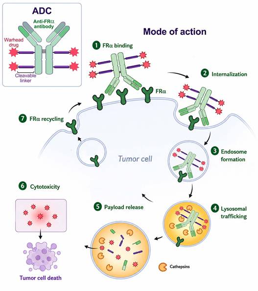
ADCs, referred to as "biological missiles” or 'magic bullets', consist of a mAb which is covalently attached via a chemical linker to a cytotoxic drug. ADCs are endowed with the important advantages of both high target specificity and potent killing, thus efficiently eliminating cancer cells with little off-target adverse effects [104,105]. Unlike anti-FRα antibodies for IHC on fixed tissues, antibodies for ADC need to bind FRα in its native conformation and facilitate receptor-mediated endocytosis (RME) for the internalization of the cytotoxic drug conjugate. A scheme illustrating the mode of action of FRα-targeted ADCs is depicted in Figure 1.
Structure and mode of action of FRα-targeted antibody-drug conjugates (ADCs). The ADC consists of an anti-FRα mAb conjugated to a cytotoxic drug, via a cleavable linker, with a drug:antibody ratio of ~ 4. 1) The ADC targets cancer cells via binding to FRα on the cell surface. 2) The binding of the mAb to FRα triggers receptor-mediated endocytosis. 3) The ADC is transported from the plasma membrane via an endosome which then fuses with a lysosome to form an endolysosome. 4) Within the acidic lysosome, FA dissociates from FRα which recycles to the plasma membrane (7). 5) Following reduction (i.e. cleavage) of the linker by glutathione or thiol reductase, and degradation of the mAb by lysosomal cathepsins, the drug payload becomes free to exit the lysosome into the cytosol. 6) The free drug exerts its cytotoxic activity, leading to cell death.
3.1.2.1. Mirvetuximab soravtansine-gynx
The FDA-approved ADC MIRV is an anti-FRα antibody conjugated through an N-succinimidyl 4-(2-pyridyldithio)-2-sulfobutanoate (sulfo-SPDB) linker to an N2′-deacetyl-N2′-(4-mercapto-4-methyl-1-oxopentyl)-maytansine (DM4) at a drug to antibody ratio (DAR) of ~ 4 [160]. During the development of MIRV, >100 murine anti-FRα antibodies were tested. The murine antibody-drug conjugates which exhibited the highest activity in vitro underwent humanization and were further tested in vivo using mice harboring highly FRα-positive KB xenografts. The in vivo experiments highlighted the superiority of one antibody, i.e., M9346A, the humanized derivative of the established anti-FRα antibody MOv19 [158]. Next, the cytotoxic activity of four linker/maytansinoid combinations of M9346A conjugates were compared using xenografts with varying FRα expression levels [160]. For comparison, the mean FRα antibody-binding sites/cell in the xenografts' source cell lines were 4.5 × 10⁶, 1.3 × 10⁶ and 4.8 × 10⁴ for KB, IGROV-1 and OVCAR-3 cells, respectively. The highly hindered disulfide hydrophilic linker sulfo-SPDB [165] was better than a thioether-based linker, as well as better than both a hindered and a highly hindered disulfide linker [166]. The M9346A-sulfo-SPDB-DM4 conjugate, i.e., MIRV, elicited the strongest anti-tumor effect, resulting in extended survival time of the murine models, especially towards the low FRα-presenting xenografts [160]. The specificity of the conjugate was further verified in vitro by competition assays using saturating excess of the M9346A antibody. While MIRV exhibited sub-nM growth inhibition IC50 values in different highly FRα-positive cell lines, its cytotoxic activity decreased by >10-fold in the presence of the free antibody. However, when MIRV was tested against relatively low FRα expressing cell lines (i.e., ≤105 FRα antibody-binding sites/cell), its IC50 values of 1-10 nM were not affected by the excess of free antibody. To explore the basis underlying the decreased sensitivity of the low FRα expressing cell lines to MIRV, the authors tested the intracellular processing capabilities of the cell lines [160]. Maytansinoids are naturally occurring heterocyclic compounds that bind to β-tubulin in the α, β -tubulin heterodimer. Since their binding site is in the interface between tubulin heterodimers, maytansinoids hinder microtubule polymerization, resulting in microtubule destabilization, consequent mitotic arrest and cell death [167-169]. When internalized, the DM4 maytansine conjugate is processed in the lysosomes, resulting in several active metabolites [168]. Following incubation of the various cells with radiolabeled MIRV, and identification of the MIRV catabolites by HPLC, the authors concluded that the amount of processed active metabolites in the cells was proportionate to the cell surface antigen number [160]. For example, KB cells processed 2.2 pmol/106 cells, while OVCAR-3 cells, that express ~ 100-fold less surface FRα, processed only 0.02 pmol/106 cells. Finally, the in vivo anti-tumor efficacy of MIRV was tested in murine models harboring NSCLC patient-derived xenografts (PDXs), mimicking clinically relevant cell surface FRα expression. MIRV treatment resulted in a substantial regression in tumor growth [160].
MIRV was further tested against biologically aggressive, type II, endometrioid and uterine serous carcinomas (USC) [170]. First, FRα expression was evaluated via IHC in a retrospective cohort of 70 USC specimens. This revealed that 41% of the specimens exhibited moderate to high FRα expression, suggesting that this gynecological cancer should respond well to MIRV. Second, 20 primary cell lines were established from freshly derived endometrioid and USC tumor biopsies and evaluated for FRα expression using flow cytometry quantification. Moderate to high FRα expression was found in 22% and 27% of endometrioid and USC cell lines, respectively. These cell lines were then used in cytotoxicity assays, comparing MIRV to non-targeting ADC control and the free M9346A antibody. In FRα-overexpressing endometrioid cells, MIRV elicited 10-14-fold higher cytotoxicity than the ADC control (p < 0.01), while no such difference was observed in low-FRα expressing cells. Moreover, the free antibody had no detectable anti-tumor activity in vitro. In FRα-overexpressing USC cells, the impact of MIRV was also substantial but less prominent being 2.7-4.6-fold over the ADC control (p < 0.05). Interestingly, by using a co-culture of low- and high-FRα expressing endometrioid cells, the authors demonstrated that MIRV has a strong in vitro bystander effect, eliciting a 10-fold increase in cytotoxicity towards low FRα expressing cells (p < 0.01). Finally, the anti-tumor activity of MIRV was evaluated in vivo, using PDX murine models of both endometrioid and USC high-FRα expressing cells. Consistently, overall survival (OS) was significantly longer in MIRV-treated mice compared to controls (p < 0.001). Remarkably, the endometrioid-derived tumors were completely eradicated by MIRV, resulting in the survival of all mice at the end of evaluation (78 days) [170]. Hence, endometrial cancer patients, and to a lesser extent USC patients, could apparently benefit from FRα-targeted MIRV treatment.
The first-in-human phase I clinical evaluation of MIRV as a monotherapy included 44 patients presenting with refractory tumors, i.e., OC >> endometrial cancer > renal cell cancer > NSCLC > cervical cancer, considered as FRα-positive (NCT01609556) [171]. Two OC patients achieved confirmed partial response (PR), whereas SD was observed in 22 patients. Furthermore, 5 patients displayed a ≥50% reduction in the serum marker CA-125 for >28 days, defined as a CA-125 response [172]. The adjusted ideal body weight (AIBW) of 6 mg/kg MIRV was chosen as the RP2D based on safety and activity [171]. Taken together, the overall clinical benefit rate, i.e., PR + SD for ≥4 months + CA-125 response, was 23%. However, in this trial, FRα expression was not validated prior to treatment. Thus, in an expansion of the above phase I trial, FRα expression was a prerequisite, and the threshold for FRα positivity was defined as ≥25% of tumor staining at ≥2+ intensity by IHC. However, the overall clinical benefit rate remained 22% [173]. These consistent findings suggested that a higher percent of FRα positive cells are required to achieve a beneficial cytotoxic effect by MIRV.
Advanced-stage epithelial OC is treated by surgery and platinum-based chemotherapy, which together achieve good initial results. However, most patients (~ 80%) relapse within several years and display platinum-resistant tumors. If disease progression occurs within 6 months of the last platinum-based regimen, the cancer is regarded as PROC [174]. PROC is currently treated by non-platinum single agent (NPSA) chemotherapy (primarily paclitaxel, pegylated liposomal doxorubicin, or topotecan) alone or in combination with bevacizumab. A phase III FORWARD I trial, compared MIRV to other NPSAs in >300 patients with FRα-positive PROC [175]. The threshold for FRα positivity by IHC was defined as ≥50% of tumor cells with any visible FRα membrane staining at ≤ ×10 microscope objective. While the required percent of FRα positive cells was higher than before, the ambiguous scoring of the level of FRα expression could present an impediment for successful MIRV therapy. Indeed, while some outcome parameters favored MIRV over the other chemotherapy agents, no statistically significant results were obtained [175].
The phase II SORAYA study utilized the VENTANA FOLR1 assay, to screen PROC patients according to their FRα expression status [81,82]. In this study, the threshold for FRα positivity was defined as ≥75% of tumor cells exhibiting ≥2+ level of membrane staining intensity using the uniform grading score of the VENTANA FOLR1 assay. Another recruitment criterion was that patients already underwent 1-3 lines of therapy, one of which was bevacizumab. The clinical trial included 105 PROC patients, of which 5 achieved complete response (CR) and 29 achieved PR, increasing the objective response rate (ORR) above 30% (p < 0.0001). The highest ORR was obtained in patients previously treated by poly ADP-ribose polymerase inhibitor (PARPi), i.e., ORR 38.0% and 27.5% in patients who underwent prior PARPi treatment (n = 51) and those who did not (n = 54), respectively [82]. Interestingly, serum sFR was proposed as a biomarker for PARPi resistance in OC patients, as high levels of FRα were associated with non-responders (p < 0.0026) [176]. A reduction in tumor size occurred in 71.4% of patients, and the disease control rate (CR, PR, or SD ≥ 12 weeks) was 51.4% [81] vs. the previously reported 22.0% [173]. Median OS was 15.0 months [82]. This promising clinical trial was followed by the phase III randomized trial MIRASOL, which included 453 PROC patients from 21 countries [83,177]. The same recruitment criteria were used as in the SORAYA study, except for the requirement for a previous bevacizumab treatment, and MIRV treatment outcome was compared to other NPSAs. The reported ORRs were 42.3% and 15.9% for the MIRV and the NPSAs groups, respectively (p < 0.001), resulting in longer OS times of the MIRV-treated patients, median, 16.46 months vs. 12.75 months (p = 0.005) [83]. The SORAYA and MIRASOL studies underscored the importance of correct patient participation criteria and FRα-positivity scoring for efficient and beneficial treatment of PROC with MIRV, and established MIRV as a preferential therapy over standard NPSAs.
MIRV was further evaluated as a third-line, or greater, treatment of patients with recurrent platinum-sensitive ovarian cancer (PSOC) [178]. The single-arm, phase II, PICCOLO trial (NCT05041257) used the same FRα positivity threshold as the SORAYA study [81] and required patients to have radiographic progressive disease (PD) following ≥2 lines of platinum-containing therapy. Initial results included an ORR of 51.9%, while OS was not mature at last follow-up. MIRV exhibited notable efficacy and tolerable safety [178]. GLORIOSA, an ongoing phase III randomized, open-label trial (NCT05445778), will evaluate the combination of MIRV with bevacizumab vs. bevacizumab alone as maintenance therapy in high-FRα PSOC patients [179]. The actively recruiting GLORIOSA study is planned to run through 2029.
3.1.2.2. MORAb-202
A second FRα targeted ADC, designated MORAb-202 (farletuzumab ecteribulin, farletuzumab-[Mal-PEG2-Val-Cit-PAB-eribulin]), includes farletuzumab conjugated to the highly potent microtubule targeting agent (MTA) eribulin via a cathepsin cleavable valine-citrulline (Val-Cit) linker [180] at a DAR of 4 [132]. Eribulin mesylate (Halaven), used in the treatment of heavily pretreated metastatic breast cancer [181] and liposarcoma [182], binds β-tubulin at the microtubule plus-end and slows/prevents elongation leading to microtubule catastrophe [183]. Importantly, while eribulin exerts functionally irreversible antimitotic effects, in addition to several non-mitotic anti-tumor effects such as tumor vascular remodeling and perfusion [184], it inflicts markedly less peripheral neuropathy compared to other MTAs, such as taxanes (e.g. paclitaxel) and Vinca alkaloids (e.g. vincristine) [185,186]. MORAb-202 exhibited high specificity and potency during in vitro cytotoxicity assays, with superb IC50 values of 0.01-0.06 nM in high-FRα expressing cells as opposed to >100 nM in FRα-null cells [132]. In moderate to low FRα expressing cell lines, the IC50 values were at the sub-nanomolar to nM range. In vivo evaluation of MORAb-202 using a moderate FRα expressing human NSCLC NCI-H2110 xenograft model, revealed that at a dose of 5 mg/kg MORAb-202 completely abolished tumor growth, thereby inducing a CR. Remarkably, at the end of the study (day 80), all mice were tumor-free with no effect on body weight. MORAb-202 was further evaluated using two PDX mouse models, the human NSCLC LXFA-737 and gastric cancer GA0055, both expressing moderate levels of FRα. Although CR was not achieved in all cases, a dramatic and durable tumor growth inhibition (TGI) was apparent [132]. Notably, since these results were obtained following a single MORAb-202 dose, additional timely injections could extend the duration of the response. Indeed, in an extensive study with PDXs of poor prognosis gynecological cancer subtypes, the authors showed the contribution of additional dosing [187]. Mice harboring low-FRα high-grade serous OC xenografts which exhibited PD after 85 days, received additional 3 weekly doses and achieved CR at least until day 120. Additional preclinical testing of MORAb-202 revealed a possible bystander effect both in vitro and in vivo [188]. The cytotoxicity IC50 value of MORAb-202 in FRα-negative acute myeloid leukemia (AML) HL-60 cells dropped a 100-fold to 2.4 nM when co-cultured with high-FRα expressing IGROV-1 cells. In the OD-BRE-0631 PDX model of triple negative breast cancer (TNBC), MORAb-202 displayed a moderate anti-tumor effect which slowed tumor growth, resulting in a ~ 2.4-fold smaller tumor at day 31 (p < 0.0001) [188]. This xenograft expresses low levels of FRα and is highly enriched in tumor stroma, both of which hinder targeted therapy [189]. The stroma develops, in part, by the interstitial transformation of normal fibroblasts into CAFs. While the former cells express low levels of alpha-smooth muscle actin (α-SMA) and FAP, CAFs overexpress these proteins [75]. Hence, using an anti-α-SMA antibody, the authors explored the status of the stroma in the OD-BRE-0631 xenograft mouse model following treatment with MORAb-202 [188]. The study showed that ablation of the CAF network was most profound in the vicinity of apoptotic tumor cells, presumably as a result of the diffusion and infiltration of released eribulin into neighboring tumor areas, resulting in a bystander killing effect. An additional TNBC PDX model was used, the high-FRα M-BRE-0563 model. In this model, MORAb-202 achieved a complete and lasting elimination of the tumors, up to the end of the experiment on day 60. Free eribulin, at an equivalent drug dose, had little to no anti-tumor effect, highlighting the advantage of targeted therapy. The serum levels of sFRα protein were monitored during MORAb-202 treatment, as a marker for the successful eradication of the OC tumor. Serum sFRα levels peaked 1-2 days after the administration of MORAb-202, suggesting the release of FRα by dead tumor cells. sFRα levels in MORAb-202 treated mice decreased back to the initial levels, while they retained a steady increase in mock treated mice, indicating the increase in tumor size. Toxicology studies in cynomolgus monkeys established bone marrow myelosuppression as the primary MORAb-202-related TEAE [188]. This off-target toxicity of MORAb-202 could stem from the instability of its linker in vivo, as was demonstrated by the reduction of its DAR from 4 to 3 during a 10-day incubation period in human and mouse sera in vitro [132], indicating the release of free eribulin into the bloodstream. At a dose range of 2-6 mg/kg, the myelosuppression was reversible with neutropenia being resolved by day 21 [188]. Elimination half-life of MORAb-202 in plasma was 135-178 hours. Hence, the suggested MTD was 6 mg/kg every 3 weeks.
A first-in-human phase I study of MORAb-202 was conducted in 22 patients with IHC-confirmed FRα-positive solid tumors who failed to respond to standard therapy (NCT03386942) [96]. Fifty five percent of patients had OC and the rest harbored other carcinomas including breast, endometrial, NSCLC, and fallopian tube cancer. Patients received low doses of 0.3-1.2 mg/kg MORAb-202 once every three weeks. The vast majority of the patients (95%) suffered from either leukopenia or neutropenia, however, all hematological adverse events were of grade 1 or 2. There were seven grade 3 and 4 TEAEs which included primarily increased levels of alanine aminotransferase and γ-glutamyl transferase, indicating liver cell damage. The serum concentrations of circulating sFRα were measured in each patient before and after the first and second dosing cycles. A dose-dependent increase in serum sFRα levels was recorded for 24 h following MORAb-202 treatment, albeit the amount of serum sFRα returned to its baseline level when cycle 2 was initiated. A positive correlation was observed between normalized serum sFRα levels and tumor shrinkage (p = 0.029). One OC patient achieved CR, nine patients attained PR and eight presented a SD; these tumor responses were seen within the entire MORAb-202 dosing range and in all types of cancer [96]. A follow up expansion study was conducted with an additional 15 NSCLC patients, where the criterion for FRα-positivity was ≥5% of tumor cells stained at any intensity by IHC [190]. Unsurprisingly, this small study was completed with modest results. As was evident from the MIRV clinical trials, FRα-positivity for successful treatment with an FRα-targeted ADC requires ≥75% of tumor cells stained with ≥2+ intensity. A phase II open-label randomized study in >100 patients with platinum-resistant high-grade serous gynecological cancers has been recently completed (NCT05613088). MORAb-202 was compared to other NPSAs, and in both groups >80% of patients displayed PD. 12% of patients in the MORAb-202 arm are still being treated vs. 6% in the NPSA arm. Given these results, an additional clinical trial is ongoing (NCT04300556).
3.1.2.3. STRO-002
Conventional ADCs are synthesized by conjugating the drug of interest to a lysine or cysteine on the antibody surface. However, since common antibodies contain >50 lysine and 8 cysteine residues in four pairs of interchain disulfide bonds, conventional synthesis results in a heterogeneous mixture of ADCs that vary qualitatively and quantitatively in conjugation sites. Hence, this conjugation produces ADCs with a range of DARs, which can affect solubility, stability and activity [191-193]. To overcome these variabilities, methods have been developed for the synthesis of homogeneous ADCs [191-193].
Although only recently developed [194], STRO-002 (luveltamab tazevibulin), a homogeneous FRα-targeted ADC, has already been evaluated in six phase I/II/III clinical trials. However, due to strategic business considerations only two trials were executed through to completion (NCT03748186, NCT06238687). The homogeneity of STRO-002 was achieved by using a patented SP8166 anti-FRα antibody (Patent WO2019055931). SP8166, discovered by ribosome display technology, is the product of the H01 anti-FRα antibody, in which the artificial amino acid para-azidomethyl-L-phenylalanine [195] was incorporated at positions Y180 and F404 within the heavy chain sequence. This allowed for a site-specific conjugation with the commercially available click chemistry linker-warhead SC239, resulting in a robust and homogenous (>97%) DAR of 3.9 [194]. For comparison, during the synthesis of MIRV, the maytansinoid per antibody ratio varied from 3.3 to 5.0 [160]. SP8166 exhibited remarkable Kd values of 2.3-9.9 nM to the extracellular domain of the human and cynomolgus monkey FRα overexpressed in cell lines, with weak, non-saturating binding to FRβ [194]. SC239 consists of a cathepsin cleavable Val-Cit linker and the hemiasterlin synthetic analogue 3-aminophenyl hemiasterlin (SC209). Hemiasterlin is a marine sponge cytotoxic tripeptide MTA, which induces mitotic arrest at sub-nanomolar concentrations [196]. Its small size, high cytotoxic activity and zwitterionic structure make hemiasterlin a good drug payload for ADCs [197]. Importantly, hemiasterlin analogues were shown to surmount P-glycoprotein (P-gp, ABCB1)-dependent multidrug resistance in vitro and in vivo [198], and total synthesis of hemiasterlin and its analogues has been previously achieved [199]. In vitro tubulin polymerization assays revealed that SC209 is more potent as an inhibitor of tubulin polymerization than DM4, used in the MIRV ADC [194]. STRO-002 exhibited 10-50-fold lower cytotoxic IC50 values than free SC209 in FRα expressing cell lines, further establishing the advantage of targeted therapy. The stability of STRO-002 was tested via its incubation in human and monkey sera in vitro as well as mouse serum in vivo, where STRO-002 maintained a DAR of 3.9 up to 20 days. For comparison, when MORAb-202 was incubated in vitro for 10 days in human and mouse sera, its DAR was reduced from 4 to 3 [132]. Following STRO-002 treatment, its stability was further demonstrated by the presence of free SC209 exclusively within the tumors of IGROV-1-xenograft bearing mice [194]. This indicates the specific release of the drug from the ADC by cathepsin cleavage within the lysosomes of FRα-targeted cells, hence suggesting minimal future off-target untoward toxicity. To ensure that the conjugation did not hinder the specificity and affinity of SP8166 to cell surface FRα, cytotoxicity assays were undertaken using FRα-positive and null cell lines. STRO-002 exhibited IC50 values of ≤0.05 nM in human FRα-positive IGROV-1 and OVSAHO cells, values that increased by 68-2000-fold in the presence of excess free SP8166. No growth inhibition was observed in FRα-null A549 and Daudi cells up to 100 nM. As seen with the ADCs described above, when Daudi cells were co-incubated with IGROV-1 cells, the former displayed growth inhibition at 0.59 nM STRO-002, indicating a strong bystander effect. These results establish the high specificity and potency of STRO-002 in vitro. The levels of circulating sFRα, specifically its extracellular domain, were quantified in serum samples from OC patients and healthy individuals. The highest level of sFRα detected in healthy controls was 1.39 ng/ml, while OC patients exhibited up to 80.46 ng/ml. To validate that STRO-002 will retain its in vivo cytotoxic potency in the presence of such high levels of circulating sFRα, the authors conducted additional cytotoxicity assays supplemented with up to 100 ng/ml recombinantly-expressed FRα extracellular domain; surpassing the highest amount found in human serum. The IC50 values of STRO-002 were not affected by the vast excess of soluble protein. In vivo evaluation of STRO-002 included several stages: Firstly, the dose-dependent anti-tumor activity of STRO-002 was tested against established (∼ 150 mm3) IGROV-1 xenografts in SCID mice. Remarkably, at single STRO-002 dose concentrations of 10 and 15 mg/kg, tumor regression was evident beyond day 30 and 50, respectively, while a dose of 5 mg/kg achieved tumor stasis until day 26. OVCAR-3-based tumors, which in this study express 7-fold less surface FRα than IGROV-1, regressed up to day 40 following a dose of 5 mg/kg STRO-002 with a total TGI of 81%. Next, in nude mice bearing a more advanced OVCAR-3-based tumor (∼ 400 mm3), TGI following a single STRO-002 dose of 10 mg/kg, developed into tumor regression up to day 60. OV-90-based xenografts in SCID mice, which harbor ≤105 FRα antibody-binding sites/cell, exhibited relative resistance to STRO-002 [194]; as was demonstrated with MIRV, ≤105 FRα antibody-binding sites/cell are the lower limit of FRα-targeted therapies [160]. In these resistant mouse models, a combination treatment of STRO-002 (5 mg/kg) with bevacizumab (5 mg/kg) achieved 96% TGI vs. STRO-002 (29% TGI, p < 0.0001) or bevacizumab (68% TGI, p < 0.0054) alone [194]. These results demonstrated the ability of STRO-002 to perform well both as monotherapy and in combination therapy for the treatment of OC, depending on the expression level of FRα. Finally, treatment with STRO-002 was evaluated in endometrial carcinoma PDX models. PDXs (n = 20) with an IHC-based FRα expression scoring of 0-4 were chosen, and mice were treated with 10 mg/kg STRO-002 weekly. A TGI >50% was observed in 53% of all FRα positive PDXs, while the response rate increased to 85% (6/7) in the highest-FRα expressing PDX group, out of which 2 exhibited complete tumor regression until the end of the experiment (day 45) [194].
Following the successful and extensive preclinical study in OC and endometrial carcinoma mouse models, STRO-002 was further evaluated for the treatment of NSCLC [65]. As stated above, non-squamous NSCLC tumors tend to overexpress FRα, and are hence considered good candidates for FRα-targeted therapy. In a study by Robert Yuan et al. [65], 29% of non-squamous NSCLC patient-derived specimens expressed moderate to high levels of FRα. NSCLC PDX mouse models were established and administered with 4 weekly doses of 10 mg/kg STRO-002; although this seems high in comparison to other ADCs, no body weight loss was observed following treatment. Of seven mice harboring PDX with ≥25% of FRα-positive tumor staining, six displayed tumor regression until study completion, one of which achieved CR. In contrast, of the six mice harboring PDX with <25% of FRα-positive tumor staining, one exhibited a SD and the rest progressed rapidly [65]. These results demonstrate the robust anti-tumor activity of STRO-002 against high-FRα expressing tumors.
Immunogenic cell death (ICD) is a type of regulated cell death, defined by damage-associated molecular patterns (DAMPs) that facilitate the recognition of dying cells by the immune system [200]. The three most common DAMPs associated with ICD are the extracellular release of the high mobility group box 1 (HMGB1) protein and adenosine triphosphate (ATP), or the cell surface exposure of calreticulin (CRT). These DAMPs attract and activate antigen-presenting cells (APCs), which initiate an inflammatory response [201]. ICD of cancer cells has a therapeutic advantage, since the release of tumor-derived antigens in the presence of DAMPs promotes their cross-presentation by APCs. This can elicit innate, adaptive, immediate and prolonged immune responses via the activation of various leukocytes, further enhancing the initial anti-tumor effect [200].
It has been demonstrated that several MTAs can induce ICD [202-204]. For example, monomethyl auristatin E (MMAE) [204], which is the antimitotic agent serving as the payload in the clinically approved ADC brentuximab vedotin (Adcetris) [205,206]. In this respect, the ability of STRO-002 to induce ICD was explored [65]. In vitro testing included both STRO-002 and the free SC209, and utilized the FRα-positive and FRα-negative human tumor cell lines KB and A549, respectively. Following treatment with both agents, KB cells exhibited the three canonical DAMPs associated with ICD, including >4-fold increase in released HMGB1 (p < 0.0001) and ~ 3-fold increase in surface expression of CRT (p < 0.0001). A549 cells responded similarly, albeit only to SC209, since the ADC could not target them. Next, the authors verified the actual induction of an immune response by STRO-002 and SC209 via co-culturing treated tumor cells with human derived PBMCs. Immune activation was validated by monitoring the expression levels of CD86 on monocytes/macrophages, as well as the phagocytosis of tumor cells by macrophages [65]. CD86 is a critical costimulatory molecule that is rapidly upregulated on the surface of activated professional APCs, like macrophages and DCs [207,208], and participates in the activation of CTLs [209] and NKs [210]. In accordance with the ICD observed following treatment of KB cells with STRO-002 and SC209, co-cultured monocytes from two different donors exhibited upregulation of surface CD86 and activation of tumor cell phagocytosis; both parameters were stronger in response to STRO-002 compared to SC209 [65]. In A549 cells, only high concentrations of SC209 elicited the upregulation of CD86 on the surface of monocytes, whereas no phagocytosis was observed. The ability of STRO-002 to induce ICD in vivo was evaluated using a syngeneic murine model, harboring murine colorectal cancer MC38 cell xenografts engineered to express human FRα (i.e., MC38-hFRα cells). Before challenging the mice with a subcutaneous injection of MC38-hFRα cells for xenograft establishment, the mice were “vaccinated” with pretreated MC38-hFRα cells. To this end, MC38-hFRα cells were exposed in vitro for 72 h to either SC209, STRO-002 or cisplatin (which does not induce ICD), achieving ~ 85%, ~ 40% and ~ 100% cell death, respectively. Treated cells (~ 106), resuspended in PBS, were injected subcutaneously into the left flank of model mice. Fourteen days later, vaccinated and naïve mice were injected in the contralateral flank with ~ 0.5 × 10⁶ untreated MC38-hFRα cells, and monitored for tumor growth. Naïve mice (n = 45) exhibited a comparable tumor-rejection rate as those vaccinated with cisplatin-treated MC38-hFRα cells (n = 25), i.e., 17.8% and 13.3%, respectively. In contrast, in mice vaccinated with STRO-002-treated cells (n = 18), 83% remained tumor-free for 39 days. Vaccination with SC209-treated cells resulted in a similar tumor rejection rate, where 76% of mice (n = 25) remained tumor-free. This demonstrated for the first time the ability of a hemiasterlin derivative to induce robust ICD, resulting in prolonged anti-tumor immune activation. Examination of MC38-hFRα-based xenografts revealed a STRO-002-dependent TIME reprograming. Specifically, STRO-002 treatment induced an increase in tumor infiltrating monocytes (p < 0.0001), increased macrophage CD86 expression (p < 0.01) and activation of CTLs (p < 0.01) [65].
The treatment of NSCLC has been dramatically transformed with the introduction of immune checkpoint inhibitors (ICIs) [211-213]. Since the anti-tumor activity of STRO-002 relies, in part, on the activation of an anti-tumor immune response, the research group of Helena Kiefel explored the possible benefit of combining STRO-002 with the FDA approved anti-PD-L1 antibody avelumab [65]. Avelumab, approved for the treatment of metastatic Merkel cell carcinoma and urothelial carcinoma [214,215], exerts its activity by blocking the interaction between the T-cell receptor programmed cell death protein 1 (PD-1) and its tumor surface ligand PD-L1, thereby circumventing immunosuppression [216]. When treating MC38-hFRα tumor (~ 150 mm3) bearing mice, STRO-002 and avelumab monotherapies achieved 44% and 67% TGI, respectively, whereas the combination of the two resulted in a staggering 94% TGI with 40% CR rates [65]. Five weeks after treatment, complete responders were re-challenged with either parental MC38 or MC38-hFRα cells (106), and monitored for tumor growth for an additional 60 days. Owing to the robust immediate and prolonged immune responses elicited by the combination therapy of STRO-002 and avelumab, mice injected with MC38-hFRα cells (n = 7) completely rejected the re-challenge and remained tumor free. Since the vaccination occurred in the presence of MC38 antigens as well, 3 of 7 mice re-challenged with FRα-null MC38 cells also remained tumor free. Furthermore, while each single agent had no effect on the number of tumor infiltrating CTLs, the combination of the two resulted in an increased density of tumor CTLs [65]. Thus, the combination of STRO-002 and avelumab appears to elicit a potent and robust anti-tumor immunity that should prevent the recurrence of the original tumor as well as prevent metastases.
A phase I, dose escalation study of STRO-002 has been completed in PROC patients without prior selection for FRα expression (NCT03748186). STRO-002 was administered once every 3 weeks, at a dose range of 0.5 to 6.4 mg/kg. Initial results showed that doses ≥ 2.9 mg/kg were clinically active, with a clinical benefit rate of 48%, i.e., 17% of evaluable patients (n = 29) achieving PR and 31% patients presenting a SD. A >50% reduction in serum CA-125 levels was found in 64% of patients. A percentage of the clinical response remained durable, with 36% and 24% for >16 and >24 weeks, respectively. The vast majority (88%) of TEAEs were of grades 1-2, however reversible grade 3-4 neutropenia occurred in 39% of the patients. Dose-limiting toxicity was reported at the highest doses of STRO-002 (6.0-6.4 mg/kg) [217]. In an expansion follow up study, participants included 44 advanced OC patients with both platinum resistance and sensitivity. FRα expression was evaluated via the VENTANA FOLR1 assay, and patients with >25% of tumor positivity (n = 35) were evaluated. STRO-002 was administered at two doses of either 4.3 mg/kg or 5.2 mg/kg, and elicited grade ≥3 neutropenia in 70.5% of the participants. However, dose delay due to TEAEs occurred in 80% of the patients, with 3 patient withdrawals, suggesting lower doses of STRO-002 should be considered. An ORR of 37.5%, with a median response duration of 5.5 months were reported [218]. Hence, further clinical trials are warranted.
3.1.2.4. AZD5335
AZD5335 is a novel FRα-targeted ADC that differs from the previously described ADCs in its drug payload; instead of an MTA, AZD5335 consists of the topoisomerase-1 inhibitor (TOP1i) (4-NH2)-Exatecan (AZ14170132) [219]. The latter is an ADC-oriented derivative of exatecan (DX-8951f) [220], which displayed superior cytotoxicity against various tumor cell lines and PDXs compared to classic TOP1is such as topotecan and the active metabolite of irinotecan, SN-38 [221]. Moreover, unlike topotecan and SN-38, the water-soluble exatecan was shown to surmount multiple mechanisms of multidrug resistance, including those based on P-gp and breast cancer resistance protein (BCRP, ABCG2) [222]. TOP1 is an essential enzyme that catalyzes the underwinding of DNA supercoils during DNA replication and transcription, hence the trapping of TOP1 in a ternary complex with DNA by TOP1is results in cell death [223]. AZD5335 consists of the patented anti-FRα mAb INT-019 (PCT WO 2023/169896 A1), generated by classical hybridoma technology, which displayed high affinity to both human and cynomolgus monkey FRα (Kd = 15.74 nM and 36.86 nM, respectively), and a ~ 250-fold lower affinity to murine FRα and human FRβ [219]. The efficient FRα-dependent cellular internalization and lysosomal trafficking of INT-019 was validated using a fluorescent conjugate. The TOP1i payload was conjugated to native interchain cysteines within the INT-019 mAB, via a protease-cleavable maleimide-PEG8-valine-alanine linker, with a DAR of ~ 8. A 7-day in vitro incubation period in human, monkey and murine sera resulted in <4% deconjugation of AZD5335, demonstrating its stability. Moreover, following conjugation, AZD5335 maintained the original affinity values of its parental INT-019 mAB. The cytotoxicity of AZD5335 was affirmed in vitro in FRα-positive tumor cells lines (IC50 values 0.5-5 nM), and so was its bystander potential. In accordance with the mechanism of action of the payload drug, AZD5335-treated cells exhibited increased levels of DNA damage markers. The plasma elimination half-life of AZD5335 was calculated in humanized FcRn mice and found to be 8 days, with serum levels returning to their lower limit 21 days after administration. For the in vivo evaluation of AZD5335, four gynecologic cancer xenograft models were established with varying FRα expression levels. KB- and IGROV-1-based tumors, displaying ≥75% of viable tumor cells with ≥2+ FRα staining intensity, were considered as high-FRα. SKOV-3 tumors with <25% of viable tumor cells with any staining intensity, were classified as ultra-low FRα. While the intermediately stained OVCAR-3 tumors were classified as low-FRα. Remarkably, a single 5 mg/kg dose of AZD5335 achieved 100% and 95.55% TGI of KB- and IGROV-1-based tumors, respectively, inducing complete and durable regression for over 90 days. In the ultra-low FRα group, AZD5335 achieved TGI of 53.37% (p = 0.0003) [219]. This AZD5335-dependent TGI effect in ultra-low-FRα SKOV-3-based xenografts was much higher than observed with MIRV [160], or with STRO-002 in comparable OV-90 tumors [194]. Indeed, a head-to-head comparison was conducted between AZD5335 and MIRV as monotherapies in three xenograft models classified as MIRV-ineligible, i.e., <75% of viable tumor cells with ≥2+ FRα staining intensity [219]. In CTG-3226- and OV0857-CIS-based xenograft models, an AZD5335 dose of 2.5 mg/kg was sufficient to induce ~ 95% TGI (p = 0.0004 and p = 0.0054), while MIRV induced a non-significant 30% and 2% TGI, respectively. In OVCAR-3-based xenograft models, the difference between the anti-tumor activity of AZD5335 and MIRV was nullified at a dose of 5 mg/kg, with both achieving ~ 68% TGI (p = 0.0007 and p = 0.0013, respectively). The authors established MIRV-resistant-OVCAR-3 tumors by continuous biweekly dosing of OVCAR-3 xenografts with 5 mg/kg MIRV. When MIRV-resistant OVCAR-3 xenograft models were re-challenged with 5 mg/kg AZD5335, 4/4 regressed following the first AZD5335 dose, and 3/4 achieved complete regression following the second dose. These results indicate that AZD5335 is more efficacious than MIRV in a wider range of FRα-expressing tumors. The in vivo anti-tumor activity of AZD5335 as a monotherapy was further evaluated in 17 PDX models, originating from primary and metastatic OC tumors [219]. According to their FRα staining, of the 17 PDXs, 4 were considered high-FRα, 11 were classified as low-FRα, and 2 were ultra-low-FRα. A single 5 mg/kg dose of AZD5335 achieved an ORR of 76% (i.e., ≥30% reduction in tumor volume from baseline). The PDX responders, half of which displayed 100% tumor regression, included the 4 high-FRα and 9 of the low-FRα, however, the two remaining low-FRα PDXs exhibited complete tumor stasis. No apparent benefit could be concluded for the AZD5335 treatment in the 2 ultra-low-FRα PDX models [219].
FONTANA, a phase I/IIa, open-label study is ongoing to assess the safety, tolerability, and preliminary clinical activity of AZD5335 in patients with solid tumors (NCT05797168). Initial results obtained from PROC patients, without prior selection for FRα expression, who received AZD5335 as monotherapy once every three weeks at a dose range of 0.8‒3.0 mg/kg have been presented [224]. While grade ≤2 nausea was a common (61%) side effect of AZD5335 treatment, grade ≥3 adverse effects were minor (18% anemia) with no dose-limiting toxicity reported. Among eight patients, with the above described high-FRα-expressing tumors, 5 exhibited an objective radiological response. In a follow up report with over 120 patients [225], dose reduction due to TEAEs occurred in 19.7% of the participants and discontinuation in 7.1%. Grade ≥3 neutropenia developed in 22.9% of the patients. ORRs of 60.7% and 47.5% were achieved in the high-FRα (n = 56) and low-FRα (n = 61) groups, respectively. Undoubtedly, these are the best response rates demonstrated thus far for low-FRα-expressing tumors with an FRα-targeted monotherapy. A remarkable clinical case study was reported on a 66-year-old patient with stage IIIC serous OC, whose tumor exhibited low FRα expression (i.e., ≥25% of viable tumor cells with ≥1+ staining intensity and <75% of viable tumor cells with ≥2+ staining intensity) [219]. The patient, receiving 2.4 mg/kg AZD5335 every 3 weeks, achieved a PR after 6 weeks, and proceeded to a CR after 8 months. The superiority of ZD5335 as a monotherapy will be evaluated in a large-scale (n > 1000) clinical trial, which is currently recruiting participants for the comparison of AZD5335 with either MIRV in high-FRα PROC or NPSAs in low-FRα PROC (NCT07218809). Additional FRα-targeted ADCs which are under evaluation are reviewed by Yi Liu et al., [226].
3.2. FA-conjugates
3.2.1. FRα-selective PROteolysis TArgeting Chimeras (PROTACs)
Proteolysis targeting chimeras (PROTACs), first pioneered and introduced by Craig Crews, Raymond Deshaies and Kathleen Sakamoto [227], are chimeric molecules that target a protein of interest (POI) for ubiquitination and degradation [228,229]. Designed to treat a spectrum of diseases, PROTACs constitute a novel modality for targeted cancer therapy [230,231]. PROTACs consist of two covalently linked ligands, one targeting a POI and the other engages an E3 ubiquitin ligase (E3) [232]. The POI ligand selectively binds the target protein and brings along the linked E3 ligand. The latter recruits an E3 in close proximity, thereby facilitating the usage of the ubiquitin proteasome system (UPS) [233] for the selective degradation of the POI. In contrast to pharmacological enzyme inhibitors which require prolonged binding to their target in order to elicit their occupancy-dependent inhibitory activity, PROTACs have an event-based mechanism of action. Specifically, PROTACs need to bind their POI for as long as it takes for the E3 to catalyze the ubiquitination process, i.e., attach a ubiquitin to a lysine site in the targeted protein. Following steady state detachment from- or degradation of the POI, the PROTAC is free to re-engage an additional substrate. Hence, this sub-stoichiometric activity along with their selective targeting, allows PROTACs to be administrated at lower drug doses, reducing possible TEAEs and highlighting them as a promising therapeutic modality.
Since many cancer POIs are intracellular and are often expressed by- and essential for healthy cells as well, a strategy was developed for the targeted internalization and activation of PROTACs selectively in cancer cells, termed caged PROTACs [234,235]. The caging approach includes the addition of a conjugated appendage group to either of the ligands within the PROTAC, hence preventing their interaction with the POI/E3 and rendering the PROTAC inert, similarly to a prodrug. Unlike the short and stable linker between the two PROTAC ligands, the caging group's linker is long and cleavable by intracellular hydrolases or proteases, allowing the release and activation of the PROTAC. In the case of folate-caged PROTACs, the FA moiety plays three functions: 1) A high affinity homing molecule which targets the PROTAC to FRα-expressing cancer cells. 2) An entry key that activates RME of the PROTAC via FRα. 3) An inactivation appendage that is enzymatically removed upon hydrolysis within the lysosome. A scheme illustrating the mode of action of FRα-caged PROTACs is depicted in Figure 2.
Structure and mode of action of folate-caged PROteolysis TArgeting Chimeras (PROTACs). The PROTAC consists of a ligand for an intracellular protein of interest (POI) that is covalently linked to an E3 ubiquitin ligase ligand. The latter is attached via a cleavable linker to folic acid (FA), that renders the PROTAC inert. 1) The folate-caged PROTAC targets cancer cells via binding to FRα on the cell surface, following which, the complex undergoes receptor-mediated endocytosis. The complex is transported from the plasma membrane via an endosome which fuses with a lysosome. 2) Under the acidic conditions within the lysosome, FA dissociates from FRα, which is then free to recycle to the plasma membrane. Depending on the specific FA linker used, the latter is cleaved by active lysosomal components to release the PROTAC; disulfide linkers are reduced by thiol reductase or glutathione, ester bonds are cleaved by esterases, and cathepsin-sensitive linkers by cathepsins. 3) The activated PROTAC exits the lysosome into the cytosol. 4) The POI ligand selectively binds the target protein and brings along the linked E3 ligand, which recruits an E3 ubiquitin ligase in close proximity, thereby forming a ternary complex. 5) The POI undergoes ubiquitination and subsequent degradation via the proteasome machinery, releasing the PROTAC to re-engage a new POI substrate.
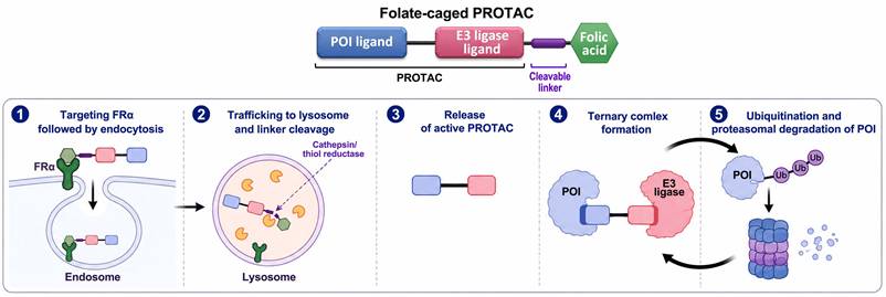
In this context, the Wenyi Wei group developed three folate-caged PROTACs [236] based on the previously described chimeras ARV-771 [237,238], MS432 [239] and MS99 [240]. These three PROTACs are based on an E3 Von Hippel-Lindau (VHL)-binding moiety [241], linked to either a bromodomain and extra-terminal (BET) protein-binding triazolo-diazepine acetamide moiety [242], a portion of the Raf-activated MAP/ERK kinase (MEK) inhibitor PD0325901 [243], or the anaplastic lymphoma kinase (ALK) inhibitor ceritinib [244], respectively. BET proteins are epigenetic readers that regulate gene transcription during various stages of cellular proliferation and differentiation [245]. BET proteins are attractive targets for cancer therapy [246,247], since their inhibition was shown to modulate the suppressive TIME [248-250] as well as abolish the self-renewal ability of pluripotent stem cells and induce their differentiation [251]. MEK1/2 are mitogen-activated kinases of serine, threonine and tyrosine, that are referred to as the “gatekeepers” of ERK activity [252]. As such, MEK1/2 are validated therapeutic targets [253], with five FDA approved inhibitors for the treatment of cancers harboring BRAF mutations like malignant melanoma and NSCLC [254]. ALK is a transmembrane tyrosine kinase receptor involved in the tumorigenesis of several human cancers [255], and hence has been the target of various FDA approved inhibitors, including for the treatment of NSCLC [256]. Thus, the three POIs chosen by the Wenyi Wei group are well-deserved degradation targets as an anti-cancer strategy. Within the folate-caged PROTACs, the FA moiety was conjugated to the E3 ligand via an ester bond, thus blocking the ability of the chimeras to mark the POIs for degradation [236]. Folate-ARV-771 was incubated in cell culture medium supplemented with 10% FBS for 16 h to evaluate its stability. Spontaneous and esterase-mediated hydrolysis released 20% of the PROTAC within this incubation period, suggesting a different cleavable linker should be used to reduce off-target toxicity. The ability of ARV-771 to induce intracellular degradation of the BET proteins bromodomain-containing protein 3 (BRD3) and BRD4 has already been verified in human prostate carcinoma cells with a remarkable DC50 (drug concentration to induce 50% protein degradation) of < 1 nM [237]. Hence, a head-to-head comparison between ARV-771 and its folate-caged counterpart was performed, using high-FRα-expressing cancer cells and non-malignant FRα-null cells [236]. In FRα positive HeLa, OVCAR-8 and T47D cells both BRD degraders exhibited comparable DC50 values, with a slight advantage for the free ARV-771. Complete BRDs degradation was observed after 1-2 h. In the FRα-null 3T3 and HK2 cells, folate-ARV-771 exhibited a minor effect on BRDs degradation, whereas in human HFF-1 fibroblasts the DC50 of folate-ARV-771 was only 10-fold higher than that of ARV-771 [236]. ARV-771 has also been shown to reduce the RNA level of c-Myc, resulting in decreased c-Myc protein levels and induction of caspase-dependent apoptosis [237]. Thus, the cytotoxicity of folate-ARV-771 was evaluated [236]. Consistent with their degradation capacity, ARV-771 and folate-ARV-771 exhibited comparable IC50 values in the FRα-expressing cells, with the lowest IC50 values observed in the highest FRα expressing cell line T47D (i.e., 13 nM vs. 18 nM, respectively). In contrast, in the FRα-null cells, ARV-771 was 6-12-fold more efficient than folate-ARV-771. The cytotoxicity elicited by the folate-caged degrader in these FRα-null cells could be the result of both the instability of the ester bond and cell targeting via FRβ. Interestingly, a cleavage-resistant analogue of folate-ARV-771 had neither degradation activity nor cytotoxicity, establishing the instability of the ester bond as the culprit, as well as validating the blocking capability of the FA moiety. Similar results were obtained with the other folate-caged PROTACs, and their targeted POIs [236]. Folate-MS99 elicited the degradation of transforming ALK-fusion proteins commonly found in cancer. These included EML4-ALK in NSCLC [257] and NPM-ALK in lymphoma [258], both of which render ALK constitutively active and can confer resistance to ALK tyrosine kinase inhibitors (TKIs) [259,260]. Folate-MS432 efficiently elicited the degradation of MEK1 and MEK2 [236]. Both caged-PROTACs degraded their target proteins and elicited cell cytotoxicity in an FRα- and proteasome-dependent manner. FA-S2-MS4048, an additional ALK-targeted folate-caged PROTAC has been synthesized and evaluated [261].
With an intention to enhance solubility, biocompatibility, stability and circulation time for in vivo experiments, the concept of caged PROTACs evolved into PEGylated PROTAC nanoparticles (NPs) [262-264], where the core chimeras are coated with polyethylene glycol (PEG) [265]. In this respect, self-assembling micelles of FA-PEG-PROTAC (MPRO) were designed for the reduction-responsive proteolysis of EGF receptor (EGFR) [264]. MPRO consists of the previously published VHL E3-based PROTAC MS39, which utilized the TKI gefitinib [266] for selective targeting and degradation of mutant EGFR in lung cancer cells [267]. Activating somatic mutations in the tyrosine kinase domain of the EGFR gene are frequent in NSCLC patients [268], mostly in exon 19 [269]. The E3 ligand, within MS39, was conjugated via a glutathione (GSH) cleavable disulfide linker to the commercially available FA-PEG3400, to generate an inert FA-caged PROTAC, which self-assembled into micelles following solvent evaporation [264]. As before, an MPRO-C negative control was also synthesized harboring a non-cleavable alkyl linker; however, the hydroxyl part of the VHL E3 ligand harbors an ester bond which could reduce the stability of the linker. MPROs displayed relatively uniform spherical structures with diameters of ≈ 50 nm, and a zeta potential of -7.1, which should contribute to stability and prevent aggregation. Indeed, MPROs retained their size during a 48 h incubation in 10% FBS and up to 10 days incubation in PBS. By utilizing MPROs loaded with a fluorescent dye, the authors demonstrated the FRα-dependent cellular internalization of the micelles [264]. MPRO treatment of the FRα expressing human NSCLC cells PC-9 and HCC827, which harbor the exon 19 delE746-A750 EGFR mutation [270,271], resulted in a dose-dependent degradation of EGFR [264]. MS39 was superior at inducing EGFR degradation compared to MPRO, and MPRO-C was the least efficient. The higher-than-expected activity of MPRO-C was probably due to the hydrolysis of its ester bond. The relative activity of the three compounds was consistently demonstrated in in vitro cytotoxicity, with MPRO exhibiting no cytotoxic effect in FRα-null cells. For in vivo evaluation, HCC827 tumor-bearing mice were administered every other day, for 16 days, with either MPRO, MPRO-C or MS39 at a dose equivalent to 15 mg/kg MS39. While MPRO and MPRO-C were dissolved in saline, the non-soluble MS39 required a solution containing DMSO, ethanol and Tween 80, demonstrating the contribution of the PEGylated micelles to the solubility and biocompatibility of the PROTAC. MS39 and MPRO-C induced 48.1% and 58.2% TGI, respectively, while MPRO achieved 79.6% TGI, accompanied by significant reduction in EGFR expression [264].
3.2.2. FRα-selective LYsosome TArgeting Chimeras (LYTACs)
An additional group of proteolysis targeting chimeras are the LYTACs which utilized the hydrolytic machinery of lysosomes for the degradation of the POI, instead of the proteasome [272]. First introduced in 2020 [273], LYTACs were designed for the targeted proteolysis of extracellular- and membrane proteins via endocytosis and consequent delivery into lysosomes [274]. The frequent increased acidification of lysosomes in cancer cells, as well as those of tumor associated immune cells, [147,275], along with the elevated levels of proteases and hydrolase activities therein, render these cells ideal targets for LYTACs. The latter are conjugates composed of a ligand for a lysosome-shuttling membrane receptor and an antibody (or ligand) against the extracellular domain of a POI [276]. The tethering of the two proteins by the LYTAC results in their co-engulfment and endocytosis, followed by their fusion-based transport into lysosomes. The low luminal pH within the lysosome induces the dissociation between the receptor and its ligand, with the former recycling back to the cell membrane ready to engage another LYTAC. In the context of the present review, FRα was exploited for the selective targeting of FRα-overexpressing cancer cells by FA-containing LYTACs, while constituting their lysosome-shuttling membrane receptor [277-279]. A scheme illustrating the mode of action of FRα-targeted LYTACs is depicted in Figure 3.
Structure and mode of action of polyvalent FRα-targeted LYsosome TArgeting Chimeras (LYTACs) -FRTACs. The FRTAC consists of a mAb against an extracellular protein of interest (POI), covalently linked to 4-8 folic acid (FA) moieties. 1) The FRTAC targets cancer cells via binding to several FA receptors on the cell surface, while the mAb binds to the membrane POI. 2) Following binding, the complex undergoes FRα-mediated-endocytosis and is transported from the plasma membrane via an endosome. 3) Following fusion with a lysosome, FA dissociates from FRα under the acidic lysosomal pH, and FRα recycles to the plasma membrane. 4) The mAb and POI are degraded by lysosomal cathepsins. The same mechanism also applies to soluble extracellular POIs.
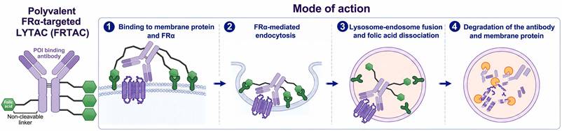
Following proof of concept experiments, establishing the ability of FA-containing LYTACs (i.e., Folate Receptor TArgeting Chimeras, FRTACs) to mediate the FRα-, FRβ- and lysosomal-dependent degradation of soluble extracellular proteins, Yaxian Zhou et al., synthesized cetuximab-containing FRTACs (Ctx-FA) [277]. Cetuximab (Ctx, Erbitux) is a recombinant chimeric mAb against EGFR, used in the treatment of squamous cell carcinoma of the head and neck and metastatic colorectal cancer [280,281]. Since all experiments in this paper [277] were performed in full growth medium, caution should be taken regarding the ability of the described FRTACs to compete with free FA for binding to FRs. In this respect, DMEM and RPMI-1640 growth media contain ~ 9 µM and 2.3 µM FA, respectively, hence far exceeding the 10-100 nM concentrations of FRTACs used in the in vitro experiments. Assessment of EGFR degradation following treatment of FRα- and FRβ-expressing squamous cell carcinoma and cervical cancer Fadu and HeLa cells with Ctx-FA revealed a dose- and time-dependent degradation of EGFR, with increased activity associated with higher degree of folate labeling [277]. Ctx linked by a 2k-PEG-linker to the highest number of ligands, i.e., 4-5, elicited the highest degree of EGFR degradation. In addition, EGFR degradation efficiency was correlated with the FR:EGFR expression ratio. Significant degradation of EGFR was noticeable after ~ 6 h, and the maximal ~ 80% degradation activity (Dmax) of Ctx-FA remained stable for at least 72 h, with DC50 values of 0.41 and 0.24 nM in Fadu and HeLa cells, respectively. Importantly, fluorescence microscopy clearly showed that following treatment with Ctx-FA, EGFR from the plasma membrane of Fadu cells translocated to the lysosomes. While Ctx-FA induced the degradation of EGFR, the levels of FRα and FRβ were not affected, indicating the lysosomal release of the receptors and consequent recycling to the plasma membrane. Two additional FRTACs were synthesized, Atz-FA and Ab2-FA for the targeted degradation of PD-L1 and CD47, respectively. Atz-FA harbors the PD-L1 targeting mAb atezolizumab (Atz, ecentriq), which is the first ICI approved for the treatment of TNBC [282], and has been approved as therapy for various cancers, including alveolar soft part sarcoma [283], hepatocellular carcinoma [284], urothelial carcinoma [285] and NSCLC [286]. Ab2-FA harbors a commercially available mouse anti-CD47 mAb. Treatment of TNBC and NSCLC cell lines MDA-MB-231 and A549, respectively, with either of the FRTACs resulted in the degradation of their cognate POI [277]. The degradation activity of Ctx-FA and Atz-FA was compared between FR-null human keratinocyte HACAT cells and FR-expressing hepatocellular carcinoma HuH7 cells, which express comparable levels of EGFR and PD-L1. The FRTACs induced negligible POI degradation in HACAT cells while dramatically degrading their POI in HuH7 cells, establishing FRTACs as a platform for the FR-dependent degradation of proteins harboring extracellular domains. For the purpose of in vivo evaluation, a mouse homologue of Atz-FA was synthesized (i.e., Ab3-FA) using a commercially available rat anti-mouse PD-L1 mAb instead of Atz [277]. This was puzzling since Atz is known to recognize murine PD-L1 with high affinity [287-289].
Although ICIs transformed therapy of various cancers, they are not without limitations. The expression of PD-L1 on healthy cells renders them targets of PD-L1 antibodies as well, thus PD-1/PD-L1 blockade is associated with multiple TEAEs, most of which are immune-related adverse events [290] and the onset of endocrine dysfunction [291]. Particularly, PD-L1 inhibitors can even provoke a rapid onset of hypothyroidism and type 1 diabetes [292]. Thus, the FR-selective PD-L1-targeting achieved by a FRTAC should alleviate this untoward toxicity. Following the validation of murine PD-L1 degradation by Ab3-FA in murine colon carcinoma and melanoma CT26 and B16F10 cells, respectively, as well as the PD-L1 translocation into lysosomes, initial in vivo pharmacokinetics were measured [277]. Ab3-FA harboring 4-5 FA moieties per antibody, which displayed the highest degradation efficiency in a cell-based assay, also exhibited the shortest circulation half-life in C57BL/6 mice, i.e., 15 h, and thus was chosen for further experiments in tumor bearing mice. Since Ab3-FA was completely cleared from the plasma within 48 h, the treatment regimen included the administration of 2.5-7.5 mg/kg free Ab3 or Ab3-FA every other day for a total of 5 doses. The in vivo testing was conducted in syngeneic mouse models, harboring established tumors (~ 100 mm3) from three cancer origins, i.e., the previously tested CT26 and B16F10 cells as well as the oral squamous cell carcinoma MOC1 cells. In all three mouse models, Ab3-FA induced a TGI which was superior to the free Ab3 (p < 0.05), with complete MOC1-based tumor stasis till the end of the experiment (day 30). Analysis of resected treated tumors confirmed the near-complete degradation of tumor PD-L1 by Ab3-FA, in addition to a dramatic increase in tumor infiltrating CTLs. These results established the enhanced anti-tumor effect of PD-L1 degradation compared to PD-L1 blockade. Spleen and lung tissue biopsies from treated mice were examined via IHC and Western blot assays, where the non-malignant cells exhibited neither morphological changes nor reduction in PD-L1 expression levels; further establishing the FR-selective activity of the Ab3-FA FRTAC [277].
A second research group synthesized FRTACs with a higher number of FA moieties per antibody, termed polyvalent FRTACs [278]. As before, EGFR and PD-L1 were targeted via the Ctx and Atz mAbs, respectively, to produce the FRTACs FR-Ctx and FR-Atz. Additionally, the human epidermal growth factor receptor 2 (HER2) was targeted via the trastuzumab (Ttz, Herceptin) mAb, which constitutes the standard first-line treatment for HER2-positive advanced breast cancer [293], to generate FR-Ttz [278]. Lastly, trophoblast cell surface antigen-2 TROP2 was targeted via the sacituzumab (Stz) mAb [294], used in the first-in-class TROP-2 targeted ADC sacituzumab govitecan, approved for the treatment of TNBC [295], to generate FR-Stz [278]. Polyvalent FRTACs with 15-20 FA conjugates per antibody induced greater degradation of the POIs at lower concentrations than their cognate FRTACs harboring two FA molecules. This is possibly due to their greater functional affinity towards FRα [296], wrongly presented in the paper as affinity [278]. While FA had a calculated Kd of 1.06 nM to FRα, which is in agreement with previous publications discussed in the introduction, the dissociation constant decreased to 0.15 nM for FR-Ctx harboring two FA moieties, and was further reduced to 0.018 pM with the conjugation of 15 FA molecules. These values demonstrate the combined effect of increased avidity and residence time achieved by polyvalent ligands [296] and erroneously misrepresented as mere “affinity” values. It should be noted that multivalent decoration of an antibody with multiple FA residues will necessarily increase the cumulative binding capacity of the conjugate without altering the mere singular interaction of each FA residue with FRα that in fact denotes its genuine Kd value. The increased functional affinity of FR-Ctx, harboring 15 FA moieties, towards FRα decreased its EGFR DC50 value (0.099 nM) by 2-4-fold compared to that of Ctx-FA, harboring 4-5 FA molecules, (0.24-0.41 nM), resulting in the same Dmax ~ 80% [278]. FR-Atz, FR-Stz and FR-Ttz at a FA:antibody ratio of 15 exhibited DC50 values of 0.08, 0.93 and 1.18 nM towards their cognate POIs, respectively. All FRTACs displayed FRα-dependent activity and, as before, had no effect on FRα protein levels.
Unless otherwise stated, the following text refers to FRTACs with a FA:antibody ratio of 15. The human lung mucoepidermoid carcinoma H292 cells (FRα+ EGFR+ PD-L1+) were used to evaluate the downstream effects of the FRTACs compared to their free mAb counterparts. FR-Ctx blocked the proliferative impact of EGF, as well as its downstream kinase signaling axis, resulting in minute levels of phosphorylated STAT3 and ERK1/2, in a manner exceeding free Ctx. Co-culturing H292 cells with activated human-derived PBMCs under treatment with FR-Atz, demonstrated the ability of FR-Atz to negate immunosuppression. The FR-Atz-dependent degradation of PD-L1 allowed for the dramatic activation of CTLs, resulting in an increased secretion of granzyme B and interferon-gamma (IFNγ), leading to tumor cell apoptosis. Free Atz had little effect on PD-L1-dependent immunosuppression. Using H292-tumor bearing mice and whole-animal body imaging, fluorescently tagged FR-Atz was shown to accumulate at the tumor site within 5 h, and retained fluorescence intensity for 5 days. In contrast, free Atz had a slow tumor accumulation rate of 1-2 days. Tumor FR-Atz fluorescence intensity far exceeded that of any healthy organ, and consistently, FR-Atz-dependent degradation of PD-L1 was achieved solely in the tumor, indicating an FRα-selective mode of action. The FR-Atz-dependent degradation of tumor PD-L1 was also demonstrated in mouse prostate cancer RM-1 cells (FRα+ PD-L1+), both in vitro and in vivo. In RM-1-tumor bearing mice, degradation of PD-L1 following FR-Atz treatment resulted in apoptosis of tumor cells and a consequent ~ 75% reduction in tumor weight (p = 0.0003). Lastly, using mouse models harboring tumors established from B16F10 melanoma cells expressing human PD-L1, the authors confirmed the FR-Atz-dependent reprograming of the immunosuppressive TIME. This included polarization of tumor associated macrophages (TAMs) from the M2 to the M1 phenotype, activation and tumor infiltration of CTLs, as well as reduction in the levels of regulatory T cells [278].
Following the promising results obtained with its published PROTACs, the Wenyi Wei group recently presented a novel design of an FRTAC developed to hijack FRα for the simultaneous degradation of a pair of complementary transmembrane POIs, i.e., EGFR/HER2 or PD-L1/VISTA [279]. EGFR and HER2, both members of the ErbB family of growth factor receptors [297], can form highly active heterodimers which instigate tumor cell proliferation [298-300]. A frequent mechanism of resistance to Ttz is EGFR overexpression [301-303], while HER2 hyper-activation was shown as a mechanism of Ctx resistance [304,305]. Hence, the simultaneous targeting of both receptors can enhance the therapeutic efficacy. In this respect, dual inhibitors have been approved for clinical use, such as the irreversible ErbB family blocker afatinib [306]. However, HER2 and EGFR are expressed in a wide variety of healthy tissues [109,307], therefore, inhibiting their signaling pathways can result in TEAEs. Indeed, like other ErbB-directed mAbs, Ctx and Ttz inflict various severe TEAEs including dermatologic toxicities and interstitial pneumonitis [308]. In fact, 70% of colorectal cancer patients treated with Ctx developed severe TEAEs [309]. The newly designed FRTAC that targets the EGFR/HER2 duo, i.e., FolTAC-dual, should overcome both problems of acquired resistance and off-target toxicity [279]. The same is true for the pair of V-domain immunoglobulin suppressor of T-cell activation (VISTA) [310] and PD-L1, both of which are negative immune checkpoint molecules, that are expressed in healthy tissues [311]. Within the TME, both tumor and immune cells frequently express the VISTA/PD-L1 duo, resulting in enhanced immune resistance [311]. Although they represent distinct immune checkpoint pathways, they can compensate for one another. Thus, following anti-PD-L1 treatment, disease progression in melanoma patients is associated with increased density of VISTA+ lymphocytes (p = 0.009) [312]. Consistently, bispecific antibodies designed on the basis of Atz and onvatilimab (CI-8993), an anti-VISTA mAb currently evaluated in clinical trials [313], for the co-inhibition of PD-L1 and VISTA, exhibited superior anti-tumor activity than either monotherapy or combination therapy [314]. The Wenyi Wei group first constructed single degrader FolTACs for each POI, harboring a farletuzumab scFv linked to either a HER2 binding affibody ligand [315], EGFR affibody ligand [316], Atz scFv, or onvatilimab scFv, with remarkable Dmax = 82-95% [279]. Then, for the assembly of the FolTAC-duals, two geometries were explored. The “knob-into-hole” geometry consisted of the two POI targeting moieties on separate sides of the construct, while in the “string” geometry both sides of the construct harbored the two targeting moieties connected one after the other. The “string” configuration was found to enhance both the binding affinity to the POIs and the degradation efficacy in both FolTAC-duals, which successfully induced the FRα- and lysosome-dependent co-degradation of their POI pairs in vitro [279].
Next, the ability of the FolTAC-duals to surmount drug resistance elicited by inhibition of their POIs was evaluated. To this end, Ttz-resistant human breast cancer SK-BR-3-pool2 cells [317] were further exposed to 0.25 µM lapatinib (Lap) for 8 weeks to establish Ttz/Lap cross-resistant cells (FRα+ EGFP+ HER2+) [279]. Lap is a small molecule TKI that blocks HER2 signaling by competitively binding the intracellular ATP-binding sites of HER-2 [318]. The EGFR/HER2 FolTAC-dual induced high apoptosis rates in the resistant cells, comparable to that in the sensitive parental cells, while normal human foreskin HFF-1 fibroblast cells were not affected [279]. In vivo experiments conducted in mouse models bearing Ttz/Lap resistant SK-BR-3-based tumors, revealed the enhanced anti-tumor activity of the FolTAC-dual compared to an anti-HER2 FolTAC. FolTAC-dual induced complete tumor stasis, while HER2-FolTAC induced a non-significant TGI. It should be conveyed that the combination of HER2-FolTAC and EGFR-FolTAC had a dramatic anti-tumor effect as well. The PD-L1/VISTA FolTAC-dual was evaluated using an MC38 syngeneic mouse model, where the MC38 cells were engineered to express human FRα and VISTA. PD-L1/VISTA FolTAC-dual induced complete degradation of both POIs only in 3/7 tumors evaluated by Western blot and this correlated with the low survival rates of the tested mouse models 3/15, although half exhibited TGI and ~ 2-fold longer survival time [279]. FRα-guided proteolysis targeting chimeras show great potential as a selective and efficient therapy mode for FRα-positive tumors, and should be further evaluated pre-clinically to establish feasible clinical value.
4. FRβ-dependent reprogramming of the suppressive TIME
As mentioned above, and detailed in our recent review [147], the predominant tumor-associated immune cell population of macrophages [319,320] is of the M2 phenotype that orchestrates cancer immune evasion and immunosuppression [321-323]. M0 monocytes can undergo polarization by naturally occurring signaling cues which have been exploited for immune research [324]. Specifically, macrophages assume the M1 configuration following stimulation by microbial agonists of toll-like receptors (TLRs), e.g., lipopolysaccharide (LPS), as well as pro-inflammatory cytokines such as IFNγ and TNF; whereas polarization to M2 macrophages is promoted by IL-4 and TGF-β. Unfortunately, the tumor associated population of M2 macrophages also increases following anti-cancer treatments, including radiotherapy [325,326] and chemotherapy based on cisplatin [327-329] and cyclophosphamide [330]. Although some ambiguity exists regarding macrophage polarization following activation with the cytokine granulocyte-macrophage colony-stimulating factor (GM-CSF) and macrophage colony stimulating factor (M-CSF), it is largely accepted that GM-CSF promotes the transition of macrophages to the M1 phenotype, whereas M-CSF favors M2 [331-333]. It has been established that by attenuating the activity of lysosomes within monocytes and macrophages, one can polarize these cells to the M1 configuration and induce an anti-tumor immune response [147]. Lysosome dysfunction can be achieved, for instance, by alkalization and lysosome membrane permeabilization (LMP) [147,334-336].
In vitro experiments, where human-derived monocytes were polarized to either the M1 or M2 phenotype, by the various factors detailed above, allowed for the characterization of “pure” macrophage populations [26]. One dramatic variable found was the differential expression of FRβ on M2 macrophages, enabling them to bind and internalize FA. Moreover, TAMs within patient-derived melanoma specimens were positive for cell surface FRβ, and so were stromal cells of lung, ovary, colon, gastric, and breast cancers [26]. Hence, the expression of FRβ on TAMs has been further explored and studied [27], revealing that active FRβ is not just a biomarker for suppressive TAMs but also necessary for their activity [337]. While FRα-targeted therapies are restricted to a small subset of tissues, the expression of FRβ on TAMs and tumor resident myeloid-derived suppressor cells (MDSC) [28] highlights it as a potential therapeutic target affecting any type of tumor. FRβ can be used for the detection of solid tumors, for targeted cytotoxicity, thereby reducing the population of immune suppressive cells, or for the targeted conversion of M2 macrophages to the M1 phenotype, which will further reprogram the TIME.
4.1. FRβ-targeted conversion of M2 macrophages
In an extensive study on the expression of FRβ in various tumor associated cells, using multiple diverse mouse tumor grafts, FRβ was found to be co-expressed with known markers of MDSCs and TAMs in 11-26% of total tumor cells [28]. This indicated the high prevalence of tumor infiltrating suppressive cells. Mice harboring syngeneic FR- MB49 bladder cancer tumors were treated with a folate-targeted photodynamic therapy (PDT) agent to eradicate FRβ-positive myeloid suppressive cells. However, full replenishment of the FRβ-positive cell population was observed within 72 h [28]. This demonstrated that using FRβ for targeted cytotoxicity of immunosuppressive cells is futile, hence reprograming these cells could be a better solution. To this end, orthotopic FR- 4T1 mammary tumor bearing mouse models were treated with a FA-conjugated TLR7 agonist (FA-TLR7a) (patent US20210170035A1) [28]. The intracellular TLR7, expressed primarily by macrophages and DCs [338], is involved in the detection of pathogen-associated molecular patterns and induction of a host immune response. Following ligand binding by TLR7, there is an induction of conformational changes and adaptor recruitment, resulting in the nuclear translocation of the central transcription factor nuclear factor kappa-light-chain-enhancer of activated B cells (NF-κB). The latter induces the expression and secretion of pro-inflammatory cytokines, such as the M1-macrophage associated IL-6 and TNF [339]. Both FA-TLR7a and free TLR7a had no cytotoxic effect on FR- 4T1 tumor cells in vitro [28]. However, the FRβ-mediated uptake of FA-TLR7a by TAMs and MDSCs in vivo resulted in significant TGI in the mouse models with no associated loss of mouse body weight. In contrast, free TLR7a had no effect on tumor growth but was highly toxic, as reflected by the drastic loss of body weight and systemic release of cytokines [28]. Indeed, TLR7 agonists have never received approval by the FDA since they activate immune cells in healthy tissues, leading to systemic toxicity and immune complications such as neuroinflammation [340], lymphopenia [341] and cytokine release syndrome [342]. The FRβ-selective targeting of TLR7 prevents a systemic reaction by confining the immune response to FRβ+ TLR7+ immune cells at the primary tumor site and any metastases. In this respect, FA-TLR7a was shown to completely circumvent the formation of metastases for 18 days (p ≤ 0.05), whereas free TLR7a had no such inhibitory effect [28]. To validate that this FA-TLR7a-induced anti-tumor effect was achieved by reprograming of immune cells to a tumoricidal M1-like phenotype, the authors isolated TAMs and MDSCs from the treated tumor-bearing mice. Analyses showed that the expression of suppressive markers by TAMs and MDSCs was reduced by >50%, accompanied by a significant shift in macrophages from an M2 towards the M1 phenotype. A scheme illustrating the repolarization of M2 TAMs by FA-TLR7a is depicted in Figure 4.
Structure and mode of action of FA-TLR7a. FA-TLR7a consists of a folic acid (FA) moiety covalently attached to a toll-like receptor 7 (TLR7) agonist (TLR7a). 1) FA-TLR7a binds to FRβ, expressed on the surface of immunosuppressive immune cells, primarily M2 tumor-associated macrophages (TAMs), following which the complex undergoes receptor-mediated endocytosis. 2) Within the endosome, the FRβ-bound FA-TLR7a binds the resident TLR7 to form a ternary complex. 3) Following binding to its agonist, TLR7 is activated and undergoes dimerization, resulting in the recruitment of its adaptor protein myeloid differentiation primary response 88 (MyD88), a key adaptor for innate immune response signaling. 4) MyD88 activates downstream signaling pathways resulting in the nuclear translocation of the transcription factors NF-κB and IRF7 and consequent transcription of pro-inflammatory cytokines and interferons. 5) The pro-inflammatory cues drive the repolarization of M2 TAMs to the M1 phenotype, resulting in secretion of anti-tumor cytokines IL-6 and TNF.
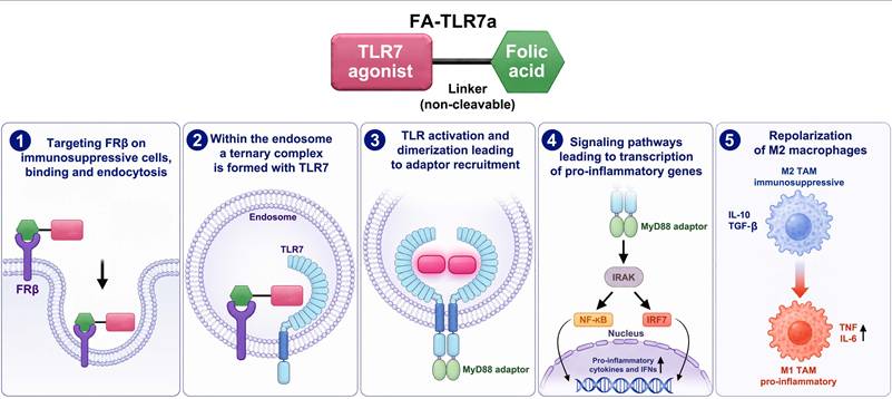
The results further revealed the consequent activation of an immune response by a ~ 10-fold increase in the number of tumor infiltrating T helper cells and CTLs (p ≤ 0.001). Finally, FA-TLR7a increased the median survival time of orthotopic 4T1 tumor-bearing mice from 27 day to 37 days (p ≤ 0.01) [28]. An additional FA-linked TLR7 agonist (FA-TLR7-1A) was designed and tested [343]. The TLR7 agonist chosen was cell-impermeable and linked to FA by a hydrophilic non-cleavable PEG spacer to prevent circulation and cell entry of free agonist. FA-TLR7-1A exhibited comparable affinities of ∼ 20 nM for both TLR7 and FRβ. The activity of FA-TLR7-1A was validated in vitro via its ability to induce the production of TNF and IL-6 by immunosuppressive M2-like macrophages, with maximal activity occurring at 10 nM. Its pharmacological safety was assessed by monitoring the weight of treated mice during five days of FA-TLR7-1A administration (60 nmol). Control and FA-TLR7-1A-treated mice gained 5% of body weight, whereas free TLR7-1A induced a drastic 10% loss of body weight [343]. These results established a therapeutic window where FA-TLR7-1A is both active and pharmacologically safe. As before, orthotopic 4T1 tumor bearing mouse models were used for in vivo evaluation, and were injected with 3 nmol/day. FA-TLR7-1A induced a ~ 50% reduction in the growth of the primary tumor (p < 0.001) and almost completely prevented the development of lung metastases (p < 0.01), with no effect on body weight for 20 days. Post treatment tumors were dissected and analyzed for pro-inflammatory and anti-inflammatory markers expressed by tumor infiltrating TAMs and MDSCs. Post-treatment immune cells exhibited a robust transition from an immunosuppressive state to an active state, accompanied by the activation of CTLs [343]. Since it appeared that FA-TLR7-1A was not potent enough as monotherapy, the authors explored its possible combination with ICI mAbs against murine PD-1 (clone: 29F.1A12) or cytotoxic T lymphocyte-associated antigen-4 (CTLA-4, clone: 9H10) [344]. T helper lymphocytes expressing high levels of CTLA-4 are reprogrammed into regulatory T lymphocytes exhibiting immunosuppressive characteristics. Anti-CTLA-4 alone induced 30% TGI in 4T1 tumor bearing mice, while the combination with FA-TLR7-1A resulted in a dramatic 90% TGI (p < 0.0001) [343]. This efficacious combination therapy reprogrammed the suppressive TIME by converting the TAMs and MDSCs to a tumoricidal phenotype, inducing an increase in tumor infiltrating activated T helper cells and CTLs. Consistently, in syngeneic MC38 colon cancer models, the combination of FA-TLR7-1A with the anti-PD-1 mAb resulted in CR. The potent immediate activation of the reprogrammed TIME also induced prolonged immunity which protected the mice upon re-challenging with MC38 tumor cells; hence preventing the establishment of new tumors [343]. A detailed demonstration of TIME reprogramming by FA-TLR7-1A has been published [345].
5. Co-targeting tumor and immune cells
Metabolic reprograming, now considered as the eighth hallmark of cancer [346], occurs in tumor cells in addition to the TME and the TIME, where it contributes to tumorigenesis, rapid cell proliferation, invasion, immune evasion, immunosuppression as well as drug resistance [347-353]. In this respect, the metabolism of the essential amino acid glutamine (Gln) is frequently enhanced in cancer [354,355], since it serves as a critical carbon and nitrogen donor for various biosynthetic processes such as lipogenesis, de novo nucleotide biosynthesis and amino acid synthesis. In addition, glutaminolysis in mitochondria allows the anaplerotic refueling of the TCA cycle via provision of alpha ketoglutarate (αKG), and αKG conversion to citrate allows adaptation to hypoxia [356]. On the other hand, Gln is also required for the proper activity of the immune system [353,355,357]; specifically, T cells rely on Gln for rapid proliferation and immune response [355,358], and Gln regulates macrophage polarization [355,359,360]. Hence selective targeted inhibition of Gln metabolism may hamper cancer cell proliferation while sparing healthy tissues and T cell populations [361].
One strategy for selective targeting of Gln metabolism utilized poly D,L-lactide-co-glycolic acid (PLGA)-PEG-FA NPs, loaded with the first-generation Gln antimetabolite 6-diazo-5-oxo-l-norleucine (DON) [362] and pH-responsive calcium carbonate (CaCO3) to generate (FA-DCNPs) [363]. DON is a two-step inhibitor of multiple glutamine-utilizing enzymes, such as glutaminase and various glutamine amidotransferases [364]. Following its competitive binding to the Gln binding active site, it forms a covalent adduct that irreversibly inhibits the enzyme. While clinical studies with DON, conducted in the 50's, were abandoned due to dose-limiting toxicity, renewed interest in the drug has arisen following the discovery of Gln-dependence in cancer [362,364]. The pH-responsive decomposition of CaCO3 was shown to facilitate drug release from PLGA NPs under acidic conditions (pH 5.5) [365], suggesting good drug release within lysosomes. Moreover, within the acidic lysosome, the carbonate ions interact with protons leading to lysosome alkalinization and calcium (Ca2+) release, both of which contribute to lysosome dysfunction and LMP [336,366]. As detailed in our recent review [147], M2 macrophages are characterized by highly acidic lysosomes with enhanced hydrolase activity, which promote augmented autophagic flux. M2 macrophages can be converted to the M1 phenotype by alkalinizing their lysosomes. In addition, tumor cells are also frequently characterized by acidic lysosomes and enhanced autophagic flux, resulting in over-processed tumor antigens which cannot be presented by MHC for the recognition and activation of immune cells [147]. Hence, by selectively binding to both FRα-positive tumor cells and their associated FRβ-positive suppressive immune cells, FA-DCNPs have the potential to disrupt the lysosomes of tumor and immune cells, inhibit tumor cell proliferation and increase immune reactivity. The size of the FA-DCNPs remained relatively stable during a 20 days incubation in cell culture medium supplemented with 10% FBS [363]. FA-DCNPs exhibited a time- and pH-dependent release of DON and Ca2+. Murine epithelial OC ID8 cells were verified for high expression of both FRα and FRβ, while both receptors were nearly undetectable in human umbilical vein HUVEC cells. As already established, M2 macrophages displayed high levels of FRβ, M0 monocytes exhibited moderate expression levels, while M1 cells expressed low levels, with all three subtypes displaying low levels of FRα. Accordingly, ID8 and M2 cells displayed high FA-DCNPs internalization rates (i.e., 96% and 88%, respectively) compared to the other cell types (p < 0.001). Specifically, the internalization rate by M1 macrophages was lower by >70% compared to M2, whereas HUVEC cells did not exhibit any internalization. In vitro cytotoxicity assays established the FR-dependent killing mechanism of FA-DCNPs, which included a 6.4-fold increase in cellular reactive oxygen species (ROS) levels [363]. Perturbation of redox homeostasis is an established consequence of disrupted Gln metabolism, following the reduction in cellular anabolism, as well as glutamate and GSH levels [356,367]. Apart from the disruption of Gln metabolism by DON, FA-DCNPs also elicited cytosolic release of Ca2+ which triggered mitochondrial Ca2+ overload and consequent dysfunction [363], thus further contributing to the oxidative stress [368]. It is surprising that the authors completely disregarded the contribution of the apparent FA-DCNPs-dependent LMP that facilitated the release of Ca2+ and ROS from lysosomes, hence triggering mitochondrial Ca2+ overload. Since mitochondrial damage was shown to induce ICD [369-372], the authors explored whether this is also relevant to FA-DCNPs. Indeed, following treatment with FA-DCNPs, ID8 cells exhibited high levels of the canonical ICD-associated DAMPs, including >10-fold increase in surface expression of CRT and a ~ 3-fold increase in HMGB1 release (p < 0.001) [363]. Given that macrophage M1 polarization can be induced by lysosomal dysfunction [147], oxidative stress-damaged mitochondria [372], deregulated Gln metabolism [373,374] and ICD [375], the ability of FA-DCNPs to rewire M2 macrophage was evaluated. M0 RAW264.7 cells polarized to M1 and M2 populations by LPS and IL-4, respectively, were analyzed for their glutamate content [363]. Following FA-DCNPs treatment, M2 macrophages exhibited a ~ 60% drop in their glutamate content and underwent robust reprograming into the M1 phenotype, presented by the co-stimulatory CD86 surface biomarker and secretion of IL-12. Importantly, treatment of RAW264.7 cells with FA-DCNPs prior to their stimulation with IL-4, skewed their polarization into M2 macrophages, consistently resulting in M1 cells.
In vivo evaluation of FA-DCNPs was conducted in ID8-based xenograft mouse models [363]. Compared to nontargeted DCNPs, FA-DCNPs exhibited a higher tumor:liver accumulation ratio after 48 h, albeit the liver accumulation was substantial. Nonetheless, blood tests demonstrated normal liver function for 28 days, and IHC showed no aberrant cellular morphology. FA-DCNPs, administered every other day for a total of 4 doses, induced a dramatic inhibition of tumor cell proliferation along with enhanced tumor cell apoptosis (p < 0.001). During the treatment, FA-DCNPs markedly prevented tumor growth (TGI ~ 85%), which was resumed following treatment cessation, resulting in a 2-fold longer survival time (p < 0.001). The anti-tumor activity of FA-DCNPs was accompanied by an increase in tumor infiltrating CTLs, mature DCs and M1-TAMs, while the number of M2-TAMs declined. Consistently, FA-DCNPs treatment resulted in increased secretion of the immunostimulatory cytokines TNF, IFN-γ and IL-12, whereas secretion of the immunosuppressive IL-10 dropped significantly (p < 0.001). To further corroborate the activation of a robust immune response, FA-DCNPs treated mice were re-challenged with subcutaneously injected ID8 cells at a distant site, to mimic metastases. This distant tumor cell inoculation was rapidly infiltrated by CTLs and its growth was dramatically attenuated. FA-DCNPs demonstrated an effective and selective co-targeting of both the tumor cells and the TIME [363].
An additional demonstration of FA-directed co-targeting NPs was published by Haoyu Xu et al., where FA-PEG decorated ginger-derived exosome-like nanoparticles (GELNs) loaded with sunitinib (FPD-GELNs/Su) were used for the treatment of renal cell carcinoma (RCC) [376]. Sunitinib is a multi-targeted TKI [377] used for the treatment of gastrointestinal and pancreatic neuroendocrine tumors as well as RCC [378]. As a hydrophobic weak-base small-molecule, sunitinib highly accumulates in lysosomes and has strong natural fluorescence, traits that have been exploited for PDT [379-381]. Sunitinib is a substrate of P-gp, and can be readily extruded from the cell by this ATP-driven efflux transporter [382,383]. The biologically active constituents of ginger, mainly 6-gingerol and 6-shogaol, have been the subject of various studies as they were found to exert anticancer activities against gastrointestinal cancer [384,385]. Isolated from fresh ginger homogenate by sucrose gradient centrifugation, GELNs are spherical bilayer membranous vesicles with a 100-200 nm diameter and a negative zeta-potential [376,386], which have been mainly studied as drug delivery systems [387,388]. However, since the composition of GELNs also consists of biologically active components, they can have intrinsic anti-tumor capabilities. Indeed, before sunitinib loading, these GELNs exhibited a dose- and time-dependent cytotoxic activity against murine and human RCC cells, RenCa, OS-RC-2 and 786-O, with little effect against normal kidney HK-2 cells [376]. In addition, GELNs inhibited colony formation, induced cell cycle arrest, and elicited anti-migratory and anti-invasive effects through inhibition of the PI3K-Akt signaling pathway [376], a mechanism previously suggested for both 6-gingerol [389] and 6-shogaol [390]. In order for the GELNs to display significant anti-tumor activity in vivo, they had to be injected into the tumor, as any other administration resulted in their rapid depletion from the circulation and the gastrointestinal track [376]. The addition of the FA-PEG coating increased the negative zeta potential of the NPs and greatly improved their biodistribution. This prevented the depletion of the NPs by spontaneous phagocytosis and allowed their intravenous injection. Following sunitinib loading, its release from the FPD-GELNs/Su was monitored. At physiological pH, the release of sunitinib occurred during the first 24 h and reached ~ 40%. At an acidic pH of 5.6, the release of sunitinib was greater and longer, achieving ~ 80% at 48 h. This suggests a good sunitinib release within lysosomes following RME. For compatibility with FPD-GELNs/Su, the expression levels of FRα and FRβ were quantified in the above cell lines. OS-RC-2 and HK-2 cells were negative for both receptors, while RenCa and 786-O cells expressed high levels of FRα [376]. The expression of FRs in macrophage subtypes has been previously discussed. In vitro cytotoxicity assays at comparable concentrations of sunitinib, revealed that FPD-GELNs/Su had the lowest IC50 value in RenCa and 786-O cells, a value that was comparable to that of GELNs + free sunitinib. Although the fast diffusion rate of free sunitinib into cells far exceeds the slower internalization rates of sunitinib loaded NPs, treatment with either FPD-GELNs/Su or GELNs + free sunitinib resulted in higher cellular sunitinib concentrations than treatment with free sunitinib alone [376]. Ginger-derived NPs have been shown to inhibit the PI3K/Akt/NF-κB signaling pathway and prevent the nuclear translocation of the central transcription factor NF-κB, thus attenuating transcription of various genes [391]. Specifically, ferulic acid, which is one of the components of GELNs, inhibited NF-κB and reduced the expression of P-gp at the RNA and protein levels, thereby restoring drug sensitivity in drug resistant cells [392]. In addition, 6-gingerol was shown to inhibit the efflux activity of P-gp and increase the cellular accumulation of P-gp substrate drugs [393]. Hence, the authors explored the impact of GELNs on P-gp expression and found that FPD-GELNs/Su induced the strongest drop in P-gp mRNA and protein levels [376], suggesting a plausible mechanism for the enhanced sunitinib accumulation. The latter, along with the cytotoxic effect of the GELNs themselves, resulted in strong synergism between GELNs and free sunitinib, which was also found for FPD-GELNs/Su, i.e., combination index (CI) [394] of 0.1-0.3 [376]. One crucial aspect of the FPD-GELNs/Su system was overlooked by the authors and must be discussed. Owing to its physicochemical properties, following the RME of FPD-GELNs/Su, sunitinib should remain sequestered within the acidic lysosomes [395-397]. Hence, we propose that GELNs destabilize the lysosomal membrane via 6-gingerol that has been shown to elicit LMP and cathepsin D release [398], thus allowing for the escape of sunitinib into the cytosol. In this respect, GELNs can also contribute to ICD through induction of LMP [200,399] and promote macrophage polarization to the M1 phenotype [147]. Both M0 and M2 macrophages exhibited a ~ 68% internalization efficiency of GELNs within 24 h, while the addition of FA-PEG markedly enhanced their efficiency to 82% and 99%, respectively (p < 0.001) [376]. FPD-GELNs also underwent a much more rapid internalization into M2 macrophages, surpassing 68% within 2 h. FPD-GELNs induced the polarization of M0 into M1 macrophages at comparable levels to the canonical LPS, and greatly surpassed it at converting M2 macrophages to the M1 phenotype. This conversion resulted, amongst others, in a >30-fold increase in IL-6 and a ~ 17-fold reduction in TGF-β (p < 0.001). Interestingly, 6-gingerol has been shown to induce the M2 to M1 polarization of macrophages in hepatocellular and lung carcinomas through the activation of NADPH oxidase 2 (NOX2) and production of ROS [400,401]. The ability of FPD-GELNs to induce robust M1 polarization was recapitulated in vivo in RenCa-based xenograft mouse models. The increase in the M1:M2 ratio within the TAMs, induced by both FPD-GELNs and FPD-GELNs/Su, was accompanied by a 4.5-7.5-fold increase in tumor infiltrating CTLs, a ~ 2.5-fold increase in T helper cells (p < 0.01), and dramatic expression of IFNγ. Sunitinib had no significant effect on tumor growth, while FPD-GELNs exhibited strong TGI (p < 0.001), however, the synergistic effect of FPD-GELNs/Su induced excessive tumor cell apoptosis resulting in near complete tumor stasis (p < 0.001) [376]. Blood tests and immunostaining revealed no drug related toxicity. Lastly, the ability of FPD-GELNs/Su to prevent RCC-related lung metastasis was also tested. Control mice developed ~ 100 lung nodules, a number that was not statistically different following sunitinib treatment, unless a much higher concentration was used. On the other hand, treatment with FPD-GELNs/Su decreased the number of metastatic lesions by ~ 80% [376]. FPD-GELNs/Su represent a multi-mechanism and synergistic therapeutic approach for the selective co-targeting of tumor cells and their TIME (Figure 5).
Co-targeting tumor and immune cells by drug-loaded Ginger Exosome-Like Nanoparticles (GELNs). A. Preparation of folic acid (FA)-decorated and sunitinib loaded GELNs (FPD-GELNs/Su). GELNs are isolated from fresh ginger homogenate by sucrose gradient centrifugation, and loaded with sunitinib, a hydrophobic weak-base tyrosine kinase inhibitor (TKI). Finally, the GELNs are decorated with FA-PEG. B. Induction of tumor cell death by FPD-GELNs/Su. 1) Renal cell carcinoma cells overexpress FRα, which binds FPD-GELNs/Su and mediates their endocytosis. 2) The endosomes fuse with lysosomes. Under the acidic lysosomal conditions, sunitinib and the ginger constituent 6-gingerol are released from the destabilized GELNs, while FA dissociates from FRα. 3) FRα recycles to the plasma membrane. 4) 6-gingerol induces lysosomal membrane permeabilization (LMP), allowing the release of the lysosomal content, including cathepsins, sunitinib, 6-gingerol and the ginger constituent 6-shogaol, into the cytosol. 5) 6-gingerol and 6-shogaol inhibit the PI3K-Akt signaling pathway, blocking the nuclear entrance of the downstream effector NF-κB. This leads to decreased transcription of NF-κB-responsive genes, including ABCB1 which encodes the ATP-driven membrane multidrug exporter, P-glycoprotein (P-gp). 6) Following the decreased expression of P-gp, its substrate sunitinib is no longer extruded out of the cell and is retained at high intracellular concentrations. Sunitinib inhibits, amongst others, the receptor tyrosine kinases vascular endothelial growth factor receptor (VEGFR) and platelet-derived growth factor receptor (PDGFR), hence further blocking their downstream PI3K-Akt signaling pathway. 7) The potent inhibition of the PI3K-Akt signaling axis abolishes tumor cell migration and invasion, preventing them from metastasizing. 8) The combined effects of LMP and the blockade of the PI3K-Akt signaling pathway lead to cell cycle arrest, decreased proliferation and enhanced apoptosis of the tumor cells. C. Polarization of tumor-associated macrophages (TAMs) to the anti-tumor M1 phenotype. 1) The immunosuppressive M2, and to a lesser degree, M0 macrophages overexpress FRβ, which binds FPD-GELNs/Su and mediates their endocytosis. 2) The endosomes fuse with lysosomes. Under the acidic lysosomal conditions, sunitinib and the ginger constituent 6-gingerol are released from the destabilized GELNs, while FA dissociates from FRβ. 3) FRβ recycles to the plasma membrane. 4) 6-gingerol induces LMP, allowing the cytosolic release of the lysosomal content, including sunitinib and 6-gingerol. 5) Following LMP, lysosomal dysfunction blocks the progression of autophagy. 6) 6-gingerol activates NADPH oxidase 2 (NOX2) which induces the production of reactive oxygen species (ROS). 7) Decreased autophagy and increased ROS promote the polarization of M0 and M2 macrophages to the M1 phenotype. M1 macrophages secrete pro-inflammatory cytokines such as IL-6, which stimulates the activity and tumor infiltration of cytotoxic T cells, and tumor necrosis factor (TNF) which triggers tumor cell death. M1 macrophages also engulf and digest cancer cells via phagocytosis. The induction of LMP within macrophages, and the release of cathepsins, sunitinib and 6-gingerol can also induce macrophage cell death, which results in the release of ROS and cytokines into the tumor microenvironment (TME), thus inducing the polarization of other immunosuppressive cells.
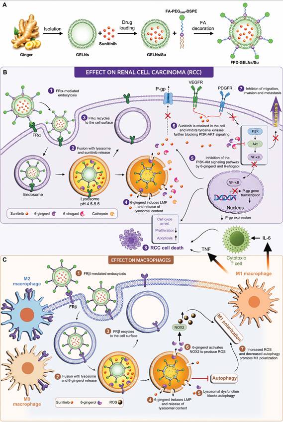
6. Summary
In conclusion, the expression pattern of FRs makes them bona fide targets for the efficacious selective delivery of either cytotoxic drugs or immune modulating factors. In case of an FRα-positive tumor, the co-targeting of the tumor cells and the surrounding TIME by a FA-tagged lysosomal disruptive agent achieved excellent pre-clinical results and should be further developed. In case of an FRα-negative tumor, reprograming of the TIME by FRβ-targeted agents can be successfully combined with validated ICIs for enhanced invigoration of the immune system. While FRα-selective ADCs were developed for tumor targeting, the ability of ADCs to induce ICD also potentiates immune cells. Hence, future designs could benefit from the combination of a homogenous ADC with an LMP-inducing payload that would elicit ICD. With regards to ADCs, special attention should be given to the latest FR-targeted ADC AZD5335, which has shown promising initial results in PROC patients with low FRα expression. This ADC could represent a real breakthrough in the treatment of FRα-expressing tumors.
Abbreviations
5-MTHF: 5-methyltetrahydrofolate; ADC: antibody-drug conjugate; ADCC: antibody-dependent cell-mediated cytotoxicity; ADCP: antibody-dependent cell-mediated phagocytosis; AIBW: adjusted ideal body weight; αKG: alpha ketoglutarate; ALK: anaplastic lymphoma kinase; AML: acute myeloid leukemia; APCs: antigen-presenting cells; α-SMA: alpha-smooth muscle actin; ATP: adenosine triphosphate; Atz: atezolizumab; BAT: basophil activation test; BCRP: breast cancer resistance protein, ABCG2; BET: bromodomain and extra-terminal; BRD3: bromodomain-containing protein 3; CAFs: cancer-associated fibroblasts; CARs: chimeric antigen receptors; CDC: complement-dependent cell death; CI: combination index; CR: complete response; CRT: calreticulin; CTLs: cytotoxic T-lymphocytes; CTLA-4: cytotoxic T lymphocyte-associated antigen-4; Ctx: Cetuximab; DAMP: damage-associated molecular pattern; DAR: drug to antibody ratio; DCs: dendritic cells; DM4: N2′-deacetyl-N2′-(4-mercapto-4-methyl-1-oxopentyl)-maytansine; DON: 6-diazo-5-oxo-l-norleucine; EGF: epidermal growth factor; EGFR: epidermal growth factor receptor; EMA: European Medicines Agency; FA: folic acid; Fab: fragment antigen-binding; FAP: fibroblast activation protein; FcγR: Fcγ receptor; FcRn: neonatal Fc receptor; FDA: US Food and Drug Administration; FFPE: formalin-fixed paraffin-embedded; FPGS: folylpoly-γ-glutamate synthetase; FR: folate receptor; FRTAC: folate receptor targeting chimera; GELNs: ginger-derived exosome-like nanoparticles; GM-CSF: granulocyte-macrophage colony-stimulating factor; GSH: glutathione; GPI: glycosyl-phosphatidylinositol; HER2: human epidermal growth factor receptor 2; HMGB1: high mobility group box 1; ICD: immunogenic cell death; ICIs: immune checkpoint inhibitors; IFNγ: interferon-gamma; IHC: immunohistochemistry; IL-4: interleukin-4; IMI: intraoperative molecular imaging; ITC: isothermal titration calorimetry; Lap: lapatinib; LMP: lysosome membrane permeabilization; LPS: lipopolysaccharide; mAbs: monoclonal antibodies; MAFs: metastasis-associated fibroblasts; M-CSF: macrophage colony stimulating factor; MDSC: myeloid-derived suppressor cells; MEK: Raf-activated MAP/ERK kinase; MIRV: mirvetuximab soravtansine-gynx, IMGN853, ELAHERE; MORAb-202: farletuzumab ecteribulin, farletuzumab-[Mal-PEG2-Val-Cit-PAB-eribulin]; MPRO: folate-PEG-S-S-PROTAC conjugate; MTA: microtubule targeting agent; MTD: maximum tolerated dose; MyD88: myeloid differentiation primary response 88; NF-κB: nuclear factor kappa-light-chain-enhancer of activated B cells; NIR: near infra-red; NK: natural killer; NOX2: NADPH oxidase 2; NPs: nanoparticles; NPSA: non-platinum single-agent; NSCLC: non-small cell lung cancer; OC: ovarian cancer; ORR: objective response rate; OS: overall survival; PARPi: poly ADP-ribose polymerase inhibitor; PBMCs: peripheral blood mononuclear cells; PCFT: proton-coupled folate transporter, SLC46A1; PD: progressive disease; PD-1: programmed cell death protein 1; PD-L1: programmed death-ligand 1; PDT: photodynamic therapy; PDXs: patient-derived xenografts; P-gp: P-glycoprotein, ABCB1; PLGA: poly D,L-lactide-co-glycolic acid; POI: protein of interest; PR: partial response; PROC: platinum-resistant epithelial ovarian cancer; PSOC: platinum-sensitive ovarian cancer; RCC: renal cell carcinoma; RD: residual disease; RFC: reduced folate carrier, SLC19A1; RME: receptor-mediated endocytosis; RP2D: recommended phase II dose; ROS: reactive oxygen species; SC209: 3-aminophenyl hemiasterlin; scFv: single-chain variable fragment; SCID: severe combined immunodeficiency; SD: stable disease; sFRα: soluble FRα; Stz: Sacituzumab; sulfo-SPDB: N-succinimidyl 4-(2-pyridyldithio)-2-sulfobutanoate; TAMs: tumor associated macrophages; TBR: tumor-to-background ratio; TEAEs: treatment emergent adverse events; TGF-α: transforming growth factor alpha; TGI: tumor growth inhibition; TIME: tumor immune microenvironment; TKIs: tyrosine kinase inhibitors; TLRs: toll-like receptors; TME: tumor microenvironment; TNF: tumor necrosis factor; TOP1i: topoisomerase-1 inhibitor; TROP2: trophoblast cell surface antigen-2; Ttz: trastuzumab; UPS: ubiquitin proteasome system; USC: uterine serous carcinoma; VATS: video-assisted thoracoscopic surgery; VEGF: vascular endothelial growth factor; VHL: Von Hippel-Lindau; VISTA: v-domain immunoglobulin suppressor of T-cell activation.
Acknowledgements
ChatGPT 5 academic was used to generate individual images within the figures.
Competing Interests
The authors have declared that no competing interest exists.
References
1. Raz S, Stark M, Assaraf YG. Folylpoly-γ-glutamate synthetase: A key determinant of folate homeostasis and antifolate resistance in cancer. Drug Resist Updat. 2016;28:43-64
2. de la Torre Guzmán SR, Pelayo-Chávez B, García-Muro AM, Soto-Reyes E, Sánchez-López JY. The Role of Folic Acid in DNA Methylation and Breast Cancer. Int J Vitam Nutr Res. 2025;95(2):26221
3. Korsmo HW, Jiang X. One carbon metabolism and early development: a diet-dependent destiny. Trends Endocrinol Metab. 2021;32(8):579-93
4. Van Winkle LJ, Ryznar R. One-Carbon Metabolism Regulates Embryonic Stem Cell Fate Through Epigenetic DNA and Histone Modifications: Implications for Transgenerational Metabolic Disorders in Adults. Front cell Dev Biol. 2019;7:300
5. Bou Ghanem A, Hussayni Y, Kadbey R, Ratel Y, Yehya S, Khouzami L. et al. Exploring the complexities of 1C metabolism: implications in aging and neurodegenerative diseases. Front Aging Neurosci. 2024;15:1322419
6. Majumder A, Bano S, Nayak KB. The Pivotal Role of One-Carbon Metabolism in Neoplastic Progression During the Aging Process. Biomolecules. 2024;14(11):1387
7. Lim EW, Metallo CM. Tracing the Diverse Paths of One-Carbon Metabolism in Cancer and Beyond. Cold Spring Harb Perspect Med. 2024;14(9):a041533
8. Zhao R, Goldman ID. Folate and thiamine transporters mediated by facilitative carriers (SLC19A1-3 and SLC46A1) and folate receptors. Mol Aspects Med. 2013;34(2-3):373-85
9. Zhang Q, Zhang X, Liu K, Zhu Y, Nie X, Ma J. et al. Molecular basis of SLC19A1-mediated folate and cyclic dinucleotide transport. Nat Commun. 2025;16(1):3146
10. Hou Z, Gangjee A, Matherly LH. The evolving biology of the proton-coupled folate transporter: New insights into regulation, structure, and mechanism. FASEB J. 2022;36(2):e22164
11. Elnakat H, Ratnam M. Distribution, functionality and gene regulation of folate receptor isoforms: Implications in targeted therapy. Adv Drug Deliv Rev. 2004;56(8):1067-84
12. Jansen G, Mauritz R, Drori S, Sprecher H, Kathmann I, Bunni M. et al. A structurally altered human reduced folate carrier with increased folic acid transport mediates a novel mechanism of antifolate resistance. J Biol Chem. 1998;273(46):30189-98
13. Goldman ID. The characteristics of the membrane transport of amethopterin and the naturally occurring folates. Ann N Y Acad Sci. 1971;186(1):400-22
14. Mahadeo K, Diop-Bove N, Shin D, Unal ES, Teo J, Zhao R. et al. Properties of the Arg376 residue of the proton-coupled folate transporter (PCFT-SLC46A1) and a glutamine mutant causing hereditary folate malabsorption. Am J Physiol - Cell Physiol. 2010;299(5):C1153
15. Qiu A, Jansen M, Sakaris A, Min SH, Chattopadhyay S, Tsai E. et al. Identification of an intestinal folate transporter and the molecular basis for hereditary folate malabsorption. Cell. 2006;127(5):917-28
16. Kamen BA, Capdevila A. Receptor-mediated folate accumulation is regulated by the cellular folate content. Proc Natl Acad Sci U S A. 1986;83(16):5983-7
17. Egorova O, Domellöf E, Ardalan M, Mallard C. Folate Receptor Alpha (FRα) and the Developing Brain: From Molecular Function to Neurodevelopmental Outcomes. Mol Neurobiol. 2026;63(1):522
18. Chen C, Ke J, Edward Zhou X, Yi W, Brunzelle JS, Li J. et al. Structural basis for molecular recognition of folic acid by folate receptors. Nature. 2013;500(7463):486-9
19. Wibowo AS, Singh M, Reeder KM, Carter JJ, Kovach AR, Meng W. et al. Structures of human folate receptors reveal biological trafficking states and diversity in folate and antifolate recognition. Proc Natl Acad Sci U S A. 2013;110(38):15180-8
20. Elwood PC, Kane MA, Portillo RM, Kolhouse JF. The isolation, characterization, and comparison of the membrane-associated and soluble folate-binding proteins from human KB cells. J Biol Chem. 1986;261(33):15416-23
21. Xin W, Feng S, Freisheim JH, Gentry LE, Ratnam M. Differential stereospecificities and affinities of folate receptor isoforms for folate compounds and antifolates. Biochem Pharmacol. 1992;44(9):1898-901
22. Aweda TA, Meares CF. Combination of isothermal titration calorimetry and time-resolved luminescence for high affinity antibody-ligand interaction thermodynamics and kinetics. Methods. 2011;56(2):145
23. Nawaz FZ, Kipreos ET. Emerging roles for folate receptor FOLR1 in signaling and cancer. Trends Endocrinol Metab. 2022;33(3):159-74
24. Liu Y, Lian T, Yao Y. A systematic review and meta-analysis of higher expression of folate receptor alpha (FOLR1) predicts poor cancer prognosis. Biomarkers. 2020;25(5):367-74
25. Chen CI, Li WS, Chen HP, Liu KW, Tsai CJ, Hung WJ. et al. High Expression of Folate Receptor Alpha (FOLR1) is Associated With Aggressive Tumor Behavior, Poor Response to Chemoradiotherapy, and Worse Survival in Rectal Cancer. Technol Cancer Res Treat. 2022;21:15330338221141795
26. Puig-Kröger A, Sierra-Filardi E, Domínguez-Soto A, Samaniego R, Corcuera MT, Gómez-Aguado F. et al. Folate receptor beta is expressed by tumor-associated macrophages and constitutes a marker for M2 anti-inflammatory/regulatory macrophages. Cancer Res. 2009;69(24):9395-403
27. Jiang D, Xiao R, Bai J, Wang X, Fang Y, Liu J. et al. FOLR2+ macrophages in cancer: allies or enemies. Cell Commun Signal. 2025;23(1):261
28. Cresswell GM, Wang B, Kischuk EM, Broman MM, Alfar RA, Vickman RE. et al. Folate Receptor Beta Designates Immunosuppressive Tumor-Associated Myeloid Cells That Can Be Reprogrammed with Folate-Targeted Drugs. Cancer Res. 2021;81(3):671-84
29. Zhu X, Chen C. FOLR2+ macrophages and CAFs: coordinating immunosuppression in gastric cancer. Int J Surg. 2026;112(4):10812-4
30. Ahmadi M, Ritter CA, von Woedtke T, Bekeschus S, Wende K. Package delivered: folate receptor-mediated transporters in cancer therapy and diagnosis. Chem Sci. 2024;15(6):1966-2006
31. Scaranti M, Cojocaru E, Banerjee S, Banerji U. Exploiting the folate receptor α in oncology. Nat Rev Clin Oncol. 2020;17(6):349-59
32. Crider KS, Devine O, Qi YP, Yeung LF, Sekkarie A, Zaganjor I. et al. Systematic Review and Bayesian Meta-analysis of the Dose-response Relationship between Folic Acid Intake and Changes in Blood Folate Concentrations. Nutrients. 2019;11(1):71
33. Ganji V, Kafai MR. Demographic, lifestyle, and health characteristics and serum B vitamin status are determinants of plasma total homocysteine concentration in the post-folic acid fortification period, 1999-2004. J Nutr. 2009;139(2):345-52
34. Lamers Y, Prinz-Langenohl R, Brämswig S, Pietrzik K. Red blood cell folate concentrations increase more after supplementation with [6S]-5-methyltetrahydrofolate than with folic acid in women of childbearing age. Am J Clin Nutr. 2006;84(1):156-61
35. Chen MY, Rose CE, Qi YP, Williams JL, Yeung LF, Berry RJ. et al. Defining the plasma folate concentration associated with the red blood cell folate concentration threshold for optimal neural tube defects prevention: A population-based, randomized trial of folic acid supplementation. Am J Clin Nutr. 2019;109(5):1452-61
36. Crider KS, Qi YP, Devine O, Tinker SC, Berry RJ. Modeling the impact of folic acid fortification and supplementation on red blood cell folate concentrations and predicted neural tube defect risk in the United States: have we reached optimal prevention? Am J Clin Nutr. 2018;107(6):1027
37. Mathias CJ, Wang S, Lee RJ, Waters DJ, Low PS, Green MA. Tumor-Selective Radiopharmaceutical Targeting via Receptor-Mediated Endocytosis of Gallium-67-Deferoxamine-Folate. J Nucl Med. 1996;37(6):1003-8
38. Wang L, Cherian C, Desmoulin SK, Polin L, Deng Y, Wu J. et al. Synthesis and antitumor activity of a novel series of 6-substituted pyrrolo[2,3-d]pyrimidine thienoyl antifolate inhibitors of purine biosynthesis with selectivity for high affinity folate receptors and the proton-coupled folate transporter over the redu…. J Med Chem. 2010;53(3):1306-18
39. Leamon CP, Reddy JA, Dorton R, Bloomfield A, Emsweller K, Parker N. et al. Impact of high and low folate diets on tissue folate receptor levels and antitumor responses toward folate-drug conjugates. J Pharmacol Exp Ther. 2008;327(3):918-25
40. Fu T, Dai LJ, Wu SY, Xiao Y, Ma D, Jiang YZ. et al. Spatial architecture of the immune microenvironment orchestrates tumor immunity and therapeutic response. J Hematol Oncol. 2021;14(1):98
41. Roh TH, Kim SH. Supramaximal Resection for Glioblastoma: Redefining the Extent of Resection Criteria and Its Impact on Survival. Brain Tumor Res Treat. 2023;11(3):166
42. Brown ZJ, Heh V, Labiner HE, Brock GN, Ejaz A, Dillhoff M. et al. Surgical resection rates after neoadjuvant therapy for localized pancreatic ductal adenocarcinoma: meta-analysis. Br J Surg. 2022;110(1):34-42
43. Reveron-Thornton RF, Teng MLP, Lee EY, Tran A, Vajanaphanich S, Tan EX. et al. Global and regional long-term survival following resection for HCC in the recent decade: A meta-analysis of 110 studies. Hepatol Commun. 2022;6(7):1813-26
44. Chen Y, Jing W, Chen M, Wang Z, Wu J, Yang J. et al. Long-term outcomes of local resection versus surgical resection for high-risk T1 colorectal cancer: a systematic review and meta-analysis. Gastrointest Endosc. 2023;97(6):1016-1030.e14
45. Bois A Du, Reuss A, Pujade-Lauraine E, Harter P, Ray-Coquard I, Pfisterer J. Role of surgical outcome as prognostic factor in advanced epithelial ovarian cancer: a combined exploratory analysis of 3 prospectively randomized phase 3 multicenter trials: by the Arbeitsgemeinschaft Gynaekologische Onkologie Studiengruppe Ovarialkarzi…. Cancer. 2009;115(6):1234-44
46. Eskander RN, Kauderer J, Tewari KS, Mannel RS, Bristow RE, O'Malley DM. et al. Correlation between Surgeon's assessment and radiographic evaluation of residual disease in women with advanced stage ovarian cancer reported to have undergone optimal surgical cytoreduction: An NRG Oncology/Gynecologic Oncology Group study. Gynecol Oncol. 2018;149(3):525-30
47. Trelis Blanes A, Lago V, Pérez Martínez R, Belloch Ripollés V, Montoliu G, Padilla-Iserte P. et al. Residual Tumour at CT Scan Based on Radiologic Peritoneal Carcinomatosis Index After Optimal Cytoreduction in Advanced Ovarian Cancer: A True Prognostic Factor. Cancers (Basel). 2025;17(5):746
48. Esbona K, Li Z, Wilke LG. Intraoperative imprint cytology and frozen section pathology for margin assessment in breast conservation surgery: a systematic review. Ann Surg Oncol. 2012;19(10):3236-45
49. Agdoğan Ö, Arslan Aİ. Examining Surgical Margins in the Excision of Basal Cell Carcinoma and Squamous Cell Carcinoma: Evaluating Pre-Excision, Post-Excision, Post-Fixation, and Pathological Margin Measurements. J Surg Oncol. 2026;133(3):295-301
50. Aziz D, Rawlinson E, Narod SA, Sun P, Lickley HLA, McCready DR. et al. The role of reexcision for positive margins in optimizing local disease control after breast-conserving surgery for cancer. Breast J. 2006;12(4):331-7
51. Parker N, Turk MJ, Westrick E, Lewis JD, Low PS, Leamon CP. Folate receptor expression in carcinomas and normal tissues determined by a quantitative radioligand binding assay. Anal Biochem. 2005;338(2):284-93
52. Mahalingam SM, Kularatne SA, Myers CH, Gagare P, Norshi M, Liu X. et al. Evaluation of Novel Tumor-Targeted Near-Infrared Probe for Fluorescence-Guided Surgery of Cancer. J Med Chem. 2018;61(21):9637-46
53. Keating JJ, Runge JJ, Singhal S, Nims S, Venegas O, Durham AC. et al. Intraoperative near-infrared fluorescence imaging targeting folate receptors identifies lung cancer in a large-animal model. Cancer. 2017;123(6):1051-60
54. Frangioni J V. In vivo near-infrared fluorescence imaging. Curr Opin Chem Biol. 2003;7(5):626-34
55. Weissleder R. A clearer vision for in vivo imaging. Nat Biotechnol. 2001;19(4):316-7
56. Hoogstins CES, Tummers QRJG, Gaarenstroom KN, De Kroon CD, Trimbos JBMZ, Bosse T. et al. A Novel Tumor-Specific Agent for Intraoperative Near-Infrared Fluorescence Imaging: A Translational Study in Healthy Volunteers and Patients with Ovarian Cancer. Clin Cancer Res. 2016;22(12):2929-38
57. Randall LM, Wenham RM, Low PS, Dowdy SC, Tanyi JL. A phase II, multicenter, open-label trial of OTL38 injection for the intra-operative imaging of folate receptor-alpha positive ovarian cancer. Gynecol Oncol. 2019;155(1):63-8
58. Tanyi JL, Randall LM, Chambers SK, Butler KA, Winer IS, Langstraat CL. et al. A Phase III Study of Pafolacianine Injection (OTL38) for Intraoperative Imaging of Folate Receptor-Positive Ovarian Cancer (Study 006). J Clin Oncol. 2023;41(2):276-84
59. Ferrari FA, Ceccaroni M, Scambia G, Fagotti A, Pavone M, Bourdel N. et al. Enhancing surgical precision in ovarian cancer with FRα-fluorescence-guided surgery. Eur J Surg Oncol. 2025;51(8):110120
60. Erdemoglu E, Langstraat CL, Kumar A, Ostby SA, Girardo ME, Giannini A. et al. Fluorescence-Guided Surgery to Detect Microscopic Disease in Ovarian Cancer: A Systematic Review with Meta-Analysis. Cancers (Basel). 2025;17(3):410
61. Fayaz M, Ahmed KAHM. Advancing intraoperative tumour detection and molecular-guided precision surgery, FDA's approval of pafolacianine injection: an editorial. Ann Med Surg. 2023;86(1):18
62. O'Shannessy DJ, Yu G, Smale R, Fu YS, Singhal S, Thiel RP. et al. Folate Receptor Alpha Expression in Lung Cancer: Diagnostic and Prognostic Significance. Oncotarget. 2012;3(4):414
63. Nunez MI, Behrens C, Woods DM, Lin H, Suraokar M, Kadara H. et al. High expression of folate receptor alpha in lung cancer correlates with adenocarcinoma histology and mutation. J Thorac Oncol. 2012;7(5):833-40
64. Cagle PT, Zhai QJ, Murphy L, Low PS. Folate receptor in adenocarcinoma and squamous cell carcinoma of the lung: potential target for folate-linked therapeutic agents. Arch Pathol Lab Med. 2013;137(2):241-4
65. Yuan R, McGeehan A, Zhou S, Cheng C, Armanini M, Smith J. et al. The Anti-FRα Antibody-Drug Conjugate Luveltamab Tazevibulin Demonstrates Efficacy in Non-Small Cell Lung Cancer Preclinical Models and Induces Immunogenic Cell Death. Mol Cancer Ther. 2025;24(9):1428-41
66. Predina JD, Newton AD, Keating J, Dunbar A, Connolly C, Baldassari M. et al. A Phase I Clinical Trial of Targeted Intraoperative Molecular Imaging for Pulmonary Adenocarcinomas. Ann Thorac Surg. 2018;105(3):901-8
67. Gangadharan S, Sarkaria IN, Rice D, Murthy S, Braun J, Kucharczuk J. et al. Multiinstitutional Phase 2 Clinical Trial of Intraoperative Molecular Imaging of Lung Cancer. Ann Thorac Surg. 2021;112(4):1150-9
68. Sarkaria IS, Martin LW, Rice DC, Blackmon SH, Slade HB, Singhal S. et al. Pafolacianine for intraoperative molecular imaging of cancer in the lung: The ELUCIDATE trial. J Thorac Cardiovasc Surg. 2023;166(6):e468-78
69. Lehane A, Polites SF, Dodd A, Goldstein SD, Lautz TB. Let it glow: Intraoperative visualization of pulmonary metastases using pafolacianine, a next-generation fluorescent agent, for young adults undergoing pulmonary metastasectomy. Pediatr Blood Cancer. 2024;71(11):e31293
70. Mukkamala R, Mar BCW, Keltner RG, Noble BA, Mahalingam SM, Shahriar I. et al. Design of a Pan-Tumor Fluorescence Imaging Cocktail for Fluorescence-Guided Surgery. Bioconjug Chem. 2026;37(1):22
71. Busek P, Mateu R, Zubal M, Kotackova L, Sedo A. Targeting fibroblast activation protein in cancer - Prospects and caveats. Front Biosci (Landmark Ed. 2018;23(10):1933-68
72. Scanlan MJ, Raj BKM, Calvo B, Garin-Chesa P, Sanz-Moncasi MP, Healey JH. et al. Molecular cloning of fibroblast activation protein alpha, a member of the serine protease family selectively expressed in stromal fibroblasts of epithelial cancers. Proc Natl Acad Sci U S A. 1994;91(12):5657-61
73. Liu Y, Sinjab A, Min J, Han G, Paradiso F, Zhang Y. et al. Conserved spatial subtypes and cellular neighborhoods of cancer-associated fibroblasts revealed by single-cell spatial multi-omics. Cancer Cell. 2025;43(5):905-924.e6
74. Tao L, Huang G, Song H, Chen Y, Chen L. Cancer associated fibroblasts: An essential role in the tumor microenvironment. Oncol Lett. 2017;14(3):2611-20
75. Pan C, Zheng L, Wang J. Cancer associated fibroblasts in tumors: focusing on solid tumors and hematological malignancies. Front Oncol. 2025;15:1596947
76. Zheng H, Liu D, Liu Z, An M, Luo Y, Chen C. et al. Roles of cancer-associated fibroblast functional heterogeneity in shaping the lymphatic metastatic landscape: new insights and therapeutic strategies. Cancer Biol Med. 2024;21(6):445-50
77. Wang Z, Liu J, Huang H, Ye M, Li X, Wu R. et al. Metastasis-associated fibroblasts: an emerging target for metastatic cancer [Internet]. Vol. 9, Biomarker Research. Biomark Res. 2021 p. 47
78. Kou Z, Liu C, Zhang W, Sun C, Liu L, Zhang Q. Heterogeneity of primary and metastatic CAFs: From differential treatment outcomes to treatment opportunities (Review). Int J Oncol. 2024;64(5):38577950
79. James RL, Sisserson T, Cai Z, Dumas ME, Inge LJ, Ranger-Moore J. et al. Development of an FRα Companion Diagnostic Immunohistochemical Assay for Mirvetuximab Soravtansine. Arch Pathol Lab Med. 2024;148(11):1226-33
80. Administration USF& D. FDA's approval. 2022. VENTANA FOLR1 (FOLR-2.1) RxDx Assay - P220006 | FDA.
81. Matulonis UA, Lorusso D, Oaknin A, Pignata S, Dean A, Denys H. et al. Efficacy and Safety of Mirvetuximab Soravtansine in Patients With Platinum-Resistant Ovarian Cancer With High Folate Receptor Alpha Expression: Results From the SORAYA Study. J Clin Oncol. 2023;41(13):2436-45
82. Coleman RL, Lorusso D, Oaknin A, Cecere SC, Denys H, Colombo N. et al. Mirvetuximab soravtansine in folate receptor alpha (FRα)-high platinum-resistant ovarian cancer: final overall survival and post hoc sequence of therapy subgroup results from the SORAYA trial. Int J Gynecol Cancer. 2024;34(8):1119-25
83. Moore KN, Angelergues A, Konecny GE, García Y, Banerjee S, Lorusso D. et al. Mirvetuximab Soravtansine in FRα-Positive, Platinum-Resistant Ovarian Cancer. N Engl J Med. 2023;389(23):2162-74
84. Heo YA. Mirvetuximab Soravtansine: First Approval. Drugs. 2023;83(3):265-73
85. Dilawari A, Shah M, Ison G, Gittleman H, Fiero MH, Shah A. et al. FDA Approval Summary: Mirvetuximab Soravtansine-Gynx for FRα-Positive, Platinum-Resistant Ovarian Cancer. Clin Cancer Res. 2023;29(19):3835-40
86. Previs RA, Strickland KC, Wallen Z, Ko H, Green M, Cooper M. et al. Analysis of real world FRα testing in ovarian, fallopian tube, and primary peritoneal cancers. Gynecol Oncol. 2025;192:102-10
87. Chen X, Jiang L, Ruan L, Yu T, Rui W, Fan Y. et al. Performance of the VENTANA FOLR1 Assay for folate receptor alpha: Real-world evidence from 313 Chinese participants. Pathol - Res Pract. 2026;279:156359
88. Chu GCW, Lazare K, Sullivan F. Serum and blood based biomarkers for lung cancer screening: a systematic review. BMC Cancer. 2018;18(1):181
89. Rajapaksha W, Khetan R, Johnson IRD, Blencowe A, Garg S, Albrecht H. et al. Future theranostic strategies: emerging ovarian cancer biomarkers to bridge the gap between diagnosis and treatment. Front drug Deliv. 2024;4:1339936
90. Tang Y, Cui Y, Zhang S, Zhang L. The sensitivity and specificity of serum glycan-based biomarkers for cancer detection. Prog Mol Biol Transl Sci. 2019;162:121-40
91. Grancher A, Cuissy S, Girot H, Gillibert A, Di Fiore F, Guittet L. Where do we stand with screening for colorectal cancer and advanced adenoma based on serum protein biomarkers? A systematic review. Mol Oncol. 2024;18(11):2629-48
92. Holm J, Hansen SI. Characterization of soluble folate receptors (folate binding proteins) in humans. Biological roles and clinical potentials in infection and malignancy. Biochim Biophys Acta - Proteins Proteomics. 2020;1868(10):140466
93. Kurosaki A, Hasegawa K, Kato T, Abe K, Hanaoka T, Miyara A. et al. Serum folate receptor alpha as a biomarker for ovarian cancer: Implications for diagnosis, prognosis and predicting its local tumor expression. Int J cancer. 2016;138(8):1994-2002
94. Bax HJ, Chauhan J, Stavraka C, Santaolalla A, Osborn G, Khiabany A. et al. Folate receptor alpha in ovarian cancer tissue and patient serum is associated with disease burden and treatment outcomes. Br J Cancer. 2023;128(2):342-53
95. Farran B, Albayrak S, Abrams J, Tainsky MA, Levin NK, Morris R. et al. Serum folate receptor α (sFR) in ovarian cancer diagnosis and surveillance. Cancer Med. 2019;8(3):920-7
96. Shimizu T, Fujiwara Y, Yonemori K, Koyama T, Sato J, Tamura K. et al. First-in-Human Phase 1 Study of MORAb-202, an Antibody-Drug Conjugate Comprising Farletuzumab Linked to Eribulin Mesylate, in Patients with Folate Receptor-α-Positive Advanced Solid Tumors. Clin Cancer Res. 2021;27(14):3905-15
97. Liu H, Sun Q, Zhang M, Zhang Z, Fan X, Yuan H. et al. Differential expression of folate receptor 1 in medulloblastoma and the correlation with clinicopathological characters and target therapeutic potential. Oncotarget. 2017;8(14):23048-60
98. O'Shannessy DJ, Somers EB, Albone E, Cheng X, Park YC, Tomkowicz BE. et al. Characterization of the human folate receptor alpha via novel antibody-based probes. Oncotarget. 2011;2(12):1227-43
99. Köhler G, Milstein C. Continuous cultures of fused cells secreting antibody of predefined specificity. Nature. 1975;256(5517):495-7
100. Paul S, Konig MF, Pardoll DM, Bettegowda C, Papadopoulos N, Wright KM. et al. Cancer therapy with antibodies. Nat Rev Cancer. 2024;24(6):399-426
101. Labanieh L, Mackall CL. CAR immune cells: design principles, resistance and the next generation. Nature. 2023;614(7949):635-48
102. Ong MZ, Kimberly SA, Lee WH, Ling M, Lee M, Tan KW. et al. FDA-approved CAR T-cell Therapy: A Decade of Progress and Challenges. Curr Pharm Biotechnol. 2023;25(11):1377-93
103. Okpasuo OJ, Olaoba OT, Bokolo P, Aleke AS, Dare A, Adelusi TI. et al. The evolving landscape of antibody-based cancer therapies: From monospecific to multi-specific and beyond. Crit Rev Oncol Hematol. 2026;217:105037
104. Fu Z, Li S, Han S, Shi C, Zhang Y. Antibody drug conjugate: the “biological missile” for targeted cancer therapy. Signal Transduct Target Ther. 2022;7(1):93
105. Veneziani AC, Sneha S, Oza AM. Antibody-Drug Conjugates: Advancing from Magic Bullet to Biological Missile. Clin Cancer Res. 2024;30(8):1434-7
106. Bussel JB, Cines DB, Blumberg RS. Neonatal Fc Receptor - Biology and Therapeutics. N Engl J Med. 2025;392(16):1621-35
107. Redman JM, Hill EM, AlDeghaither D, Weiner LM. Mechanisms of action of therapeutic antibodies for cancer. Mol Immunol. 2015;67(2):28-45
108. Rodríguez-Nava C, Ortuño-Pineda C, Illades-Aguiar B, Flores-Alfaro E, Leyva-Vázquez MA, Parra-Rojas I. et al. Mechanisms of Action and Limitations of Monoclonal Antibodies and Single Chain Fragment Variable (scFv) in the Treatment of Cancer. Biomedicines. 2023;11(6):1610
109. Tito C, Masciarelli S, Colotti G, Fazi F. EGF receptor in organ development, tissue homeostasis and regeneration. J Biomed Sci. 2025;32(1):24
110. Zhang M, Zhu J, Bao Y, Ao Q, Mao X, Qiu Z. et al. Bevacizumab in ovarian cancer therapy: current advances, clinical challenges, and emerging strategies. Front Bioeng Biotechnol. 2025;13:1589841
111. Chitoran E, Rotaru V, Stefan DC, Gullo G, Simion L. Blocking Tumoral Angiogenesis VEGF/VEGFR Pathway: Bevacizumab-20 Years of Therapeutic Success and Controversy. Cancers (Basel). 2025;17(7):1126
112. Chin DS, Lim CSY, Nordin F, Arifin N, Jun TG. Antibody-Dependent Cell-Mediated Cytotoxicity Through Natural Killer (NK) Cells: Unlocking NK Cells for Future Immunotherapy. Curr Pharm Biotechnol. 2022;23(4):552-78
113. Cao X, Chen J, Li B, Dang J, Zhang W, Zhong X. et al. Promoting antibody-dependent cellular phagocytosis for effective macrophage-based cancer immunotherapy. Sci Adv. 2022;8(11):eabl9171
114. Vivier E, Rebuffet L, Narni-Mancinelli E, Cornen S, Igarashi RY, Fantin VR. Natural killer cell therapies. Nature. 2024;626(8000):727-36
115. Taylor RP, Lindorfer MA. Cytotoxic mechanisms of immunotherapy: Harnessing complement in the action of anti-tumor monoclonal antibodies. Semin Immunol. 2016;28(3):309-16
116. Bordron A, Bagacean C, Tempescul A, Berthou C, Bettacchioli E, Hillion S. et al. Complement System: a Neglected Pathway in Immunotherapy. Clin Rev Allergy Immunol. 2020;58(2):155-71
117. Li SY, Guo YL, Tian JW, Zhang HJ, Li RF, Gong P. et al. Anti-Tumor Strategies by Harnessing the Phagocytosis of Macrophages. Cancers (Basel). 2023;15(10):2717
118. Sadreddini S, Baradaran B, Aghebati-Maleki A, Sadreddini S, Shanehbandi D, Fotouhi A. et al. Immune checkpoint blockade opens a new way to cancer immunotherapy. J Cell Physiol. 2019;234(6):8541-9
119. Rolfo C, Giovannetti E, Martinez P, McCue S, Naing A. Applications and clinical trial landscape using Toll-like receptor agonists to reduce the toll of cancer. NPJ Precis Oncol. 2023;7(1):26
120. Suzuki R, Takizawa T, Maruyama K. Cloning of a tumor-associated antigen : MOv18 and MOv19 antibodies recognize a folate-binding protein. Cancer Res. 1991;51(6):6125-32
121. Miotti S, Canevari S, Ménard S, Mezzanzanica D, Porro G, Pupa SM. et al. Characterization of human ovarian carcinoma-associated antigens defined by novel monoclonal antibodies with tumor-restricted specificity. Int J cancer. 1987;39(3):297-303
122. Rettig WJ, Cordon-Cardo C, Koulos JP, Lewis JL, Oettgen HF, Old LJ. Cell surface antigens of human trophoblast and choriocarcinoma defined by monoclonal antibodies. Int J cancer. 1985;35(4):469-75
123. Ebel W, Routhier EL, Foley B, Jacob S, McDonough JM, Patel RK. et al. Preclinical evaluation of MORAb-003, a humanized monoclonal antibody antagonizing folate receptor-alpha. Cancer Immun a J Acad Cancer Immunol. 2007;7:6
124. Nicolaides NC, Ebel W, Kline B, Chao Q, Routhier E, Sass PM. et al. Morphogenics as a tool for target discovery and drug development. Ann N Y Acad Sci. 2005;1059:86-96
125. Konner JA, Bell-McGuinn KM, Sabbatini P, Hensley ML, Tew WP, Pandit-Taskar N. et al. Farletuzumab, a humanized monoclonal antibody against folate receptor alpha, in epithelial ovarian cancer: a phase I study. Clin Cancer Res. 2010;16(21):5288-95
126. Smith-Jones PM, Pandit-Taskar N, Cao W, O'Donoghue J, Philips MD, Carrasquillo J. et al. Preclinical radioimmunotargeting of folate receptor alpha using the monoclonal antibody conjugate DOTA-MORAb-003. Nucl Med Biol. 2008;35(3):343-51
127. Kim KH, Jelovac D, Armstrong DK, Schwartz B, Weil SC, Schweizer C. et al. Phase 1b safety study of farletuzumab, carboplatin and pegylated liposomal doxorubicin in patients with platinum-sensitive epithelial ovarian cancer. Gynecol Oncol. 2016;140(2):210-4
128. Vergote I, Armstrong D, Scambia G, Teneriello M, Sehouli J, Schweizer C. et al. A Randomized, Double-Blind, Placebo-Controlled, Phase III Study to Assess Efficacy and Safety of Weekly Farletuzumab in Combination With Carboplatin and Taxane in Patients With Ovarian Cancer in First Platinum-Sensitive Relapse. J Clin Oncol. 2016;34(19):2271-8
129. Herzog TJ, Pignata S, Ghamande SA, Rubio MJ, Fujiwara K, Vulsteke C. et al. Randomized phase II trial of farletuzumab plus chemotherapy versus placebo plus chemotherapy in low CA-125 platinum-sensitive ovarian cancer. Gynecol Oncol. 2023;170:300-8
130. Wu M, Lv F, Jin Y, Wang S, Meng L, Tong J. et al. Dynamic changes of the immune microenvironment in ovarian cancer following neoadjuvant chemotherapy. Cell death Discov. 2026;12(1):130
131. Zhu W, Li J, Sun Y, Lin Q, Sun Y, Xue M. et al. Neoadjuvant chemotherapy-induced immune remodeling in ovarian cancer: implications for TIL dynamics and combination immunotherapy. Front Immunol. 2026;17:1757366
132. Cheng X, Li J, Tanaka K, Majumder U, Milinichik AZ, Verdi AC. et al. MORAb-202, an Antibody-Drug Conjugate Utilizing Humanized Anti-human FRα Farletuzumab and the Microtubule-targeting Agent Eribulin, has Potent Antitumor Activity. Mol Cancer Ther. 2018;17(12):2665-75
133. Gould HJ, Mackay GA, Karagiannis SN, O'toole CM, Marsh PJ, Daniel BE. et al. Comparison of IgE and IgG antibody-dependent cytotoxicity in vitro and in a SCID mouse xenograft model of ovarian carcinoma. Eur J Immunol. 1999;29(11):3527-37
134. Sutton BJ, Davies AM, Bax HJ, Karagiannis SN. IgE Antibodies: From Structure to Function and Clinical Translation. Antibodies (Basel, Switzerland). 2019;8(1):19
135. Jensen-Jarolim E, Achatz G, Turner MC, Karagiannis S, Legrand F, Capron M. et al. AllergoOncology: the role of IgE-mediated allergy in cancer. Allergy. 2008;63(10):1255
136. Di Gioacchino M, Della Valle L, Allegra A, Pioggia G, Gangemi S. AllergoOncology: Role of immune cells and immune proteins. Clin Transl Allergy. 2022;12(3):e12133
137. Hogarth PM, Pietersz GA. Fc receptor-targeted therapies for the treatment of inflammation, cancer and beyond. Nat Rev Drug Discov 2012 114. 2012;11(4):311-31
138. Wu L, Xu Y, Zhao H, Zhou Y, Chen Y, Yang S. et al. FcγRIIB potentiates differentiation of myeloid-derived suppressor cells to mediate tumor immunoescape. Theranostics. 2022;12(2):842
139. Shin JS, Greer AM. The role of FcεRI expressed in dendritic cells and monocytes. Cell Mol Life Sci. 2015;72(12):2349-60
140. Brüggemann M, Williams GT, Bindon CI, Clark MR, Walker MR, Jefferis R. et al. Comparison of the effector functions of human immunoglobulins using a matched set of chimeric antibodies. J Exp Med. 1987;166(5):1351-61
141. Karagiannis SN, Wang Q, East N, Burke F, Riffard S, Bracher MG. et al. Activity of human monocytes in IgE antibody-dependent surveillance and killing of ovarian tumor cells. Eur J Immunol. 2003;33(4):1030-40
142. Karagiannis SN, Bracher MG, Beavil RL, Beavil AJ, Hunt J, McCloskey N. et al. Role of IgE receptors in IgE antibody-dependent cytotoxicity and phagocytosis of ovarian tumor cells by human monocytic cells. Cancer Immunol Immunother. 2008;57(2):247-63
143. Acharya M, Borland G, Edkins AL, MacLellan LM, Matheson J, Ozanne BW. et al. CD23/FcεRII: molecular multi-tasking. Clin Exp Immunol. 2010;162(1):12-23
144. Karagiannis SN, Bracher MG, Hunt J, McCloskey N, Beavil RL, Beavil AJ. et al. IgE-antibody-dependent immunotherapy of solid tumors: cytotoxic and phagocytic mechanisms of eradication of ovarian cancer cells. J Immunol. 2007;179(5):2832-43
145. Josephs DH, Bax HJ, Dodev T, Georgouli M, Nakamura M, Pellizzari G. et al. Anti-Folate Receptor-α IgE but not IgG Recruits Macrophages to Attack Tumors via TNFα/MCP-1 Signaling. Cancer Res. 2017;77(5):1127-41
146. Dombrowicz D, Quatannens B, Papin JP, Capron A, Capron M. Expression of a functional Fc epsilon RI on rat eosinophils and macrophages. J Immunol. 2000;165(3):1266-71
147. Stark M, Assaraf YG. Targeting Lysosomes for Enhanced Anti-Cancer Therapeutics and Immune Response. Int J Biol Sci. 2026;22(4):1693-716
148. Bertani FR, Mozetic P, Fioramonti M, Iuliani M, Ribelli G, Pantano F. et al. Classification of M1/M2-polarized human macrophages by label-free hyperspectral reflectance confocal microscopy and multivariate analysis. Sci Rep. 2017;7(1):8965
149. Pellizzari G, Hoskin C, Crescioli S, Mele S, Gotovina J, Chiaruttini G. et al. IgE re-programs alternatively-activated human macrophages towards pro-inflammatory anti-tumoural states. EBioMedicine. 2019;43:67-81
150. Nakamura M, Souri EA, Osborn G, Laddach R, Chauhan J, Stavraka C. et al. IgE Activates Monocytes from Cancer Patients to Acquire a Pro-Inflammatory Phenotype. Cancers (Basel). 2020;12(11):1-19
151. Pellizzari G, Bax HJ, Josephs DH, Gotovina J, Jensen-Jarolim E, Spicer JF. et al. Harnessing Therapeutic IgE Antibodies to Re-educate Macrophages against Cancer. Trends Mol Med. 2020;26(6):615-26
152. Osborn G, López-Abente J, Adams R, Laddach R, Grandits M, Bax HJ. et al. Hyperinflammatory repolarisation of ovarian cancer patient macrophages by anti-tumour IgE antibody, MOv18, restricts an immunosuppressive macrophage:Treg cell interaction. Nat Commun 2025 161. 2025;16(1):2903
153. Karagiannis SN, Josephs DH, Karagiannis P, Gilbert AE, Saul L, Rudman SM. et al. Recombinant IgE antibodies for passive immunotherapy of solid tumours: from concept towards clinical application. Cancer Immunol Immunother. 2012;61(9):1547-64
154. Spicer J, Basu B, Montes A, Banerji U, Kristeleit R, Miller R. et al. Safety and anti-tumour activity of the IgE antibody MOv18 in patients with advanced solid tumours expressing folate receptor-alpha: a phase I trial. Nat Commun. 2023;14(1):4180
155. Stavraka C, Chauhan J, Crescioli S, McSweeney SM, Pope A, Gillett C. et al. Non-Allergic Urticarial Skin Reactions Associated With MOv18 IgE, a First-In-Class IgE Antibody Recognising Folate Receptor Alpha. Allergy. 2025;80(8):2225-39
156. Smith AE, Pinkney M, Piggott NH, Calvert H, Milton ID, Lunec J. A novel monoclonal antibody for detection of folate receptor alpha in paraffin-embedded tissues. Hybridoma (Larchmt). 2007;26(5):281-8
157. Grebenciucova E, VanHaerents S. Interleukin 6: at the interface of human health and disease. Front Immunol. 2023;14:1255533
158. Frigerio B, Montermini M, Silvana C, Figini M. Role of antibody engineering in generation of derivatives starting from MOv19 MAb: 40 years of biological/therapeutic tools against folate receptor alfa. Antib Ther. 2022;5(4):301-10
159. Coney LR, Mezzanzanica D, Sanborn D, Casalini P, Colnaghi MI, Zurawski VR. Chimeric murine-human antibodies directed against folate binding receptor are efficient mediators of ovarian carcinoma cell killing - PubMed. Cancer Res. 1994;54(9):2448-55
160. Ab O, Whiteman KR, Bartle LM, Sun X, Singh R, Tavares D. et al. IMGN853, a Folate Receptor-α (FRα)-Targeting Antibody-Drug Conjugate, Exhibits Potent Targeted Antitumor Activity against FRα-Expressing Tumors. Mol Cancer Ther. 2015;14(7):1605-13
161. Figini M, Obici L, Mezzanzanica D, Griffiths A, Colnaghi MI, Winter G. et al. Panning phage antibody libraries on cells: isolation of human Fab fragments against ovarian carcinoma using guided selection - PubMed. Cancer Res. 1998;58(5):991-6
162. Song DG, Ye Q, Carpenito C, Poussin M, Wang LP, Ji C. et al. In vivo persistence, tumor localization, and antitumor activity of CAR-engineered T cells is enhanced by costimulatory signaling through CD137 (4-1BB). Cancer Res. 2011;71(13):4617-27
163. Song DG, Ye Q, Poussin M, Liu L, Figini M, Powell DJ. A fully human chimeric antigen receptor with potent activity against cancer cells but reduced risk for off-tumor toxicity. Oncotarget. 2015;6(25):21533-46
164. Song DG, Ye Q, Poussin M, Chacon JA, Figini M, Powell DJ. Effective adoptive immunotherapy of triple-negative breast cancer by folate receptor-alpha redirected CAR T cells is influenced by surface antigen expression level. J Hematol Oncol. 2016;9(1):56
165. Zhao RY, Wilhelm SD, Audette C, Jones G, Leece BA, Lazar AC. et al. Synthesis and evaluation of hydrophilic linkers for antibody-maytansinoid conjugates. J Med Chem. 2011;54(10):3606-23
166. Gao WC, Tian J, Shang YZ, Jiang X. Steric and stereoscopic disulfide construction for cross-linkage via N-dithiophthalimides. Chem Sci. 2020;11(15):3903
167. Prota AE, Bargsten K, Diaz JF, Marsh M, Cuevas C, Liniger M. et al. A new tubulin-binding site and pharmacophore for microtubule-destabilizing anticancer drugs. Proc Natl Acad Sci U S A. 2014;111(38):13817-21
168. Lopus M, Oroudjev E, Wilson L, Wilhelm S, Widdison W, Chari R. et al. Maytansine and cellular metabolites of antibody-maytansinoid conjugates strongly suppress microtubule dynamics by binding to microtubules. Mol Cancer Ther. 2010;9(10):2689-99
169. Zafar S, Armaghan M, Khan K, Hassan N, Sharifi-Rad J, Habtemariam S. et al. New insights into the anticancer therapeutic potential of maytansine and its derivatives. Biomed Pharmacother. 2023;165:115039
170. Altwerger G, Bonazzoli E, Bellone S, Egawa-Takata T, Menderes G, Pettinella F. et al. In Vitro and In Vivo Activity of IMGN853, an Antibody-Drug Conjugate Targeting Folate Receptor Alpha Linked to DM4, in Biologically Aggressive Endometrial Cancers. Mol Cancer Ther. 2018;17(5):1003-11
171. Moore KN, Borghaei H, O'Malley DM, Jeong W, Seward SM, Bauer TM. et al. Phase 1 dose-escalation study of mirvetuximab soravtansine (IMGN853), a folate receptor α-targeting antibody-drug conjugate, in patients with solid tumors. Cancer. 2017;123(16):3080-7
172. Rustin GJS, Vergote I, Eisenhauer E, Pujade-Lauraine E, Quinn M, Thigpen T. et al. Definitions for response and progression in ovarian cancer clinical trials incorporating recist 1.1 and CA 125 agreed by the gynecological cancer intergroup (GCIG). Int J Gynecol Cancer. 2011;21(2):419-23
173. Martin LP, Konner JA, Moore KN, Seward SM, Matulonis UA, Perez RP. et al. Characterization of folate receptor alpha (FRα) expression in archival tumor and biopsy samples from relapsed epithelial ovarian cancer patients: A phase I expansion study of the FRα-targeting antibody-drug conjugate mirvetuximab soravtansine. Gynecol Oncol. 2017;147(2):402
174. Eskander RN, Moore KN, Monk BJ, Herzog TJ, Annunziata CM, O'Malley DM. et al. Overcoming the challenges of drug development in platinum-resistant ovarian cancer. Front Oncol. 2023;13:1258228
175. Moore KN, Oza AM, Colombo N, Oaknin A, Scambia G, Lorusso D. et al. Phase III, randomized trial of mirvetuximab soravtansine versus chemotherapy in patients with platinum-resistant ovarian cancer: primary analysis of FORWARD I. Ann Oncol. 2021;32(6):757-65
176. Kulbe H, Kassuhn WN, Ringel F, Welsch G, Treffkorn P, Taube ET. et al. Elucidating resistance mechanism to PARP inhibitors for the development of novel therapeutic approaches in high-grade serous ovarian cancer. J Clin Oncol. 2020;38(15_suppl):6078-6078
177. Van Gorp T, Moore KN, Konecny GE, Leary A, García-García Y, Banerjee S. et al. Patient-reported outcomes from the MIRASOL trial evaluating mirvetuximab soravtansine versus chemotherapy in patients with folate receptor α-positive, platinum-resistant ovarian cancer: a randomised, open-label, phase 3 trial. Lancet Oncol. 2025;26(4):503-15
178. Alvarez Secord A, Lewin SN, Murphy CG, Cecere SC, Barquín A, Gálvez-Montosa F. et al. The efficacy and safety of mirvetuximab soravtansine in FRα-positive, third-line and later, recurrent platinum-sensitive ovarian cancer: the single-arm phase II PICCOLO trial. Ann Oncol. 2025;36(3):321-30
179. O'Malley DM, Myers T, Wimberger P, Van Gorp T, Redondo A, Cibula D. et al. Maintenance with mirvetuximab soravtansine plus bevacizumab vs bevacizumab in FRα-high platinum-sensitive ovarian cancer. Future Oncol. 2024;20(32):2423-36
180. Watanabe T, Arashida N, Fujii T, Shikida N, Ito K, Shimbo K. et al. Exo-Cleavable Linkers: Enhanced Stability and Therapeutic Efficacy in Antibody-Drug Conjugates. J Med Chem. 2024;67(20):18124-38
181. Menis J, Twelves C. Eribulin (Halaven): a new, effective treatment for women with heavily pretreated metastatic breast cancer. Breast cancer (Dove Med Press. 2011;3:101-11
182. Setola E, Noujaim J, Benson C, Chawla S, Palmerini E, Jones RL. Eribulin in advanced liposarcoma and leiomyosarcoma. Expert Rev Anticancer Ther. 2017;17(8):717-23
183. Doodhi H, Prota AE, Rodríguez-García R, Xiao H, Custar DW, Bargsten K. et al. Termination of Protofilament Elongation by Eribulin Induces Lattice Defects that Promote Microtubule Catastrophes. Curr Biol. 2016;26(13):1713-21
184. Cortes J, Schöffski P, Littlefield BA. Multiple modes of action of eribulin mesylate: Emerging data and clinical implications. Cancer Treat Rev. 2018;70:190-8
185. Benbow SJ, Wozniak KM, Kulesh B, Savage A, Slusher BS, Littlefield BA. et al. Microtubule-Targeting Agents Eribulin and Paclitaxel Differentially Affect Neuronal Cell Bodies in Chemotherapy-Induced Peripheral Neuropathy. Neurotox Res. 2017;32(1):151-62
186. Genualdi C, Feinstein SC, Wilson L, Jordan MA, Stagg NJ. Assessing the utility of in vitro microtubule assays for studying mechanisms of peripheral neuropathy with the microtubule inhibitor class of cancer chemotherapy. Chem Biol Interact. 2020;315:108906
187. Nesic K, Rybinski K, Ratnayake G, Ho G, Lim R, Radke M. et al. Farletuzumab ecteribulin and MORAb-109, folate receptor alpha and mesothelin targeting antibody-drug conjugates, show activity in poor prognosis gynaecological cancer models. Clin Transl Med. 2025;15(3):e70274
188. Furuuchi K, Rybinski K, Fulmer J, Moriyama T, Drozdowski B, Soto A. et al. Antibody-drug conjugate MORAb-202 exhibits long-lasting antitumor efficacy in TNBC PDx models. Cancer Sci. 2021;112(6):2467-80
189. Valkenburg KC, De Groot AE, Pienta KJ. Targeting the tumour stroma to improve cancer therapy. Nat Rev Clin Oncol. 2018;15(6):366
190. Yoshida T, Kozuki T, Ikezawa H, Yusa W, Miura T, Otake Y. et al. Safety and Antitumor Activity of Farletuzumab Ecteribulin in Patients With NSCLC: Phase I Expansion Results. JTO Clin Res reports. 2026;7(4):100962
191. Kline T, Steiner AR, Penta K, Sato AK, Hallam TJ, Yin G. Methods to Make Homogenous Antibody Drug Conjugates. Pharm Res. 2014;32(11):3480
192. Behrens CR, Ha EH, Chinn LL, Bowers S, Probst G, Fitch-Bruhns M. et al. Antibody-Drug Conjugates (ADCs) Derived from Interchain Cysteine Cross-Linking Demonstrate Improved Homogeneity and Other Pharmacological Properties over Conventional Heterogeneous ADCs. Mol Pharm. 2015;12(11):3986-98
193. Fan Q, Chen H, Wei G, Wei D, Wang Z, Zhang L. et al. A review of conjugation technologies for antibody drug conjugates. Antib Ther. 2025;8(2):157
194. Li X, Zhou S, Abrahams CL, Krimm S, Smith J, Bajjuri K. et al. Discovery of STRO-002, a Novel Homogeneous ADC Targeting Folate Receptor Alpha, for the Treatment of Ovarian and Endometrial Cancers. Mol Cancer Ther. 2023;22(2):155-67
195. Zimmerman ES, Heibeck TH, Gill A, Li X, Murray CJ, Madlansacay MR. et al. Production of site-specific antibody-drug conjugates using optimized non-natural amino acids in a cell-free expression system. Bioconjug Chem. 2014;25(2):351-61
196. Anderson HJ, Coleman JE, Andersen RJ, Roberge M. Cytotoxic peptides hemiasterlin, hemiasterlin A and hemiasterlin B induce mitotic arrest and abnormal spindle formation. Cancer Chemother Pharmacol. 1997;39(3):223-6
197. Charoenpattarapreeda J, Walsh SJ, Carroll JS, Spring DR. Expeditious Total Synthesis of Hemiasterlin through a Convergent Multicomponent Strategy and Its Use in Targeted Cancer Therapeutics. Angew Chem Int Ed Engl. 2020;59(51):23045-50
198. Loganzo F, Discafani CM, Annable T, Beyer C, Musto S, Hari M. et al. HTI-286, a Synthetic Analogue of the Tripeptide Hemiasterlin, Is a Potent Antimicrotubule Agent that Circumvents P-Glycoprotein-mediated Resistance in Vitro and in Vivo 1. Cancer Res. 2003;63(8):1838-45
199. Nieman JA, Coleman JE, Wallace DJ, Piers E, Lim LY, Roberge M. et al. Synthesis and antimitotic/cytotoxic activity of hemiasterlin analogues. J Nat Prod. 2003;66(2):183-99
200. Arimoto KI, Miyauchi S, Liu M, Zhang DE. Emerging role of immunogenic cell death in cancer immunotherapy. Front Immunol. 2024;15:1390263
201. Chen R, Zou J, Liu J, Kang R, Tang D. DAMPs in the immunogenicity of cell death. Mol Cell. 2025;85(20):3874-89
202. Lau TS, Chan LKY, Man GCW, Wong CH, Lee JHS, Yim SF. et al. Paclitaxel Induces Immunogenic Cell Death in Ovarian Cancer via TLR4/IKK2/SNARE-Dependent Exocytosis. Cancer Immunol Res. 2020;8(8):1099-111
203. Liu J, Zhang K, Cheng L, Zhu H, Xu T. Progress in Understanding the Molecular Mechanisms Underlying the Antitumour Effects of Ivermectin. Drug Des Devel Ther. 2020;14:285-96
204. Heiser RA, Cao AT, Zeng W, Ulrich M, Younan P, Anderson ME. et al. Brentuximab Vedotin-Driven Microtubule Disruption Results in Endoplasmic Reticulum Stress Leading to Immunogenic Cell Death and Antitumor Immunity. Mol Cancer Ther. 2024;23(1):68-83
205. Senter PD, Sievers EL. The discovery and development of brentuximab vedotin for use in relapsed Hodgkin lymphoma and systemic anaplastic large cell lymphoma. Nat Biotechnol. 2012;30(7):631-7
206. Fabbri A, Cencini E, Gozzetti A, Schiattone L, Bocchia M. Therapeutic Use of Brentuximab Vedotin in CD30+ Hematologic Malignancies. Anticancer Agents Med Chem. 2017;17(7):886-95
207. Van Gool SW, Vandenberghe P, Boer M de, Ceuppens JL. CD80, CD86 and CD40 provide accessory signals in a multiple-step T-cell activation model. Immunol Rev. 1996;153(1):47-83
208. Sansom DM, Manzotti CN, Zheng Y. What's the difference between CD80 and CD86? Trends Immunol. 2003;24(6):313-8
209. Freeman GJ, Gribben JG, Boussiotis VA, Ng JW, Restivo VA, Lombard LA. et al. Cloning of B7-2: a CTLA-4 counter-receptor that costimulates human T cell proliferation. Science. 1993;262(5135):909-11
210. Wilson JL, Charo J, Martín-Fontecha A, Dellabona P, Casorati G, Chambers BJ. et al. NK Cell Triggering by the Human Costimulatory Molecules CD80 and CD86. J Immunol. 1999;163(8):4207-12
211. Vansteenkiste J, Wauters E, Reymen B, Ackermann CJ, Peters S, De Ruysscher D. Current status of immune checkpoint inhibition in early-stage NSCLC. Ann Oncol. 2019;30(8):1244-53
212. Guo X, Du H, Li J, Yang M, Xiong A, Zhang H. et al. Efficacy of ICIs on patients with oncogene-driven non-small cell lung cancer: a retrospective study. Cancer drug Resist (Alhambra, Calif). 2022;5(1):15-24
213. Pasello G, Pavan A, Attili I, Bortolami A, Bonanno L, Menis J. et al. Real world data in the era of Immune Checkpoint Inhibitors (ICIs): Increasing evidence and future applications in lung cancer. Cancer Treat Rev. 2020;87:102031
214. Carson K, Mahmoudpour SH, Ike C, Monzon S, Fragkogianni S, Kearney M. Outcomes with Avelumab Maintenance Treatment for Advanced Urothelial Cancer in a US Patient Cohort. Curr Oncol. 2026;33(3):138
215. Freitag A, Lan Z, Moradian H, Rutherford M, Kwon C, Kearney M. Avelumab real-world use in advanced Merkel cell carcinoma: a systematic review and non-comparative meta-analysis. Future Oncol. 2026;22(7):853-66
216. Collins JM, Gulley JL. Product review: avelumab, an anti-PD-L1 antibody. Hum Vaccin Immunother. 2019;15(4):891-908
217. Naumann R, Braiteh F, Diaz J, Hamilton E, Diab S, Schilder R. et al. 138 Phase 1 dose-escalation study of STRO-002, an anti-folate receptor alpha (FRα) antibody drug conjugate (ADC), in patients with advanced platinum-resistant/refractory epithelial ovarian cancer (OC). 2020;A61.1-A61.
218. Oaknin A, Fariñas-Madrid L, García-Duran C, Martin LP, O'Malley DM, Schilder RJ. et al. Luveltamab tazevibulin (STRO-002), an anti-folate receptor alpha (FolRα) antibody drug conjugate (ADC), safety and efficacy in a broad distribution of FolRα expression in patients with recurrent epithelial ovarian cancer (OC): Update of STRO-002-GM1 phas…. J Clin Oncol. 2023;41(16_suppl):5508-5508
219. Zoeller JJ, Tammali R, Dodd RB, Patel N V, Andoni A, Bisha I. et al. Derivation of AZD5335, a Novel FRα-Targeted TOP1i-Loaded ADC, for the Treatment of FRα-Expressing Cancers. Clin Cancer Res. 2025;31(24):5261-75
220. Nomoto T, Nishio K, Ishida T, Mori M, Saijo N. Characterization of a human small-cell lung cancer cell line resistant to a new water-soluble camptothecin derivative, DX-8951f. Jpn J Cancer Res. 1998;89(11):1179-86
221. Hexacyclic A, Analog C, Rowinsky EK. Preclinical and Clinical Development of Exatecan (DX-951f). In: Cancer Drug Discovery and Development. Humana Press. 2005 p. 317-41
222. Ishii M, Iwahana M, Mitsui I, Minami M, Imagawa S, Tohgo A. et al. Growth inhibitory effect of a new camptothecin analog, DX-8951f, on various drug-resistant sublines including BCRP-mediated camptothecin derivative-resistant variants derived from the human lung cancer cell line PC-6. Anticancer Drugs. 2000;11(5):353-62
223. Thomas A, Pommier Y. Targeting Topoisomerase I in the Era of Precision Medicine. Clin Cancer Res. 2019;25(22):6581
224. Shapira-Frommer R, Sudo K, Harano K, Mileshkin L, Perets R, Song M. et al. 754P Initial results from a first-in-human study of AZD5335, a folate receptor α (FRα)-targeted antibody-drug conjugate, in patients (pts) with platinum-resistant recurrent ovarian cancer (PRROC). Ann Oncol. 2024;35:S572-3
225. Oaknin A, Perets R, Shapira-Frommer R, Oza AM, Ang JE, Sudo K. et al. 1065MO First-in-human study of AZD5335, a folate receptor α (FRα)-targeted antibody-drug conjugate, in patients with platinum-resistant recurrent ovarian cancer. Ann Oncol. 2025;36:S669-70
226. Liu Y, Chen X, Evan T, Esapa B, Chenoweth A, Cheung A. et al. Folate receptor alpha for cancer therapy: an antibody and antibody-drug conjugate target coming of age. MAbs. 2025;17(1):2470309
227. Sakamoto KM, Kim KB, Kumagai A, Mercurio F, Crews CM, Deshaies RJ. Protacs: chimeric molecules that target proteins to the Skp1-Cullin-F box complex for ubiquitination and degradation. Proc Natl Acad Sci U S A. 2001;98(15):8554-9
228. Li K, Crews CM. PROTACs: past, present and future. Chem Soc Rev. 2022;51(12):5214-36
229. Graham H. The mechanism of action and clinical value of PROTACs: A graphical review. Cell Signal. 2022;99:110446
230. Wang C, Zhang Y, Chen W, Wu Y, Xing D. New-generation advanced PROTACs as potential therapeutic agents in cancer therapy. Mol Cancer. 2024;23(1):110
231. Zhang J, Li Y, Jin H, Yang P, Min H, Song J. et al. New-generation advanced Nano-PROTACs as potential therapeutic agents in cancer therapy. J Control Release. 2026;392:114673
232. Yang Q, Zhao J, Chen D, Wang Y. E3 ubiquitin ligases: styles, structures and functions. Mol Biomed. 2021;2(1):23
233. Sahu I, Glickman MH. Proteasome in action: substrate degradation by the 26S proteasome. Biochem Soc Trans. 2021;49(2):629-44
234. Naro Y, Darrah K, Deiters A. Optical Control of Small Molecule-Induced Protein Degradation. J Am Chem Soc. 2020;142(5):2193-7
235. Kounde CS, Shchepinova MM, Saunders CN, Muelbaier M, Rackham MD, Harling JD. et al. A caged E3 ligase ligand for PROTAC-mediated protein degradation with light. Chem Commun (Camb). 2020;56(41):5532-5
236. Liu J, Chen H, Liu Y, Shen Y, Meng F, Kaniskan HÜ. et al. Cancer Selective Target Degradation by Folate-Caged PROTACs. J Am Chem Soc. 2021;143(19):7380-7
237. Raina K, Lu J, Qian Y, Altieri M, Gordon D, Rossi AMK. et al. PROTAC-induced BET protein degradation as a therapy for castration-resistant prostate cancer. Proc Natl Acad Sci U S A. 2016;113(26):7124-9
238. Sun B, Fiskus W, Qian Y, Rajapakshe K, Raina K, Coleman KG. et al. BET protein proteolysis targeting chimera (PROTAC) exerts potent lethal activity against mantle cell lymphoma cells. Leukemia. 2018;32(2):343-52
239. Wei J, Hu J, Wang L, Xie L, Jin MS, Chen X. et al. Discovery of a First-in-Class Mitogen-Activated Protein Kinase Kinase 1/2 Degrader. J Med Chem. 2019;62(23):10897-911
240. Kang CH, Lee DH, Lee CO, Du Ha J, Park CH, Hwang JY. Induced protein degradation of anaplastic lymphoma kinase (ALK) by proteolysis targeting chimera (PROTAC). Biochem Biophys Res Commun. 2018;505(2):542-7
241. Diehl CJ, Ciulli A. Discovery of small molecule ligands for the von Hippel-Lindau (VHL) E3 ligase and their use as inhibitors and PROTAC degraders. Chem Soc Rev. 2022;51(19):8216
242. Filippakopoulos P, Knapp S. Targeting bromodomains: epigenetic readers of lysine acetylation. Nat Rev Drug Discov. 2014;13(5):337-56
243. Barrett SD, Bridges AJ, Dudley DT, Saltiel AR, Fergus JH, Flamme CM. et al. The discovery of the benzhydroxamate MEK inhibitors CI-1040 and PD 0325901. Bioorganic Med Chem Lett. 2008;18(24):6501-4
244. Marsilje TH, Pei W, Chen B, Lu W, Uno T, Jin Y. et al. Synthesis, structure-activity relationships, and in vivo efficacy of the novel potent and selective anaplastic lymphoma kinase (ALK) inhibitor 5-chloro-N2-(2-isopropoxy-5-methyl-4-(piperidin-4-yl)phenyl)-N4-(2-(isopropylsulfonyl)phenyl)pyrimidine-2,4-dia…. J Med Chem. 2013;56(14):5675-90
245. Taniguchi Y. The Bromodomain and Extra-Terminal Domain (BET) Family: Functional Anatomy of BET Paralogous Proteins. Int J Mol Sci. 2016;17(11):1849
246. Shorstova T, Foulkes WD, Witcher M. Achieving clinical success with BET inhibitors as anti-cancer agents. Br J Cancer. 2021;124(9):1478-90
247. Trojer P. Targeting BET Bromodomains in Cancer. Annu Rev Cancer Biol. 2022 6(Volume 6, 2022):313-36
248. Tian S, Gu S, Sun S, Wang Z, Zhao Y, Yuan B. et al. BRD2 is a transcriptional coactivator of Smad3 in mediating TGF-β tumor suppressive responses. Oncogene. 2026;45(21):1988-98
249. Geanes ES, Greening G, Aggelakopoulou M, Truong LH, Khanal S, LeMaster C. et al. Bromodomain and extra-terminal protein inhibitors modulate natural killer cell function and differentiation. Sci Rep. 2026;16(1):3465
250. Sharma D, Bushnell GG, Kalman AP, Hutchens CM, Burness ML. Bromodomain and extra-terminal proteins in solid tumors: regulators of immune microenvironment and emerging therapeutic targets. Front Immunol. 2026;16:1727365
251. Jothi M, Devakrishnan AK, Haridhasapavalan KK. Bromodomain-Driven Regulation of Stem Cells: A Potential Target for Cancer Therapeutic Intervention. Stem cell Rev reports. 2026;22(2):767-802
252. Ram T, Singh AK, Kumar A, Singh H, Pathak P, Grishina M. et al. MEK inhibitors in cancer treatment: structural insights, regulation, recent advances and future perspectives. RSC Med Chem. 2023;14(10):1837-57
253. Barbosa R, Acevedo LA, Marmorstein R. The MEK/ERK Network as a Therapeutic Target in Human Cancer. Mol Cancer Res. 2021;19(3):361-74
254. Chan WY, Pires da Silva I, Long G V, Rizos H. Targeting MEK in cancer and beyond: mechanistic insights and therapeutic opportunities. Lancet. 2026;407(10539):1639-56
255. Della Corte CM, Viscardi G, Di Liello R, Fasano M, Martinelli E, Troiani T. et al. Role and targeting of anaplastic lymphoma kinase in cancer. Mol Cancer. 2018;17(1):30
256. Kiełbowski K, Żychowska J, Becht R. Anaplastic lymphoma kinase inhibitors-a review of anticancer properties, clinical efficacy, and resistance mechanisms. Front Pharmacol. 2023;14:1285374
257. Soda M, Choi YL, Enomoto M, Takada S, Yamashita Y, Ishikawa S. et al. Identification of the transforming EML4-ALK fusion gene in non-small-cell lung cancer. Nature. 2007;448(7153):561-6
258. Morris SW, Kirstein MN, Valentine MB, Dittmer KG, Shapiro DN, Saltman DL. et al. Fusion of a kinase gene, ALK, to a nucleolar protein gene, NPM, in non-Hodgkin's lymphoma. Science. 1994;263(5151):1281-4
259. Lin JJ, Zhu VW, Yoda S, Yeap BY, Schrock AB, Dagogo-Jack I. et al. Impact of EML4-ALK Variant on Resistance Mechanisms and Clinical Outcomes in ALK-Positive Lung Cancer. J Clin Oncol. 2018;36(12):1199-206
260. Sharma GG, Mota I, Mologni L, Patrucco E, Gambacorti-Passerini C, Chiarle R. Tumor Resistance against ALK Targeted Therapy-Where It Comes From and Where It Goes. Cancers (Basel). 2018;10(3):62
261. Chen H, Liu J, Kaniskan HÜ, Wei W, Jin J. Folate-Guided Protein Degradation by Immunomodulatory Imide Drug-Based Molecular Glues and Proteolysis Targeting Chimeras. J Med Chem. 2021;64(16):12273-85
262. Wang S, Feng Z, Qu C, Yu S, Zhang H, Deng R. et al. Novel Amphiphilic PROTAC with Enhanced Pharmacokinetic Properties for ALK Protein Degradation. J Med Chem. 2024;67(12):9842-56
263. Fu Y, Rathod D, Patel K. Protein kinase C inhibitor anchored BRD4 PROTAC PEGylated nanoliposomes for the treatment of vemurafenib-resistant melanoma. Exp Cell Res. 2020;396(1):112275
264. Ma J, Fang L, Sun Z, Li M, Fan T, Xiang G. et al. Folate-PEG-PROTAC Micelles for Enhancing Tumor-Specific Targeting Proteolysis In Vivo. Adv Healthc Mater. 2024;13(20):e2400109
265. Park H, Otte A, Park K. Evolution of drug delivery systems: From 1950 to 2020 and beyond. J Control Release. 2022;342:53-65
266. Kumar P, Mangla B, Javed S, Ahsan W, Musyuni P, Ahsan A. et al. Gefitinib: An Updated Review of its Role in the Cancer Management, its Nanotechnological Interventions, Recent Patents and Clinical Trials. Recent Pat Anticancer Drug Discov. 2023;18(4):448-69
267. Cheng M, Yu X, Lu K, Xie L, Wang L, Meng F. et al. Discovery of Potent and Selective Epidermal Growth Factor Receptor (EGFR) Bifunctional Small-Molecule Degraders. J Med Chem. 2020;63(3):1216-32
268. Midha A, Dearden S, McCormack R. EGFR mutation incidence in non-small-cell lung cancer of adenocarcinoma histology: a systematic review and global map by ethnicity (mutMapII). Am J Cancer Res. 2015;5(9):2892
269. Lynch TJ, Bell DW, Sordella R, Gurubhagavatula S, Okimoto RA, Brannigan BW. et al. Activating mutations in the epidermal growth factor receptor underlying responsiveness of non-small-cell lung cancer to gefitinib. N Engl J Med. 2004;350(21):2129-39
270. Arao T, Fukumoto H, Takeda M, Tamura T, Saijo N, Nishio K. Small in-frame deletion in the epidermal growth factor receptor as a target for ZD6474. Cancer Res. 2004;64(24):9101-4
271. Righi L, Cuccurullo A, Vatrano S, Cappia S, Giachino D, Giuli P De. et al. Detection and characterization of classical and “uncommon” exon 19 Epidermal Growth Factor Receptor mutations in lung cancer by pyrosequencing. BMC Cancer. 2013;13:114
272. Li YY, Yang Y, Zhang RS, Ge RX, Xie SB. Targeted degradation of membrane and extracellular proteins with LYTACs. Acta Pharmacol Sin. 2025;46(1):1-7
273. Banik SM, Pedram K, Wisnovsky S, Ahn G, Riley NM, Bertozzi CR. Lysosome-targeting chimaeras for degradation of extracellular proteins. Nature. 2020;584(7820):291-7
274. Ma YM, Xia HF, Xiao YL, Yu ZL. Unlocking the “undruggable”: current landscape and emerging frontiers in lysosomal receptor-mediated protein degradation. Drug Deliv. 2026;33(1):2636323
275. Webb BA, Aloisio FM, Charafeddine RA, Cook J, Wittmann T, Barber DL. pHLARE: a new biosensor reveals decreased lysosome pH in cancer cells. Mol Biol Cell. 2021;32(2):131-42
276. Ramadas B, Kumar Pain P, Manna D. LYTACs: An Emerging Tool for the Degradation of Non-Cytosolic Proteins. ChemMedChem. 2021;16(19):2951-3
277. Zhou Y, Li C, Chen X, Zhao Y, Liao Y, Huang P. et al. Development of folate receptor targeting chimeras for cancer selective degradation of extracellular proteins. Nat Commun. 2024;15(1):8695
278. Xiao D, Dong J, Xie F, Feng X, Wang J, Xu X. et al. Polyvalent folate receptor-targeting chimeras for degradation of membrane proteins. Nat Chem Biol. 2025;21(11):1731-41
279. Wang Z, Li Z, Högström J, Inuzuka H, Jing R, Yan P. et al. Dual membrane receptor degradation via folate receptor targeting chimera. Nat Commun. 2025;16(1):8804
280. Bou-Assaly W, Mukherji S. Cetuximab (erbitux). AJNR Am J Neuroradiol. 2010;31(4):626-7
281. Saoudi González N, Ros J, Baraibar I, Salvà F, Rodríguez-Castells M, Alcaraz A. et al. Cetuximab as a Key Partner in Personalized Targeted Therapy for Metastatic Colorectal Cancer. Cancers (Basel). 2024;16(2):412
282. Kwapisz D. Pembrolizumab and atezolizumab in triple-negative breast cancer. Cancer Immunol Immunother. 2021;70(3):607-17
283. Bergsma EJ, Elgawly M, Mancuso D, Orr R, Vuskovich T, Seligson ND. Atezolizumab as the First Systemic Therapy Approved for Alveolar Soft Part Sarcoma. Ann Pharmacother. 2024;58(4):407-15
284. Casak SJ, Donoghue M, Fashoyin-Aje L, Jiang X, Rodriguez L, Shen YL. et al. FDA Approval Summary: Atezolizumab Plus Bevacizumab for the Treatment of Patients with Advanced Unresectable or Metastatic Hepatocellular Carcinoma. Clin Cancer Res. 2021;27(7):1836-41
285. Ning YM, Suzman D, Maher VE, Zhang L, Tang S, Ricks T. et al. FDA Approval Summary: Atezolizumab for the Treatment of Patients with Progressive Advanced Urothelial Carcinoma after Platinum-Containing Chemotherapy. Oncologist. 2017;22(6):743-9
286. Mathieu LN, Larkins E, Sinha AK, Mishra-Kalyani PS, Jafri S, Kalavar S. et al. FDA Approval Summary: Atezolizumab as Adjuvant Treatment following Surgical Resection and Platinum-Based Chemotherapy for Stage II to IIIA NSCLC. Clin Cancer Res. 2023;29(16):2973-8
287. Ludwig SD, Meksiriporn B, Tan J, Kureshi R, Mishra A, Kaeo KJ. et al. Multiparatopic antibodies induce targeted downregulation of programmed death-ligand 1. Cell Chem Biol. 2024;31(5):904-919.e11
288. Lesniak WG, Chatterjee S, Gabrielson M, Lisok A, Wharram B, Pomper MG. et al. PD-L1 Detection in Tumors Using [64Cu]Atezolizumab with PET. Bioconjug Chem. 2016;27(9):2103
289. Magiera-Mularz K, Kocik J, Musielak B, Plewka J, Sala D, Machula M. et al. Human and mouse PD-L1: similar molecular structure, but different druggability profiles. iScience. 2020;24(1):101960
290. Naidoo J, Page DB, Li BT, Connell LC, Schindler K, Lacouture ME. et al. Toxicities of the anti-PD-1 and anti-PD-L1 immune checkpoint antibodies. Ann Oncol Off J Eur Soc Med Oncol. 2015;26(12):2375-91
291. Barroso-Sousa R, Barry WT, Garrido-Castro AC, Hodi FS, Min L, Krop IE. et al. Incidence of Endocrine Dysfunction Following the Use of Different Immune Checkpoint Inhibitor Regimens: A Systematic Review and Meta-analysis. JAMA Oncol. 2018;4(2):173-82
292. Yun K, Daniels G, Gold K, McCowen K, Patel SP. Rapid onset type 1 diabetes with anti-PD-1 directed therapy. Oncotarget. 2020;11(28):2740-6
293. Wang L, Wang Y, Li Y, Zhou L, Du J, Wang J. et al. Resistance mechanisms and prospects of trastuzumab. Front Oncol. 2024;14:1389390
294. Ferrao R, Zhang J, Chou CC, Nieto A, Langeslay D, Chatterjee M. et al. The therapeutic antibody sacituzumab induces trophoblast cell-surface antigen-2 conformational rearrangement. Structure. 2025;34(2):264-272.e3
295. Tolaney SM, Cardillo TM, Chou CC, Dornan C, Faris M. The Mode of Action and Clinical Outcomes of Sacituzumab Govitecan in Solid Tumors. Clin Cancer Res. 2025;31(8):1390-9
296. Vauquelin G, Charlton SJ. Exploring avidity: understanding the potential gains in functional affinity and target residence time of bivalent and heterobivalent ligands. Br J Pharmacol. 2013;168(8):1771
297. Drago JZ, Ferraro E, Abuhadra N, Modi S. Beyond HER2: Targeting the ErbB receptor family in breast cancer. Cancer Treat Rev. 2022;109:102436
298. Goldman R, Levy R Ben, Peles E, Yarden Y. Heterodimerization of the erbB-1 and erbB-2 receptors in human breast carcinoma cells: a mechanism for receptor transregulation. Biochemistry. 1990;29(50):11024-8
299. Bai X, Sun P, Wang X, Long C, Liao S, Dang S. et al. Structure and dynamics of the EGFR/HER2 heterodimer. Cell Discov. 2023;9(1):18
300. Ma K, Ma X, Shida JF, Niu Z, Lin YK, Mandal S. et al. ErbB family receptor dimerization dynamics and dysregulation via long-term single-molecule imaging. Cell. 2026;189(11):3413-3431.e19
301. Kim J, Fox C, Peng S, Pusung M, Pectasides E, Matthee E. et al. Preexisting oncogenic events impact trastuzumab sensitivity in ERBB2-amplified gastroesophageal adenocarcinoma. J Clin Invest. 2014;124(12):5145-58
302. Dua R, Zhang J, Nhonthachit P, Penuel E, Petropoulos C, Parry G. EGFR over-expression and activation in high HER2, ER negative breast cancer cell line induces trastuzumab resistance. Breast Cancer Res Treat. 2010;122(3):685-97
303. Sanchez-Vega F, Hechtman JF, Castel P, Ku GY, Tuvy Y, Won H. et al. EGFR and MET Amplifications Determine Response to HER2 Inhibition in ERBB2-Amplified Esophagogastric Cancer. Cancer Discov. 2019;9(2):199-209
304. Quesnelle KM, Grandis JR. Dual kinase inhibition of EGFR and HER2 overcomes resistance to cetuximab in a novel in vivo model of acquired cetuximab resistance. Clin Cancer Res. 2011;17(18):5934-44
305. Iida M, Brand TM, Starr MM, Huppert EJ, Luthar N, Bahrar H. et al. Overcoming acquired resistance to cetuximab by dual targeting HER family receptors with antibody-based therapy. Mol Cancer. 2014;13(1):242
306. Wind S, Schnell D, Ebner T, Freiwald M, Stopfer P. Clinical Pharmacokinetics and Pharmacodynamics of Afatinib. Clin Pharmacokinet. 2017;56(3):235-50
307. Press MF, Cordon-Cardo C, Slamon DJ. Expression of the HER-2/neu proto-oncogene in normal human adult and fetal tissues. Oncogene. 1990;5(7):953-62
308. Baldo BA. Immune- and Non-Immune-Mediated Adverse Effects of Monoclonal Antibody Therapy: A Survey of 110 Approved Antibodies. Antibodies (Basel, Switzerland). 2022;11(1):17
309. Zhang D, Ye J, Xu T, Xiong B. Treatment related severe and fatal adverse events with cetuximab in colorectal cancer patients: a meta-analysis. J Chemother. 2013;25(3):170-5
310. Zhang RJ, Kim TK. VISTA-mediated immune evasion in cancer. Exp Mol Med. 2024;56(11):2348-56
311. Martin AS, Molloy M, Ugolkov A, von Roemeling RW, Noelle RJ, Lewis LD. et al. VISTA expression and patient selection for immune-based anticancer therapy. Front Immunol. 2023;14:1086102
312. Kakavand H, Jackett LA, Menzies AM, Gide TN, Carlino MS, Saw RPM. et al. Negative immune checkpoint regulation by VISTA: A mechanism of acquired resistance to anti-PD-1 therapy in metastatic melanoma patients. Mod Pathol. 2017;30(12):1666-76
313. Wu C, Cao X, Zhang X. VISTA inhibitors in cancer immunotherapy: a short perspective on recent progresses. RSC Med Chem. 2021;12(10):1672-9
314. Bielski P, Barczyński J, Mikitiuk M, Myrcha M, Rykała K, Boon L. et al. The bispecific antibody targeting VISTA and PD-L1 shows enhanced tumor inhibitory activity in pancreatic, endometrial and breast cancers compared to mono- and combination immune checkpoint blockade. Front Immunol. 2025;16:1486799
315. Wikman M, Steffen AC, Gunneriusson E, Tolmachev V, Adams GP, Carlsson J. et al. Selection and characterization of HER2/neu-binding affibody ligands. Protein Eng Des Sel. 2004;17(5):455-62
316. Friedman M, Orlova A, Johansson E, Eriksson TLJ, Höidén-Guthenberg I, Tolmachev V. et al. Directed Evolution to Low Nanomolar Affinity of a Tumor-Targeting Epidermal Growth Factor Receptor-Binding Affibody Molecule. J Mol Biol. 2008;376(5):1388-402
317. Huang X, Gao L, Wang S, McManaman JL, Thor AD, Yang XH. et al. Heterotrimerization of the growth factor receptors erbB2, erbB3, and insulin-like growth factor-i receptor in breast cancer cells resistant to herceptin. Cancer Res. 2010;70(3):1204-14
318. Voigtlaender M, Schneider-Merck T, Trepel M. Lapatinib. In: Small Molecules in Oncology. Springer New York LLC. 2018 p. 19-44
319. Thorsson V, Gibbs DL, Brown SD, Wolf D, Bortone DS, Ou Yang TH. et al. The Immune Landscape of Cancer. Immunity. 2018;48(4):812-830.e14
320. Wang Z, Katsaros D, Wang J, Biglio N, Hernandez BY, Fei P. et al. Machine learning-based cluster analysis of immune cell subtypes and breast cancer survival. Sci Rep. 2023;13(1):18962
321. Sica A, Schioppa T, Mantovani A, Allavena P. Tumour-associated macrophages are a distinct M2 polarised population promoting tumour progression: Potential targets of anti-cancer therapy. Eur J Cancer. 2006;42(6):717-27
322. Boutilier AJ, Elsawa SF. Macrophage polarization states in the tumor microenvironment. Int J Mol Sci. 2021;22(13):6995
323. Jung M, Bonavida B. Immune Evasion in Cancer Is Regulated by Tumor-Asociated Macrophages (TAMs): Targeting TAMs. Crit Rev Oncog. 2024;29(4):1-17
324. Saeed AF. Tumor-Associated Macrophages: Polarization, Immunoregulation, and Immunotherapy. Cells. 2025;14(10):741
325. Xu J, Escamilla J, Mok S, David J, Priceman S, West B. et al. CSF1R signaling blockade stanches tumor-infiltrating myeloid cells and improves the efficacy of radiotherapy in prostate cancer. Cancer Res. 2013;73(9):2782-94
326. Li B, Tan Y, Lei JH, Deng M, Yu X, Wang X. et al. Alkaline Adjuvant Regulates Proteolytic Activity of Macrophages for Antigen Cross-Presentation and Potentiates Radioimmunotherapy. Adv Mater. 2025;37(12):e2416690
327. Ma J, Ma R, Zeng X, Zhang L, Liu J, Zhang W. et al. Lysosome blockade induces divergent metabolic programs in macrophages and tumours for cancer immunotherapy. J Exp Clin Cancer Res. 2023;42(1):192
328. Yu CC, Chien CT, Chang TC. M2 macrophage polarization modulates epithelial-mesenchymal transition in cisplatin-induced tubulointerstitial fibrosis. BioMedicine. 2016;6(1):29-34
329. Dijkgraaf EM, Heusinkveld M, Tummers B, Vogelpoel LTC, Goedemans R, Jha V. et al. Chemotherapy alters monocyte differentiation to favor generation of cancer-supporting M2 macrophages in the tumor microenvironment. Cancer Res. 2013;73(8):2480-92
330. Hughes R, Qian BZ, Rowan C, Muthana M, Keklikoglou I, Olson OC. et al. Perivascular M2 Macrophages Stimulate Tumor Relapse after Chemotherapy. Cancer Res. 2015;75(17):3479-91
331. Castro-Dopico T, Fleming A, Dennison TW, Ferdinand JR, Harcourt K, Stewart BJ. et al. GM-CSF Calibrates Macrophage Defense and Wound Healing Programs during Intestinal Infection and Inflammation. Cell Rep. 2020;32(1):107857
332. Yamane K, Leung KP. Rabbit M1 and M2 macrophages can be induced by human recombinant GM-CSF and M-CSF. FEBS Open Bio. 2016;6(9):945-53
333. Hamilton TA, Zhao C, Pavicic PG, Datta S. Myeloid colony-stimulating factors as regulators of macrophage polarization. Front Immunol. 2014;5:554
334. Wang F, Gómez-Sintes R, Boya P. Lysosomal membrane permeabilization and cell death. Traffic. 2018;19(12):918-31
335. Radulovic M, Yang C, Stenmark H. Lysosomal membrane homeostasis and its importance in physiology and disease. Nat Rev Mol Cell Biol. 2026;27(1):71-87
336. Gavini J, Dommann N, Jakob MO, Keogh A, Bouchez LC, Karkampouna S. et al. Verteporfin-induced lysosomal compartment dysregulation potentiates the effect of sorafenib in hepatocellular carcinoma. Cell Death Dis. 2019;10(10):749
337. Zhang F, Al-Amin MY, Utturkar S, Jiang R, Cresswell G, Alfar R. et al. Folate receptor β performs an immune checkpoint function in activated macrophages. Front Immunol. 2025;16:1638907
338. Dajon M, Iribarren K, Petitprez F, Marmier S, Lupo A, Gillard M. et al. Toll like receptor 7 expressed by malignant cells promotes tumor progression and metastasis through the recruitment of myeloid derived suppressor cells. Oncoimmunology. 2018;8(1):e1505174
339. Dowling JK, Mansell A. Toll-like receptors: the swiss army knife of immunity and vaccine development. Clin Transl Immunol. 2016;5(5):e85
340. Butchi NB, Pourciau S, Du M, Morgan TW, Peterson KE. Analysis of the neuroinflammatory response to TLR7 stimulation in the brain: comparison of multiple TLR7 and/or TLR8 agonists. J Immunol. 2008;180(11):7604-12
341. Perkins H, Khodai T, Mechiche H, Colman P, Burden F, Laxton C. et al. Therapy with TLR7 agonists induces lymphopenia: correlating pharmacology to mechanism in a mouse model. J Clin Immunol. 2012;32(5):1082-92
342. Yoo C, Fabregat-Franco C, Lin CC, Qvortrup C, Oh DY, Yau T. et al. First-in-human phase 1 study of RO7119929, an oral TLR7 agonist prodrug, in patients with advanced primary or metastatic liver cancers. J Immunother Cancer. 2026;14(3):e012783
343. Napoleon J V, Alfar R, Zhang F, Raine EA, Wolt TA, Low PS. A Folate Receptor β-Targeted TLR7 Agonist Significantly Augments Checkpoint Inhibitor Potencies by Reprogramming Tumor-Associated Macrophages and Myeloid-Derived Suppressor Cells. J Med Chem. 2026;69(5):5901-12
344. Wojtukiewicz MZ, Rek MM, Karpowicz K, Górska M, Polityńska B, Wojtukiewicz AM. et al. Inhibitors of immune checkpoints-PD-1, PD-L1, CTLA-4-new opportunities for cancer patients and a new challenge for internists and general practitioners. Cancer Metastasis Rev. 2021;40(3):949
345. Zhang F, Huang B, Utturkar SM, Luo W, Cresswell G, Herr SA. et al. Tumor-specific activation of folate receptor beta enables reprogramming of immune cells in the tumor microenvironment. Front Immunol. 2024;15:1354735
346. Hanahan D. Hallmarks of Cancer: New Dimensions. Cancer Discov. 2022;12(1):31-46
347. Liu S, Zhang X, Wang W, Li X, Sun X, Zhao Y. et al. Metabolic reprogramming and therapeutic resistance in primary and metastatic breast cancer. Mol Cancer. 2024;23(1):261
348. Mao Y, Xia Z, Xia W, Jiang P. Metabolic reprogramming, sensing, and cancer therapy. Cell Rep. 2024 43(12)
349. Lin J, Rao D, Zhang M, Gao Q. Metabolic reprogramming in the tumor microenvironment of liver cancer. J Hematol Oncol. 2024;17(1):6
350. Zhang F, Ma Y, Li D, Wei J, Chen K, Zhang E. et al. Cancer associated fibroblasts and metabolic reprogramming: unraveling the intricate crosstalk in tumor evolution. J Hematol Oncol. 2024;17(1):80
351. Nicolini A, Ferrari P. Involvement of tumor immune microenvironment metabolic reprogramming in colorectal cancer progression, immune escape, and response to immunotherapy. Front Immunol. 2024;15:1353787
352. Wang X, Zhang S, Xue D, Neculai D, Zhang J. Metabolic reprogramming of macrophages in cancer therapy. Trends Endocrinol Metab. 2025;36(7):660-76
353. Liu X, Ren B, Ren J, Gu M, You L, Zhao Y. The significant role of amino acid metabolic reprogramming in cancer. Cell Commun Signal. 2024;22(1):380
354. Jin J, Byun JK, Choi YK, Park KG. Targeting glutamine metabolism as a therapeutic strategy for cancer. Exp Mol Med. 2023;55(4):706-15
355. Nan D, Yao W, Huang L, Liu R, Chen X, Xia W. et al. Glutamine and cancer: metabolism, immune microenvironment, and therapeutic targets. Cell Commun Signal. 2025;23(1):45
356. Yang L, Venneti S, Nagrath D. Glutaminolysis: A Hallmark of Cancer Metabolism. Annu Rev Biomed Eng. 2017;19:163-94
357. Johnson MO, Siska PJ, Contreras DC, Rathmell JC. Nutrients and the microenvironment to feed a T cell army. Semin Immunol. 2016;28(5):505-13
358. Nakaya M, Xiao Y, Zhou X, Chang JH, Chang M, Cheng X. et al. Inflammatory T cell responses rely on amino acid transporter ASCT2 facilitation of glutamine uptake and mTORC1 kinase activation. Immunity. 2014;40(5):692-705
359. Hu X, Ma Z, Xu B, Li S, Yao Z, Liang B. et al. Glutamine metabolic microenvironment drives M2 macrophage polarization to mediate trastuzumab resistance in HER2-positive gastric cancer. Cancer Commun (London, England). 2023;43(8):909-37
360. Kieler M, Hofmann M, Schabbauer G. More than just protein building blocks: how amino acids and related metabolic pathways fuel macrophage polarization. FEBS J. 2021;288(12):3694-714
361. Ma G, Zhang Z, Li P, Zhang Z, Zeng M, Liang Z. et al. Reprogramming of glutamine metabolism and its impact on immune response in the tumor microenvironment. Cell Commun Signal. 2022;20(1):114
362. Novotná K, Tenora L, Slusher BS, Rais R. Therapeutic resurgence of 6-diazo-5-oxo-L-norleucine (DON) through tissue-targeted prodrugs. Adv Pharmacol. 2024;100:157-80
363. Chen S, Jiang Y, Zheng J, Li P, Liu M, Zhu Y. et al. Folate-targeted nanoparticles for glutamine metabolism inhibition enhance anti-tumor immunity and suppress tumor growth in ovarian cancer. J Control Release. 2025;379:89-104
364. Lemberg KM, Vornov JJ, Rais R, Slusher BS. We're Not “DON” Yet: Optimal Dosing and Prodrug Delivery of 6-Diazo-5-oxo-L-norleucine. Mol Cancer Ther. 2018;17(9):1824-32
365. Zhu Y, Yang Z, Dong Z, Gong Y, Hao Y, Tian L. et al. CaCO3-Assisted Preparation of pH-Responsive Immune-Modulating Nanoparticles for Augmented Chemo-Immunotherapy. Nano-micro Lett. 2020;13(1):29
366. Anand A, Liu B, Giacobini JD, Maeda K, Rohde M, Jaattela M. Cell death induced by cationic amphiphilic drugs depends on lysosomal Ca2+ release and cyclic AMP. Mol Cancer Ther. 2019;18(9):1602
367. Gong T, Zheng C, Ou X, Zheng J, Yu J, Chen S. et al. Glutamine metabolism in cancers: Targeting the oxidative homeostasis. Front Oncol. 2022;12:994672
368. Bhatti JS, Bhatti GK, Reddy PH. Mitochondrial dysfunction and oxidative stress in metabolic disorders - A step towards mitochondria based therapeutic strategies. Biochim Biophys Acta - Mol Basis Dis. 2017;1863(5):1066-77
369. Zheng P, Ding B, Jiang Z, Xu W, Li G, Ding J. et al. Ultrasound-Augmented Mitochondrial Calcium Ion Overload by Calcium Nanomodulator to Induce Immunogenic Cell Death. Nano Lett. 2021;21(5):2088-93
370. Jiang Q, Zhang C, Wang H, Peng T, Zhang L, Wang Y. et al. Mitochondria-Targeting Immunogenic Cell Death Inducer Improves the Adoptive T-Cell Therapy Against Solid Tumor. Front Oncol. 2019;9:1196
371. Oresta B, Pozzi C, Braga D, Hurle R, Lazzeri M, Colombo P. et al. Mitochondrial metabolic reprogramming controls the induction of immunogenic cell death and efficacy of chemotherapy in bladder cancer. Sci Transl Med. 2021;13(575):6110
372. Huang C, Lin B, Chen C, Wang H, Lin X, Liu J. et al. Synergistic Reinforcing of Immunogenic Cell Death and Transforming Tumor-Associated Macrophages Via a Multifunctional Cascade Bioreactor for Optimizing Cancer Immunotherapy. Adv Mater. 2022;34(51):e2207593
373. Menga A, Serra M, Todisco S, Riera-Domingo C, Ammarah U, Ehling M. et al. Glufosinate constrains synchronous and metachronous metastasis by promoting anti-tumor macrophages. EMBO Mol Med. 2020;12(10):e11210
374. Du B, Jiao Q, Bai Y, Yu M, Pang M, Zhao M. et al. Glutamine Metabolism-Regulated Nanoparticles to Enhance Chemoimmunotherapy by Increasing Antigen Presentation Efficiency. ACS Appl Mater Interfaces. 2022;14(7):8753-65
375. Fang K, Yuan S, Zhang X, Zhang J, Sun SL, Li X. Regulation of immunogenic cell death and potential applications in cancer therapy. Front Immunol. 2025;16:1571212
376. Xu H, Hu D, Liu S, Yang L, Li J, Bai Y. et al. Folic Acid-Modified Ginger-Derived Exosome-Like Nanoparticles Co-Delivering Sunitinib Suppress Renal Cell Carcinoma via PI3K-Akt Pathway Inhibition, P-gp Downregulation, and Macrophage Reprogramming. Adv Sci. 2025;13(6):e12563
377. AboulMagd AM, Abdelwahab NS. Analysis of sunitinib malate, a multi-targeted tyrosine kinase inhibitor: A critical review. Microchem J. 2021;163:105926
378. Jin J, Xie Y, Zhang JS, Wang JQ, Dai SJ, He W fang. et al. Sunitinib resistance in renal cell carcinoma: From molecular mechanisms to predictive biomarkers. Drug Resist Updat. 2023;67:100929
379. Nowak-Sliwinska P, Weiss A, van Beijnum JR, Wong TJ, Kilarski WW, Szewczyk G. et al. Photoactivation of lysosomally sequestered sunitinib after angiostatic treatment causes vascular occlusion and enhances tumor growth inhibition. Cell Death Dis. 2015;6(2):e1641
380. Yamamoto S, Nakayama T, Seki H, Kawada C, Fukuhara H, Karashima T. et al. Sunitinib with photoirradiation-mediated reactive oxygen species generation induces apoptosis of renal cell carcinoma cells. Photodiagnosis Photodyn Ther. 2021;35:102427
381. Xu W, Yang M, Du X, Peng H, Yang Y, Wang J. et al. Multifunctional Nanoplatform Based on Sunitinib for Synergistic Phototherapy and Molecular Targeted Therapy of Hepatocellular Carcinoma. Micromachines. 2023;14(3):613
382. Kunimatsu S, Mizuno T, Fukudo M, Katsura T. Effect of P-glycoprotein and breast cancer resistance protein inhibition on the pharmacokinetics of sunitinib in rats. Drug Metab Dispos. 2013;41(8):1592-7
383. Tan DQ. Influence of ABCB1 (1236C > T, 2677G > T and 3435C > T) polymorphisms on the transport ability of P-gp-mediated sunitinib in Caco-2 cell line. Xenobiotica. 2020;50(3):346-53
384. Prasad S, Tyagi AK. Ginger and its constituents: role in prevention and treatment of gastrointestinal cancer. Gastroenterol Res Pract. 2015;2015:142979
385. Chen GQ, Nan Y, Huang SC, Ning N, Du YH, Lu DD. et al. Research progress of ginger in the treatment of gastrointestinal tumors. World J Gastrointest Oncol. 2023;15(11):1835-51
386. Zhu H, He W. Ginger: a representative material of herb-derived exosome-like nanoparticles. Front Nutr. 2023;10:1223349
387. Man F, Meng C, Liu Y, Wang Y, Zhou Y, Ma J. et al. The Study of Ginger-Derived Extracellular Vesicles as a Natural Nanoscale Drug Carrier and Their Intestinal Absorption in Rats. AAPS PharmSciTech. 2021;22(6):206
388. Liu H, Deng Y, Li J, Lin W, Liu C, Yang X. et al. Ginger-derived exosome-like nanoparticles: a representative of plant-based natural nanostructured drug delivery system. Front Bioeng Biotechnol. 2025;13:1569889
389. Wang S, Sun X, Jiang L, Liu X, Chen M, Yao X. et al. 6-Gingerol induces autophagy to protect HUVECs survival from apoptosis. Chem Biol Interact. 2016;256:249-56
390. Hung JYU, Hsu YAL, Li C Te, Ko YC, Ni WC, Huang MS. et al. 6-Shogaol, an active constituent of dietary ginger, induces autophagy by inhibiting the AKT/mTOR pathway in human non-small cell lung cancer A549 cells. J Agric Food Chem. 2009;57(20):9809-16
391. Tri Wirattami A, Handono K, Handoko E, Nugrahenny D. Ginger-derived nanoparticles targeting NF-κB mediated inflammatory pathways: a systematic review of in vitro and in vivo evidence. Futur Sci OA. 2026;12(1):2657522
392. Muthusamy G, Gunaseelan S, Prasad NR. Ferulic acid reverses P-glycoprotein-mediated multidrug resistance via inhibition of PI3K/Akt/NF-κB signaling pathway. J Nutr Biochem. 2019;63:62-71
393. Nabekura T, Kamiyama S, Kitagawa S. Effects of dietary chemopreventive phytochemicals on P-glycoprotein function. Biochem Biophys Res Commun. 2005;327(3):866-70
394. Foucquier J, Guedj M. Analysis of drug combinations: current methodological landscape. Pharmacol Res Perspect. 2015;3(3):e00149
395. Gotink KJ, Broxterman HJ, Labots M, De Haas RR, Dekker H, Honeywell RJ. et al. Lysosomal sequestration of sunitinib: A novel mechanism of drug resistance. Clin Cancer Res. 2011;17(23):7337-46
396. Zhitomirsky B, Assaraf YG. Lysosomal sequestration of hydrophobic weak base chemotherapeutics triggers lysosomal biogenesis and lysosome-dependent cancer multidrug resistance. Oncotarget. 2015;6(2):1143-56
397. Wu S, Huang L, Shen R, Bernard-Cacciarella M, Zhou P, Hu C. et al. Drug resistance-related sunitinib sequestration in autophagolysosomes of endothelial cells. Int J Oncol. 2020;56(1):113-22
398. Yang G, Wang S, Zhong L, Dong X, Zhang W, Jiang L. et al. 6-Gingerol induces apoptosis through lysosomal-mitochondrial axis in human hepatoma G2 cells. Phytother Res. 2012;26(11):1667-73
399. Iulianna T, Kuldeep N, Eric F. The Achilles' heel of cancer: targeting tumors via lysosome-induced immunogenic cell death. Cell Death Dis. 2022;13(5):509
400. Yao J, Du Z, Li Z, Zhang S, Lin Y, Li H. et al. 6-Gingerol as an arginase inhibitor prevents urethane-induced lung carcinogenesis by reprogramming tumor supporting M2 macrophages to M1 phenotype. Food Funct. 2018;9(9):4611-20
401. Li Q, Wang M, Huang X, Wang S, Li C, Li P. et al. 6-Gingerol, an active compound of ginger, attenuates NASH-HCC progression by reprogramming tumor-associated macrophage via the NOX2/Src/MAPK signaling pathway. BMC Complement Med Ther. 2025;25(1):154
Author contact
Corresponding author: Yehuda G. Assaraf: assarafac.il.