Impact Factor ISSN: 1449-2288




Int J Biol Sci 2026; 22(13):7191-7208. doi:10.7150/ijbs.135795 This issue Cite
Research Paper
AMOTL2 inhibits lymph node metastasis in gastric cancer by suppressing TGF-β/Smad signaling and is destabilized by the E3 ubiquitin ligase SMURF1
Huizhen Wang1#, Bo Yang1#, Changqing Lin1#, Yigao Wang1#, Yida Lu1,2, Jing Li1,3, Jianhui Li1,4, Kexun Yu1, Mingliang Wang1  , Pengpeng Liu1 , Yuexiang Wang5 , Yongxiang Li1
1. Department of General Surgery, the First Affiliated Hospital of Anhui Medical University, Hefei, 230022, China,
2. Department of General Surgery, the Second Affiliated Hospital of Anhui Medical University, 678 Furong Road, Hefei, 230601, China,
3. Department of General Surgery, Hunan Provincial People's Hospital (the First Affiliated Hospital of Hunan Normal University), Changsha, 410005, China,
4. Department of Gastrointestinal Surgery, Henan Provincial People's Hospital, Zhengzhou, 450003, China,
5. Shanghai Institute of Nutrition and Health, Chinese Academy of Sciences, Shanghai 200031, China.
# These authors contributed equally to this study.
Received 2026-4-7; Accepted 2026-7-19; Published 2026-8-11
Abstract
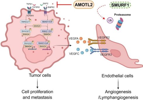
Angiomotin-like protein 2 (AMOTL2) regulates cell polarity and cytoskeletal regulation, but its role in gastric cancer (GC) remains undefined. Here, we show that AMOTL2 is frequently downregulated in GC tissues and cell lines, and its low expression is associated with deeper tumor invasion, lymph node metastasis, and shorter overall survival. Gain- and loss-of-function assays demonstrated that AMOTL2 suppresses GC cell proliferation, migration, and invasion. AMOTL2 overexpression also impaired endothelial and lymphatic-endothelial tube formation and reduced the expression of angiogenic and lymphangiogenic factors. In the subcutaneous, orthotopic, and popliteal lymph node metastasis models, AMOTL2 inhibited tumor growth and lymphatic dissemination. Mechanistically, AMOTL2 suppressed TGF-β/Smad signaling, as evidenced by reduced BMP2 and TGF-β1 expression, decreased Smad1/5/9 and Smad2/3 phosphorylation, and impaired Smads nuclear accumulation. Notably, pharmacological Smad1/5/9 activation by SB4 partially reversed the suppressive phenotypes of AMOTL2 in vitro and in vivo. Co-immunoprecipitation, GST pull-down, and ubiquitination assays identified SMURF1 as an AMOTL2-interacting E3 ubiquitin ligase that binds through its WW domains and promotes AMOTL2 ubiquitination and proteasomal degradation. AMOTL2 restoration counteracted SMURF1-driven malignant phenotypes. Collectively, these findings define AMOTL2 as a potential suppressor of GC progression that is destabilized by SMURF1, suggesting that the SMURF1-AMOTL2-Smad signaling axis may contribute to GC progression and lymphatic dissemination, representing a candidate therapeutic target that warrants further validation.
Keywords: Gastric cancer, AMOTL2, SMURF1, TGF-β/Smad pathway
Introduction
Angiomotin (AMOT) was originally identified as an angiostatin-binding protein mediating anti-angiogenic effects [1]. Subsequently, AMOTL1 and AMOTL2 were identified as related members of the angiomotin family, which is characterized by conserved coiled-coil domains and C-terminal PDZ-binding motifs [2-4]. AMOTL2 functions as a cytoplasmic scaffold that restrains the Hippo pathway effectors YAP and TAZ by sequestering them in the cytoplasm [5]. It also binds AKT and impairs its membrane translocation, and activates LATS2-dependent YAP phosphorylation [6, 7]. AMOTL2 activity is fine-tuned by post-translational modifications: mTORC2-mediated phosphorylation at Ser760 disrupts the interaction between AMOTL2 and YAP and attenuates its growth-suppressive activity [8]. In contrast, mono-ubiquitination at K347 and K408 enhances LATS binding and YAP phosphorylation [9, 10], whereas USP9X-mediated deubiquitination at K437 stabilizes AMOTL2 and strengthens YAP/TAZ restraint [9, 11]. Beyond the Hippo signaling, AMOTL2 also contributes to cytoskeletal organization through interaction with LL5β [12].
The biological functions of AMOTL2 are context dependent. Tumor-suppressive effects have been described in lung adenocarcinoma, non-small-cell lung cancer, medulloblastoma, and glioma [13-16]. In these malignancies, AMOTL2 inhibits cell growth and motility through AKT, JUN, or Wnt/β-catenin signaling pathways. Conversely, AMOTL2 has been reported to promote p38-dependent tumorigenicity in luminal breast cancer and to facilitate ERK-dependent endothelial cell migration and angiogenesis [17-19]. AMOTL2 is also detectable in glioma stem cells, but its cellular functions in this context remain poorly understood [20]. Similarly, its role in gastric cancer (GC) has not been explored to date.
SMURF1 is a HECT-type E3 ubiquitin ligase containing a C2 domain, two WW domains, and a catalytic HECT domain. It is a well-established regulator of TGF-β/BMP signaling and can target receptor-regulated Smads (Smad1/5/9), inhibitory Smads, and TGF-β receptor complexes for ubiquitin-dependent turnover [21-24]. SMURF1 is frequently increased in various cancers and promotes malignant progression through ubiquitination of substrates including Kindlin-2, ARHGAP26, RHOA, and FOXA2 [25-35]. However, context-dependent tumor-suppressive effects have also been reported, wherein SMURF1 exerts suppressive function in hepatocellular carcinoma through promoting degradation of several pro-tumorigenic substrates TRIB2 and MCAM [36-38]. In GC tissues, SMURF1 is frequently increased, and associated with shorter overall survival [25, 27, 39]. Its knockdown suppresses proliferation, migration, tumor growth, and liver metastasis, potentially through PI3K/AKT signaling [25]. Moreover, SMURF1 is negatively regulated by tumor-suppressive microRNAs, including miR-1254 and miR-424 [27, 39, 40]. Unexpectedly, SMURF1 also exerts as a suppressive factor in GC by degradation of MEKK2, leading to suppression of MEK/ERK signaling, this process was promoted by Stk38 and repressed by Kir2.1 [41, 42].
Accumulating evidence indicates that multiple Smad proteins, including Smad2/3 and Smad1/5/9, have been implicated in GC progression. The TGF-β2/TGF-βR/Smad2/3 axis can upregulate NDRG1 and promote cell migration and epithelial-mesenchymal transition (EMT) [43], whereas METTL3-dependent regulation of Smad3 expression can enhance pathway activation [44]. Within the tumor microenvironment, reciprocal TGF-β1/Smad2 signaling between GC cells and bone marrow mesenchymal stem cells (BMSCs) can promote cancer-associated fibroblast differentiation and tumor-cell EMT [45]. BMP-Smad signaling also contributes to gastric tumorigenesis: mTOR can potentiate BMP-Smad1 activity [46], GLIS3 has been linked to the TGF-β1/TGF-βR1/Smad1/5 pathway [47], and BMP4 activates Smad1/5/9-ID1 to promote EMT and metastasis [48]. Collectively, these observations suggest that both Smad2/3 and Smad1/5/9 pathways promote malignancy via distinct molecular axes, reflecting their functional complexity and context-dependent roles in gastric tumorigenesis.
In this study, we investigated the clinical relevance and functional role of AMOTL2 in GC, with particular emphasis on tumor progression, lymphatic dissemination, and TGF-β/Smad signaling. We also sought to identify an E3 ubiquitin ligase that controls AMOTL2 protein stability. To this end, clinical tissue analyses were integrated with gain- and loss-of-function experiments, endothelial and lymphatic-endothelial assays, protein-interaction studies, and several xenograft models. The study was designed to explore the putative AMOTL2-Smad regulatory axis and to preliminarily assess its prognostic and therapeutic relevance in GC, findings that require further validation.
Materials and methods
Cell culture
Gastric epithelial cells GES-1 and GC cell lines (AGS, SGC7901, MGC803, HGC27, SNU-1, MKN-45, MKN-74, and NCI-N87) were purchased from Shanghai Cell Bank, Chinese Academy of Sciences. Cell identity was authenticated by short tandem repeat profiling. Cells were cultured in RPMI 1640 medium (10-040-CVR, Corning, USA) supplemented with 10% fetal bovine serum (FBS, FB25015, Clark Bioscience, USA) and 1% penicillin/streptomycin solution (P/S, SV30010, HyClone, USA). Human endothelial cell lines (HUVECs, CL-0675, Procell, China; HLECs, VCH00407, ViCell, China) were maintained in endothelial cell medium (1001, ScienCell, USA) supplemented with FBS (0025, ScienCell, USA), endothelial cell growth supplement (ECGS, 1052, ScienCell, USA), and P/S (0503, ScienCell, USA). All cells were maintained at 37°C in a cell incubator containing 5% CO2 (HERACELL 150i, Thermo, USA).
Patient information
Two tissue microarrays (TMAs) described in our previous studies were analyzed [49, 50]. The first TMA comprised samples from 129 GC patients with pathologically confirmed GC who underwent radical gastrectomy at the First Affiliated Hospital of Anhui Medical University (December 2006-May 2008) [49]. The second TMA comprised 107 GC samples collected between October 2012 and December 2013 [50]. None of the patients received preoperative chemotherapy or radiotherapy. Tumors were staged according to the American Joint Committee on Cancer criteria. Clinicopathological characteristics are summarized in Table 1 and Supplementary Table S1. In addition, 28 paired fresh GC and adjacent non-tumor tissues were collected for RT-qPCR. This study was approved by the Ethics Committee of the First Affiliated Hospital of Anhui Medical University (2023450), was conducted in accordance with the Declaration of Helsinki, and included written informed consent from all participants.
Association between AMOTL2 expression (0-4 vs. 6-12) and the clinicopathological parameters in the GC cohort.
| Clinicopathological parameters | Total | AMOTL2 | χ² | p value | |
|---|---|---|---|---|---|
| High expression (6-12) | Low expression (0-4) | ||||
| Gender | 0.488 | 0.485 | |||
| Male | 101 | 65 | 36 | ||
| Female | 28 | 20 | 8 | ||
| Age (years) | 1.680 | 0.195 | |||
| < 60 | 63 | 45 | 18 | ||
| ≥ 60 | 66 | 40 | 26 | ||
| Tumor location | 0.474 | 0.495 | |||
| Upper | 67 | 46 | 21 | ||
| Middle/lower | 62 | 39 | 23 | ||
| Tumor size (cm) | 0.017 | 0.896 | |||
| < 6 | 86 | 57 | 29 | ||
| ≥ 6 | 43 | 28 | 15 | ||
| Tumor differentiation | 0.017 | 0.897 | |||
| Well/moderate | 42 | 28 | 14 | ||
| Poor/undifferentiated | 87 | 57 | 30 | ||
| Tumor invasion depth | |||||
| T1/T2 | 26 | 22 | 4 | 5.080 | 0.024 |
| T3/T4 | 103 | 63 | 40 | ||
| Lymph node metastasis | |||||
| N0 | 34 | 28 | 6 | 5.567 | 0.018 |
| N1-3 | 95 | 57 | 38 | ||
| pTNM stage | 1.350 | 0.245 | |||
| I/II | 53 | 38 | 15 | ||
| III/IV | 76 | 47 | 29 | ||
Immunohistochemistry assay
AMOTL2 expression was assessed by immunohistochemistry (IHC) in the first GC TMA. Staining was independently evaluated by two pathologists blinded to clinical data. The IHC scores were calculated by multiplying the staining intensity score (0: negative; 1: light yellow; 2: brownish yellow; and 3: dark brown) by the percentage of positive cells (0: 0%; 1: 1%-25%; 2: 26%-50%; 3: 51%-75%; and 4: 76%-100%). Scores were defined as high (≥ 6) or low (0-4); a score of 5 cannot arise from this scoring system. Correlations with clinicopathological parameters and overall survival were analyzed as described below using GraphPad Prism. SMURF1 expression was evaluated in the second TMA using the same scoring system.
RT-qPCR assay
Total RNA was extracted using TRIzol (15596018, Thermo, USA) and quantified with a BioPhotometer® D30 (Eppendorf, Germany). cDNA was synthesized using cDNA Synthesis SuperMix (11141ES10, Yeasen, China), and real-time quantitative PCR was performed with SYBR Green Master Mix (11202ES03, Yeasen, China) on a QuantStudioTM 3 system (Thermo, USA). Relative expression was calculated using the 2-ΔΔCt method using GAPDH as an internal control. Primer sequences were as follows: AMOTL2-F: 5'-GGGAGCAGAAGTATTTGGAGGAACG-3'; AMOTL2-R: 5'-GAATGTCGGATGAGAGTGGTGTCAC-3'; GAPDH-F: 5'-CTCTGCTCCTCCTGTTCGAC-3'; GAPDH-R: 5'-ACGACCAAATCCGTTGACTC-3'.
Plasmid construction and viral infection
Full-length human AMOTL2 (NM_016201) was amplified and cloned into the Ubi-MCS-SV40-Neomycin vector through the AgeI and NheI sites. For knockdown, shRNA-1 (5'-GTTGAGTGAACGGCTCCTTCA-3') and shRNA-3 (5'-AGGAGATGGAAAGCAGGTTAA-3') were inserted into pLKO.1-TRC vector. Full-length human SMURF1 (NM_020429.3) was cloned into the pLVX-puro vector through the XhoI and XbaI sites. All constructs were verified by DNA sequencing. Lentivirus particles were produced by co-transfecting HEK293T cells with the target vector, psPAX2, and pMD2.G using PEI 40K (G1802, Servicebio, China). The PEI-to-DNA ratio was 4 μL:1 μg, and the mass ratio of psPAX2:pMD2.G:target plasmid was 1:1.5:2. Virus supernatants were collected 48-72 h post-transfection, filtered, and used to infect AGS and SGC7901 cells in the presence of polybrene (C0351, Beyotime, China). Stable cells were selected with G418 (MA0321, Meilun Bio, China) or puromycin (ST551, Beyotime, China) and maintained under the corresponding selection thereafter.
EdU assay
DNA replication was assessed using the EdU Cell Proliferation Kit with Alexa Fluor 594 (C0078S, Beyotime, China). AMOTL2-overexpressing cells, AMOTL2 knockdown cells, and their corresponding controls were seeded on glass coverslips (BS-14-RC, Biosharp, China) in 12-well plates. On the following day, cells were incubated with 10 μM EdU for 2 h, then exposed to the click additive solution for 30 min at room temperature in the dark. Nuclei were counterstained with Hoechst 33342 (1:1000 in PBS) for 10 min. Images were captured randomly using a DMi1 microscope (Leica, Germany), and EdU-positive cells were analyzed using ImageJ software.
Cell migration and invasion assay
Cell invasion and migration were assessed in Transwell inserts with 8.0-µm-pore (3422, Corning, USA). Cells were serum-starved for 24 h before analysis. For invasion assays, the upper chamber was pre-coated with 100 μL of 1 mg/mL Matrigel (1:10 in serum-free medium) (356234, Corning, USA) and incubated for 5 h; this coating step was omitted for migration assays. AGS cells (8 × 10⁴) or SGC7901 cells (1 × 10⁵) were seeded into the upper chamber in serum-free medium, and 650 μL medium with 20% FBS was added to the lower chamber. After 22 h for migration or 28 h for invasion, non-migrated cells were removed from the upper surface. Cells on the lower surface were fixed with 4% paraformaldehyde, stained with 1% crystal violet. Images were captured using a microscope and analyzed using ImageJ.
Wound healing assay
AGS or SGC7901 cells were seeded in 6-well plates and grown to 100% confluence. A linear scratch was generated using a sterile 10 μL pipette tip, and detached cells were removed by PBS washing. Cells were cultured in serum-free medium and images were captured at 0, 24, and 48 h using a Celldiscover 7 microscope (Zeiss, Germany). Wound closure was quantified using ImageJ.
Matrigel-induced tube formation assay
To evaluate the effect of tumor-cell AMOTL2 on endothelial tube formation, HUVECs or HLECs were co-cultured with SGC7901-AMOTL2 or control cells in 0.4-µm-pore Transwell chambers (3450, Corning, USA). For tube formation, 48-well plates were pre-coated with 50 μL Matrigel per well and incubated at 37 °C for 60 min. Endothelial cells were detached with 0.25% trypsin/EDTA (SH30042.1, HyClone, USA), centrifuged, resuspended in serum-free medium, seeded onto polymerized Matrigel at 6 × 10⁴ cells in 200 μL per well, and incubated for 5 h. Random fields were imaged using a Celldiscover 7 microscope and quantified with ImageJ.
Periodic acid-Schiff (PAS) and CD31 double staining assay
Paraffin-embedded tumor sections were incubated with anti-CD31 antibody (1:50; ab28364, Abcam, UK), followed by secondary antibody (PV-6000, Zsbio, China) for 20 min at 37 °C. Immunoreactivity was visualized with DAB (ZLI-9018, Zsbio, China). Sections were then stained with a PAS Stain Kit (G1280, Solarbio, China), including incubation with Schiff reagent for 10-15 min and hematoxylin counterstaining (Baso, China). Slides were dehydrated, cleared, mounted, cover-slipped, and scanned using CaseViewer (3DHISTECH Ltd, Hungary). CD31-negative/PAS-positive channels were interpreted as vasculogenic mimicry structures, whereas CD31-positive channels were considered endothelial vessels.
Western blot analysis
Cells were lysed with M-PER buffer (78501, Thermo, USA) with protease and phosphatase inhibitors (K1007, K1015, APExBIO, USA) on ice for 30 min. Nuclear and cytoplasmic fractions were separated using a Nuclear and Cytoplasmic Protein Extraction Kit (P0028, Beyotime, China). After electrophoretic separation and transfer, membranes were blocked with 5% non-fat milk in TBST for 1 h at room temperature, then incubated overnight at 4°C with the primary antibodies listed in Supplementary Table S3. After three TBST washes, membranes were incubated with HRP-conjugated secondary antibodies (Sangon Biotech, China) for 1 h at room temperature. Signals were visualized using a 5200 Multi imaging system (Tanon, China) with an ECL kit (D045-2, Bridgen, China), and quantified using ImageJ.
Co-immunoprecipitation and mass spectrometry (MS)
To identification and validation of AMOTL2-interacting proteins, SGC7901-AMOTL2 and control cells were lysed in M-PER buffer with inhibitors. Lysates were centrifuged at 12,000 × g for 10 min at 4 °C, and 50 μL of each supernatant was saved as input. The remaining lysate was incubated overnight at 4 °C with protein A/G magnetic beads (B23201, Selleck, China) pre-bound to mouse IgG (sc-2025, Santa Cruz, USA), anti-HA (AE008, ABclonal, China), or anti-Flag antibody (AE005, ABclonal, China). After washing beads with TBST, the bound proteins were eluted with M-PER buffer, and boiled in 5× loading buffer at 95 °C before western blot or MS analysis.
For MS, immunoprecipitated samples were separated by SDS-PAGE, excised, and subjected to in-gel digestion. Peptides were separated on a C18 column using an EASY-nLC™ 1200 system coupled to a Q-Exactive mass spectrometer (Thermo, USA). Spectra were searched against in a human UniProt-Homo database (v.2.4). Candidate AMOTL2-interacting proteins were identified after exclusion of proteins detected in the IgG control.
Cell-based ubiquitination assay
HEK293T cells were co-transfected with plasmids encoding HA-tagged AMOTL2 (AMOTL2-HA), 3 × Flag tagged SMURF1 (SMURF1-3F), and His-tagged ubiquitin variants (wild-type, K6/K11/K27/K29/K33/K48/K63; each containing only the indicated lysine residue with other lysine mutated to arginine). Before collection, cells were treated with 10 μM MG132 (HY-13259, MCE, China) for 8 h. Lysates were immunoprecipitated with an anti-HA antibody, and ubiquitin signals in immunoprecipitated proteins were detected by immunoblotting with an anti-ubiquitin antibody.
Molecular docking analysis
Structural models for AMOTL2 (Q9Y2J4) and SMURF1 (Q9HCE7) were obtained using the AlphaFold3 server (https://alphafoldserver.com/), which was also used to predict a putative AMOTL2-SMURF1 complex. Predicted models were ranked according to the platform output, with the top ten highest-ranked models were retained for further analysis. Molecular representations were generated using PyMOL (Schrödinger, LLC).
GST pull-down assay
GST pull-down assays were performed to test whether AMOTL2 and SMURF1 interact directly in vitro. His-tagged AMOTL2, GST-tagged full-length SMURF1, and GST-tagged SMURF1 lacking the WW regions were purified. Purified His-AMOTL2 was incubated overnight at 4 °C with GST-SMURF1-WT, GST-SMURF1-ΔWWs, or GST control. Glutathione agarose beads (HY-K0211, MCE, China) were then added and incubated for additional 3 h at 4 °C. After extensive washing, bound proteins were eluted and analyzed by western blot using anti-His and anti-GST antibodies.
Dual-luciferase reporter assay
18 pathway-reporter constructs were generated in the psiCHECK-2 backbone using core response elements for AP-1, E2F1, ELK1, HIF1, Myc, NF-κB, RBP-Jκ, Smad, TCF/LEF, p53, ATF2/3/4, ATF6, CREB, EGR1, NFAT, Nrf1/2, STAT1/2, and STAT3. Reporter sequences are listed in Supplementary Table S4. HEK293T cells were co-transfected with an AMOTL2 overexpression plasmid or empty vector and the individual reporter. After 48 h, luciferase activities were measured using the Dual-Lumi™ Luciferase Assay Kit (RG088S, Beyotime, China) on an EnSpire Multimode Plate Reader.
Animal models
All animal experiments were approved by the Animal Ethics Committee of Anhui Medical University (LLSC20231297). Four- to five-week-old male BALB/c nude mice (GemPharmatech, China) were used.
For the subcutaneous model, mice were randomly divided into groups (n = 6 per group). SGC7901-AMOTL2 cells or control cells (5 × 10⁶ in 100 μL PBS) were injected subcutaneously into the right flank. Tumor length and width were measured every 2 days. Mice were then euthanized when tumors reached predetermined volume, and tumors were harvested and weighed.
For gastric orthotopic model, luciferase-expressing SGC7901-AMOTL2 (SGC7901-AMOTL2-Flu) cells or control cells (5 × 10⁶ in 50 μL PBS; n = 4 per group) were orthotopically injected into the gastric subserosa. Tumor growth was monitored by bioluminescence imaging (AmiX, SI Imaging, USA) after administration of D-luciferin (150 mg/kg, Biolite, China). Mice were then euthanized at the study endpoint, and tumors were collected for histologic analysis.
For the popliteal lymph node metastasis model, SGC7901-AMOTL2-Flu cells or control cells (5 × 10⁶ in 50 μL PBS) were injected into the left footpad. Primary-tumor growth and enlargement of the popliteal lymph node were monitored. Bioluminescence imaging was performed before euthanasia, after which footpad tumors and popliteal lymph nodes were dissected. In addition, to adjust for inter-animal variations in primary tumor growth, the ratio of popliteal lymph node bioluminescence to the total signal (lymph node plus footpad) was calculated and used to quantify relative lymph node metastatic index.
For pharmacologic rescue, SB4 (5 mg/kg in 100 μL 10% DMSO/90% PBS) [51, 52] was administered intraperitoneally every 2 days beginning on day 10, for a total of six doses. This regimen was applied in the subcutaneous and popliteal lymph node metastasis models.
For SMURF1 overexpression models, SGC7901-SMURF1-Flu cells or control cells were implanted subcutaneously or injected into the footpad according to the protocols described above. Tumor growth and lymph node metastasis were monitored by bioluminescence imaging. Harvested tissues were evaluated by H&E staining, IHC for Ki67, AMOTL2, or SMURF1, and CD31/PAS double staining, as indicated.
Statistical analysis
Statistical analyses were performed using SPSS version 27.0 (IBM, USA) and GraphPad Prism (GraphPad Software, USA). All data are presented as mean ± SD from at least three independent experiments. The unpaired or paired Student's t-test were used for two-group comparisons, and two-way ANOVA was applied for cell-growth-curve analyses, as specified in the figure legends. The associations between AMOTL2 or SMURF1 expression and clinicopathological parameters were analyzed with the chi-square test. Overall survival curves were estimated by the Kaplan-Meier method and compared with the log-rank (Mantel-Cox) test. Cox proportional hazards regression models were used for univariate and multivariate survival analyses. P < 0.05 was considered statistically significant. Significance is denoted as *p < 0.05, **p < 0.01, ***p < 0.001, ****p < 0.0001.
Results
Reduced AMOTL2 expression was associated with aggressive clinicopathological features and shorter survival
AMOTL2 expression was evaluated in the first GC TMA employed in our previous study [49]. Survival analysis showed that patients with low AMOTL2 expression had shorter overall survival than those with high expression (log-rank test, p = 0.0002; median survival time, 30 months vs. not reached; Figure 1A). Moreover, AMOTL2 IHC scores were lower in tumors with advanced T stage, lymph node metastasis, and higher TNM stage (Figure 1B-D). Consistently, in 28 paired samples, AMOTL2 mRNA levels were also reduced in GC tissues relative to adjacent non-tumor tissues (relative expression, 0.67 vs. 1, p < 0.0001; Figure 1E). Representative IHC images and paired scoring further showed lower AMOTL2 expression in tumor tissues (p = 0.0033, Figure 1F), and this low expression was more frequent in cases with advanced pT stage or lymph node metastatic cases (Figure 1G-H).
Reduced AMOTL2 expression is associated with aggressive clinicopathological features and shorter survival. Kaplan-Meier analysis of overall survival according to AMOTL2 IHC category in the first GC TMA (high, n = 85; low, 44) (A); AMOTL2 IHC scores according to tumor invasion (B), lymph-node status (C), and pTNM stage (D); AMOTL2 mRNA in 28 paired GC and adjacent non-tumor tissues (E, n = 28); representative IHC images and paired AMOTL2 scores in GC and adjacent non-tumor tissues (F, n = 14); distribution of high and low AMOTL2 expression according to T stage and lymph-node status (G-H). Data are presented as mean ± SD. The reported analyses used the log-rank (Mantel-Cox) test for overall survival analysis (A); the unpaired Student's t-test for B-D; the paired Student's t-test for E-F; and the chi-square test for G-H. *p < 0.05, **p < 0.01, ***p < 0.001, ****p < 0.0001.
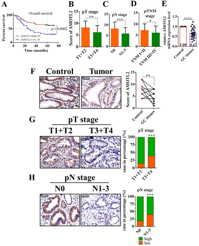
In the dichotomized IHC analysis, low AMOTL2 expression was associated with deeper tumor invasion (pT, p = 0.024) and lymph node metastasis (pN, p = 0.018) (Table 1). In univariate Cox regression analysis, pT, pN, pTNM stage, differentiation, and AMOTL2 expression were correlated with overall survival (all p ≤ 0.001) (Table 2). In multivariate analysis, lymph node metastasis (p = 0.016) and AMOTL2 expression (p = 0.024) remained independently associated with survival. These data suggest that AMOTL2 is a candidate prognostic indicator, although independent validation is required.
Univariate and multivariate analyses assessing the correlation between the clinicopathological features, AMOTL2 expression, and overall survival.
| Multivariate analysis | Univariate analysis | Multivariate analysis | ||
|---|---|---|---|---|
| HR (95%CI) | p value | HR (95%CI) | p value | |
| Gender (male vs. female) | 1.256 (0.655~2.410) | 0.493 | ||
| Age (≥ 60 vs. < 60 years) | 1.089 (0.664~1.786) | 0.735 | ||
| Tumor location (upper vs. middle/lower) | 1.137 (0.691~1.869) | 0.613 | ||
| Tumor size (≥ 6 vs. < 6 cm) | 1.627 (0.984~2.691) | 0.058 | ||
| Differentiation (poor/undifferentiated vs. well/moderate) | 4.949 (2.352~10.415) | < 0.001 | ||
| Tumor invasion depth (T3/T4 vs.T1/T2) | 11.093 (2.709~45.432) | < 0.001 | ||
| Lymph node metastasis (N1-3 vs. N0) | 3.684 (1.677~8.092) | 0.001 | 2.698 (1.203~6.053) | 0.016 |
| pTNM stage (III/IV vs. I/II) | 6.800 (3.343~13.830) | < 0.001 | ||
| AMOTL2 expression (high vs. low) | 0.412 (0.251~0.677) | < 0.001 | 0.556 (0.333~0.927) | 0.024 |
AMOTL2 inhibited GC cell proliferation, migration, and invasion in vitro
AMOTL2 protein abundance was lower in most tested GC cell lines than in GES-1 cells (Figure 2A). AMOTL2 overexpression using Ubi-MCS-SV40-AMOTL2 lentivirus (Figure 2B) significantly suppressed cell proliferation, as shown by cell growth curve and EdU incorporation (AGS, 46% to 32%, p = 0.0325; SGC7901, 45% to 35%, p = 0.0222; Figure 2C-D). AMOTL2 overexpression also inhibited cell invasion and migration. The number of invaded cells decreased from 1365 to 762 in AGS cells (p = 0.0002), and from 1468 to 449 in SGC7901 cells (p = 0.0002) (Figure 2E-F). Wound healing assays showed a similar reduction in migration (Figure 2G). Nuclear-cytoplasmic fractionation and immunofluorescence further showed AMOTL2 promoted YAP1 Ser127 phosphorylation and inhibited nuclear YAP1 accumulation, consistent with reduced YAP activity (Figure 2H-I).
AMOTL2 inhibits cell proliferation, migration, and invasion in vitro. AMOTL2 protein abundance in GES-1 and GC cell lines (A, n = 3); validation of AMOTL2 overexpression in AGS and SGC7901 cells (B); cell-growth curves and EdU incorporation after AMOTL2 overexpression (C-D, n = 3); Transwell invasion (E, n = 3), migration (F, n = 3), and wound healing assays (G, n = 3); nuclear-cytoplasmic fractionation and immunofluorescence assessing Ser127 phosphorylation and nuclear accumulation of YAP1 in AMOTL2-overexpression cells and control cells (H-I); validation of AMOTL2 knockdown with two independent shRNAs (J); cell growth (K, n = 3), Transwell invasion/migration (L, n = 3), and wound healing capacity after AMOTL2 knockdown (M, n = 3). Data are presented as mean ± SD. The reported analyses used two-way ANOVA for cell growth curves (C, K); the unpaired Student's t-test for A, D-G, and L-M. *p < 0.05, **p < 0.01, ***p < 0.001, ****p < 0.0001.
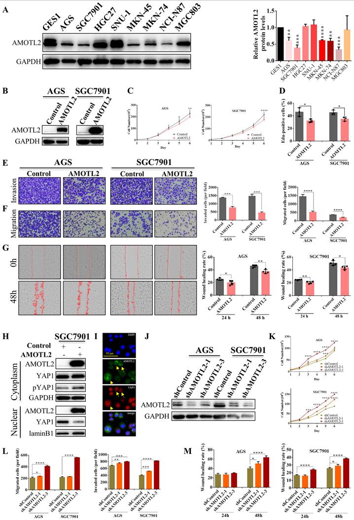
Additionally, AMOTL2 knockdown with two independent shRNAs (Figure 2J) enhanced cell proliferation, migration, and invasion (Figure 2K-M). In SGC7901 cells, AMOTL2 knockdown significantly increased the number of invaded cells from 400 to 823 (p < 0.0001), and the number of migrated cells from 218 to 558 (p < 0.0001) (Figure 2L). At 48 h, the wound healing rate also increased from 40.02% to 63.45% in AGS cells (p < 0.0001), and from 25.67% to 38.31% in SGC7901 cells (p < 0.0001) (Figure 2M). Collectively, these gain- and loss-of-function results indicate that AMOTL2 suppresses proliferative and metastatic phenotypes in tested cell models.
AMOTL2 was inversely associated with VM and reduced tube formation
Vasculogenic mimicry (VM) was defined as CD31-negative/PAS-positive tumor cell channels (red arrows, CD31-/PAS+), whereas CD31-positive channels were classified as endothelial vessels (black arrows, CD31+) (Figure 3A). In a reanalysis of our previously reported cohort [49], VM-positive tumors were associated with shorter overall survival (median, 36 months vs. not reached; p < 0.0001), deeper tumor invasion (p = 0.0069), lymph-node metastasis (p = 0.0337), and advanced TNM stage (p = 0.0072) (Figure 3B-C). AMOTL2 IHC scores were lower in VM-positive than in VM-negative tumors (6.15 vs. 7.20; p = 0.0484), and low AMOTL2 expression was more frequent in VM-positive cases (p = 0.0014) (Figure 3D).
AMOTL2 is inversely associated with VM and reduces tube formation. Representative CD31/PAS double staining showing CD31-/PAS+ VM channels (red arrows) and CD31+ endothelial vessels (black arrows) (A); overall survival according to VM (B); association of VM with tumor invasion, lymph-node status, and pTNM stage (C); AMOTL2 IHC scores and expression according to VM status (D); schematic of SGC7901/endothelial cell Transwell co-culture system (E); HUVECs and HLECs growth curves during co-culture (F, n = 3), representative tube-formation images and quantitative results in HUVECs and HLECs co-cultured with SGC7901-AMOTL2 or control cells (G-H, n = 3); immunoblotting of angiogenic and lymphangiogenic proteins in tumor cells and co-cultured HUVECs and HLECs (I). Data are presented as mean ± SD. The reported tests were log-rank (Mantel-Cox) test (B); the chi-square test (C, lower panel of D); the unpaired Student's t-test (upper panel of D, G, and H), and two-way ANOVA (F). *p < 0.05, **p < 0.01, ***p < 0.001, ****p < 0.0001.
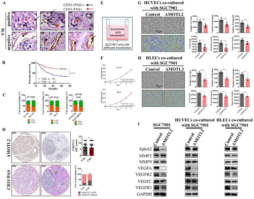
In the Transwell co-culture system, HUVECs and HLECs exposed to SGC7901-AMOTL2 cells exhibited impaired tube formation compared with those exposed to control cells, with significant reductions in nodes, junctions, segments, total length, branching length, and segment length (p < 0.05 for all), whereas cell growth curves were not detectably altered (Figure 3E-H). AMOTL2 overexpression downregulated EphA2, VEGFA, VEGFR2, VEGFC, and VEGFR3 in SGC7901 cells, whereas MMP2 and MMP9 remained unchanged. VEGFA and VEGFR2 were reduced in co-cultured HUVECs, and VEGFC and VEGFR3 were reduced in co-cultured HLECs (Figure 3I). Thus, AMOTL2 expression was inversely associated with VM in clinical tissues and attenuated angiogenic and lymphangiogenic tube-formation phenotypes in co-cultured endothelial cells.
AMOTL2 overexpression suppressed tumor growth and lymph-node metastasis in vivo
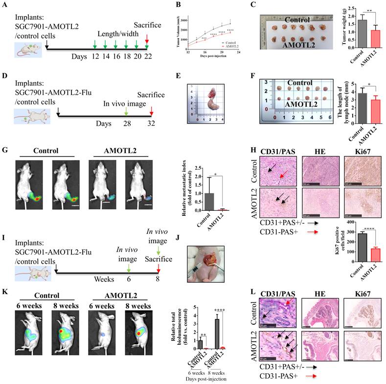
The in vivo effects of AMOTL2 were evaluated using three xenograft models. In the subcutaneous xenograft model, AMOTL2-overexpressing tumors showed smaller tumor volumes and had lower weights compared with control tumors (Figure 4A-C). In the popliteal lymph node metastasis model, AMOTL2-overexpressing tumors exhibited smaller primary footpad tumors and reduced lymphatic metastasis to the popliteal lymph nodes, as evidenced by lymph node metastatic index (defined as the ratio of isolated popliteal lymph node signal to the total signal from both lymph node and footpad) and lymph node length (Figure 4D-G). Ki67 staining and CD31/PAS analysis of footpad tumors showed lower proliferative activity and fewer VM structures in AMOTL2 overexpression group (Figure 4H). In the orthotopic model, AMOTL2 overexpression suppressed orthotopic tumor growth, as shown by bioluminescence at the measured time points (Figure 4I-K). Histologic analyses showed lower Ki67 staining and fewer VM structures (Figure 4L). Collectively, three in vivo models provided complementary evidence corroborating the inhibitory role of AMOTL2 in xenograft growth and lymph-node dissemination.
AMOTL2 overexpression reduces xenograft growth and lymph-node dissemination. Flowchart, growth curves, and terminal weights of subcutaneous xenografts generated with SGC7901-AMOTL2 or control cells (A-C, n = 6); Flowchart, representative specimens, the length of popliteal lymph node, the metastatic index (the ratio of bioluminescence signal in isolated lymph node to the total signal (lymph node and footpad)), and H&E, Ki67, and CD31/PAS staining (D-H, n = 6); panel G (left), images 1 and 2 are from the same control mouse, and images 3 and 4 from the same AMOTL2-overexpressing mouse. Images 1 and 3 show the total signal (lymph node and footpad), whereas images 2 and 4 represent the isolated popliteal lymph node signal; gastric orthotopic model, including flowchart, operative image, serial bioluminescence, and H&E, Ki67, and CD31/PAS staining (I-L, n = 4). Data are presented as mean ± SD. The reported analyses used two-way ANOVA for the subcutaneous growth curve (B); the unpaired Student's t-test for C, F, G, H, and K. *p < 0.05, **p < 0.01, ***p < 0.001, ****p < 0.0001.
AMOTL2 suppressed the TGF-β/Smad pathway
mRNA sequencing of SGC7901-AMOTL2 and control cells identified 590 upregulated and 848 downregulated genes (Figure 5A-E). GO and KEGG enrichment analyses implicated cell adhesion, focal adhesion, ECM-receptor interaction, cytokine signaling, PI3K-AKT, and MAPK signaling (Figure 5F-H). Additionally, immunoprecipitation-mass spectrometry identified SMURF1 as a candidate AMOTL2-interacting protein (Figure 5I-K).
AMOTL2 suppresses the TGF-β/Smad signaling and affects multiple signaling pathways. mRNA sequencing overview comparing SGC7901-AMOTL2 and control cells, including differential-expression and sample-level analyses (A-E); GO and KEGG enrichment analyses (F-H); immunoprecipitation-mass spectrometry candidate AMOTL2-binding proteins, including SMURF1 (I-K); 18-pathway reporter screen after AMOTL2 overexpression (L, n = 3); immunoblotting of BMP/TGF-β-Smad, Hippo-YAP, EMT, ERK, Src, Wnt, and hypoxia-related proteins (M). Data are presented as mean ± SD. The reported analysis used the unpaired Student's t-test for L. *p < 0.05, **p < 0.01, ***p < 0.001, ****p < 0.0001.
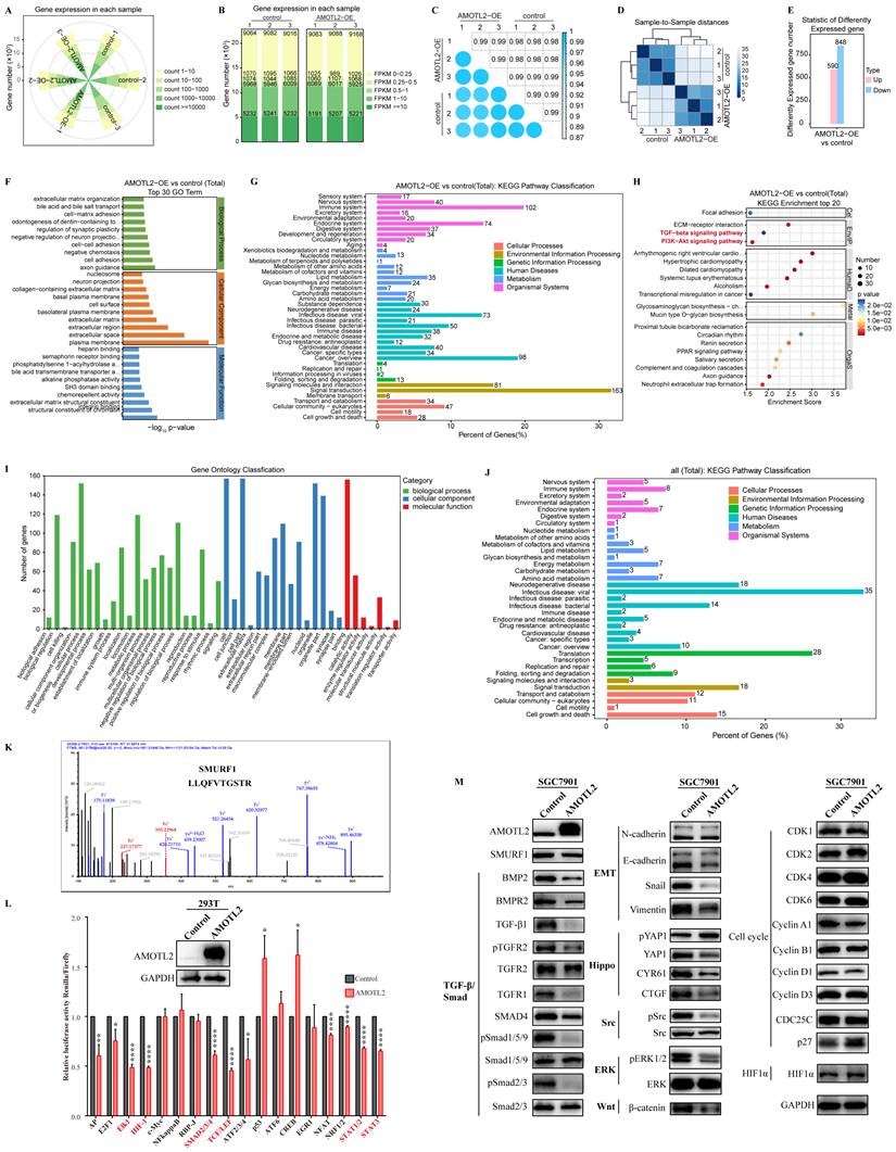
Pathway-reporter screening showed lower activity of several reporters after AMOTL2 overexpression, including reporters related to MAPK/ERK, hypoxia, TGF-β/Smad, Wnt, STAT1, and STAT3 signaling (Figure 5L; reporter sequences are provided in Supplementary Table S4).
Moreover, western blot analysis showed that AMOTL2 overexpression suppressed the TGF-β/Smad axis, as evidenced by reduced BMP2, BMPR2, TGF-β1, and TGF-β receptor I (TGFBR1), and Smad4 abundance, and decreased phosphorylation of TGFBR2, Smad1/5/9, and Smad2/3 (Figure 5M). In parallel, AMOTL2 promoted YAP1 Ser127 phosphorylation and reduced its downstream targets CYR61 and CTGF. EMT markers N-cadherin, Snail, and Vimentin were decreased, whereas E-cadherin was not detectably altered. Additionally, ERK and Src phosphorylation and β-catenin abundance were reduced, whereas the tested cell-cycle and hypoxia-associated proteins were minimally affected. Collectively, these results suggest that AMOTL2 broadly modulates multiple signaling pathways, with TGF-β/Smad signaling serving as a primary mediator, while concurrent suppression of ERK/Src and Wnt pathways may cooperatively reinforce its tumor-suppressive function.
The Smad1/5/9 agonist SB4 partially reversed AMOTL2-mediated tumor suppression
To investigate the functional contribution of Smad signaling, SGC7901-AMOTL2 cells were treated with Smad1/5/9 agonist SB4, a small-molecule BMP-signaling agonist that stabilizes or enhances Smad1/5/9 activation [52]. SB4 treatment (1 μM, 24 h) largely restored cell migration, invasion, and HLEC tube formation in the AMOTL2-overexpression setting (Figure 6A-B). SB4 increased the phosphorylation of Smad1/5/9, whereas Smad2/3 phosphorylation was not clearly elevated. Moreover, SB4 increased VEGFC and VEGFR3 abundance in co-cultured HLECs (Figure 6C).
The Smad1/5/9 agonist SB4 partially reverses AMOTL2-mediated tumor suppression. Transwell migration and invasion after treatment with SB4 or vehicle in SGC7901-AMOTL2-overexpression setting (A, n = 3); HLECs tube formation upon co-culture with the indicated SGC7901 cells and treatments (B, n = 3); immunoblotting of Smad-pathway proteins in SGC7901 cells and VEGFC and VEGFR3 in co-cultured HLECs (C); the subcutaneous model, including flowchart, terminal tumor weight, tumor-growth curve, mice body weight, and H&E, Ki67, and CD31/PAS staining (D-G, n = 6); the popliteal lymph node metastasis model, including experiment workflow, lymph node length, total bioluminescence, and H&E staining, IHC for Ki67, AMOTL2, and LYVE-1 (H-L, n = 6). Black asterisks denote comparisons between AMOTL2 and control groups, red asterisks denote comparisons between SB4 and vehicle in the AMOTL2 setting (A, B, E, F, I, and J). Data are presented as mean ± SD. The reported analyses used the unpaired Student's t-test for A, B, E, I (middle panel), and J; and two-way ANOVA for the tumor growth and body weight curves in F and I (right panel). *p < 0.05, **p < 0.01, ***p < 0.001, ****p < 0.0001.
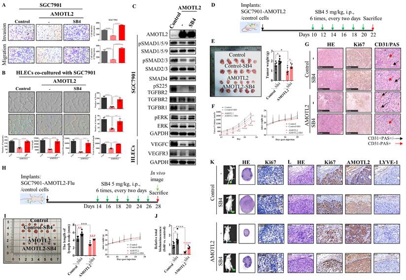
In the subcutaneous model, mice bearing SGC7901-AMOTL2 tumors were administered intraperitoneally with SB4 (5 mg/kg) every 2 days from day 10 to 20 (Figure 6D). SB4 largely increased tumor weight (p = 0.0204) and volume relative to vehicle-treated AMOTL2-overexpressing tumors (Figure 6E-F). Ki67 and CD31/PAS staining were also increased, consistent with partial restoration of proliferation and VM-associated structures (Figure 6G).
In the popliteal lymph node metastasis model, SB4 increased primary-tumor and lymph-node dissemination in the AMOTL2-overexpression setting, including popliteal lymph-node length (p = 0.0001) and total bioluminescence (p < 0.0001) (Figure 6H-J). H&E staining and IHC for Ki67 and LYVE-1 in footpad tumors consistently revealed increased proliferation and lymphatic-vessel density (Figure 6K-L). Furthermore, using a human-specific anti-Ki67 antibody that does not recognize murine cells in the popliteal lymph nodes, we observed that AMOTL2 overexpression reduced the proliferative index of the disseminated human gastric cancer cells, an effect that was reversed by SB4. Collectively, these pharmacologic rescue data indicate that Smad1/5/9 contributes to tumor-suppressive phenotypes of AMOTL2.
SMURF1 interacted with AMOTL2 through its WW domains and promoted its ubiquitination and turnover
Cycloheximide-chase and MG132 assays showed that the decrease in AMOTL2 protein levels was attenuated by proteasome inhibition, consistent with proteasome-dependent turnover (Figure 7A). SMURF1 overexpression reduced AMOTL2 protein abundance in a dose-dependent manner, whereas SMURF1 knockdown increased its abundance (Figure 7B). SMURF1 also accelerated the decrease in AMOTL2 protein levels in CHX chase assays (Figure 7C).
SMURF1 interacts with AMOTL2 through its WW domains and promotes its ubiquitination and turnover. Cycloheximide-chase analysis of AMOTL2 with or without MG132 (A, n = 3); AMOTL2 protein abundance after SMURF1 overexpression or knockdown (B); cycloheximide-chase analysis with or without SMURF1 overexpression (C); reciprocal co-immunoprecipitation of tagged proteins and endogenous AMOTL2-SMURF1 co-immunoprecipitation (D-F); AlphaFold3 prediction of a putative AMOTL2-SMURF1 complex (G); SMURF1 domain map and truncation analysis implicating the WW domains in AMOTL2 binding (H-I); GST pull-down with His-tagged AMOTL2, full-length SMURF1, a WW domains deletion construct, and GST control (J); cell-based ubiquitination assays using the indicated ubiquitin variants (K); nuclear-cytoplasmic distribution of phosphorylated and total Smad proteins after AMOTL2 overexpression (L). Black stars denote comparisons between SGC7901-AMOTL2 and control cells with sole CHX treatment, brick-red stars denote comparisons under combined treatment with CHX and MG132 (A). Data are presented as mean ± SD. The reported analysis used two-way ANOVA in A. *p < 0.05, **p < 0.01, ***p < 0.001, ****p < 0.0001.
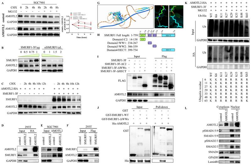
Reciprocal co-immunoprecipitation of tagged proteins in HEK293T cells and endogenous co-immunoprecipitation supported an interaction between AMOTL2 and SMURF1 (Figures 7D-F). AlphaFold3 complex prediction provided a putative structural model, and truncation analysis revealed that the WW domains of SMURF1 are critical for its binding to AMOTL2 (Figure 7G-I). GST pull-down further supported the direct in vitro interaction and demonstrated that the WW domains are required for this binding (Figure 7J). Cell-based ubiquitination assays showed increased K33-, K48-, and K63-linked ubiquitin signals associated with AMOTL2 in the presence of SMURF1 (Figure 7K). Nuclear-cytoplasmic fractionation additionally revealed that AMOTL2 reduces nuclear accumulation of phosphorylated and total Smad1/5/9 and Smad2/3, as well as total Smad (Figure 7L).
SMURF1 was associated with adverse clinical features and promoted malignant phenotypes
In the second GC TMA, elevated SMURF1 expression was associated with shorter overall survival (median, 51 vs. 66 months; p = 0.0418) and larger tumor size (p = 0.0039) (Figure 8A). SMURF1 IHC scores were higher in GC tissues than in adjacent non-tumor tissues (3.95 vs. 1.86; p = 0.0085), and this high expression was more frequent in cases with larger size tumor (p = 0.0120) or lymph node metastatic cases (p = 0.0353) (Figure 8B-D). In the dichotomized analysis, SMURF1 expression was positively associated with larger tumor size (p = 0.045) (Supplementary Table S1). The reported multivariate model identified SMURF1 expression (p = 0.041) and lymph node metastasis (p = 0.025) as independent prognostic factors (Supplementary Table S2).
SMURF1 is associated with adverse clinical features and promotes malignant phenotypes in cell and mouse models. Overall survival, tumor-size correlation, tumor/non-tumor IHC, and lymph-node-status association in the second GC TMA (A-D); validation of SMURF1 overexpression and effects on cell proliferation, EdU incorporation, Transwell migration and invasion, and wound healing (E-I, n = 3); in vitro rescue by AMOTL2 co-expression (J-M, n = 3); the subcutaneous xenograft model, including the flowchart, tumor growth, terminal tumor weight, total bioluminescence, H&E staining, and IHC for Ki67, SMURF1, and AMOTL2 (N-R, n = 4); the popliteal lymph node metastasis model, including the flowchart, lymph-node length, total tumor burden and metastatic index in lymph node, H&E staining, and IHC for Ki67, SMURF1, and LYVE-1 (S-X, n = 6). Black asterisks denote comparisons between SMURF1 and control cells; brick-red asterisks denote comparisons with and without AMOTL2 co-overexpression in SMURF1-overexpressing setting (J-M). Data are presented as mean ± SD. The reported analyses used the log-rank (Mantel-Cox) test for left panel of A; the unpaired Student's t-test for the middle and right panels of A, B, G-I, K-M, P, Q, and T-V; the chi-square test for C and D; and two-way ANOVA for growth curves in F, J, and O. *p < 0.05, **p < 0.01, ***p < 0.001, ****p < 0.0001
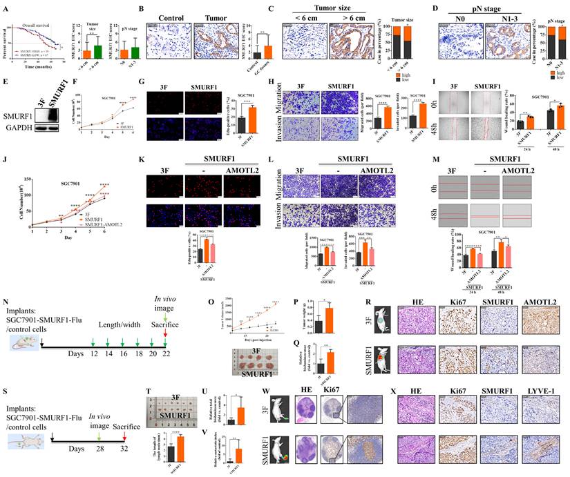
SMURF1 overexpression in GC cell lines enhanced cell proliferation, EdU incorporation, Transwell invasion and migration, and wound healing (all p < 0.05) (Figure 8E-I). Co-expression of AMOTL2 suppressed these SMURF1-driven phenotypes (Figure 8J-M). In the subcutaneous xenografts, SMURF1 overexpression resulted in larger tumor volume (p < 0.0001), higher terminal tumor weight (p = 0.0162), stronger bioluminescence (p = 0.0066), and increased Ki67 staining, along with reduced AMOTL2 staining (Figure 8N-R). In the popliteal lymph node metastasis model, SMURF1 overexpression increased total tumor burden (lymph node plus footpad) and metastatic index in lymph node (p = 0.0247 and p = 0.0080, respectively), as well as popliteal lymph node length (4.38 vs. 2.67 mm, p < 0.0001) (Figure 8S-V). H&E, Ki67, and LYVE-1 staining in the primary footpad tumors consistently indicated increased proliferation and lymphatic-vessel density (Figure 8W-X). In the popliteal lymph nodes, immunostaining with a human-specific anti-Ki67 antibody that does not cross-react with murine cells further demonstrated that SMURF1 overexpression significantly increased the proliferative index of the metastatic human gastric cancer cells. Notably, we observed that the distribution patterns of SMURF1 in xenograft tumors, including both subcutaneous and footpad models, diverged substantially from those detected in human GC clinical tissues. Collectively, these results establish SMURF1 as an oncogenic driver in GC cell lines and mouse models and show that AMOTL2 restoration can counteract SMURF1-driven phenotypes in vitro.
Discussion
In this study, we define AMOTL2 as a novel tumor suppressor in GC. AMOTL2 expression was clinically reduced in GC tissues, and its low expression was associated with deeper invasion, lymph-node metastasis, and shorter overall survival. Functionally, AMOTL2 suppressed cell proliferation, migration, and invasion, and impaired endothelial and lymphatic-endothelial tube formation, accompanied by downregulation of VEGFA/VEGFR2 and VEGFC/VEGFR3. Xenograft models further demonstrated that AMOTL2 overexpression reduced tumor growth and lymphatic dissemination. Mechanistically, our findings indicate that AMOTL2 attenuates BMP/TGF-β/Smad signaling, while SMURF1 interacts with AMOTL2 and promotes its ubiquitination and turnover. Furthermore, using a human-specific anti-Ki67 antibody, we demonstrated that AMOTL2 overexpression reduced the proliferative index of metastatic human GC cells in popliteal lymph nodes. This suppressive effect was abrogated by SB4-mediated Smad1/5/9 activation, providing in vivo evidence that AMOTL2 inhibits lymph node colonization partially through Smad signaling, especially Smad1/5/9 branch.
Other angiomotin-family members AMOT and AMOTL1 have been studied in GC. Reduced AMOT-p130 is associated with aggressive features, and restoration of AMOT-p130 can limit cell migration and invasion by inhibiting EMT [53, 54]. AMOT knockdown impedes cell migration by dissociating YAP from ZO-1, affecting tight-junction organization and migration [55]. In contrast, increased AMOTL1 is linked to poor prognosis, and enhances YAP1 stability to promote gastric oncogenesis [56]. These previous studies underscore that individual angiomotin-family proteins have distinct and context-dependent functions. In addition, we have published a review detailing the cellular localization and specific effects of AMOTL2 in tumor and normal cells [57]. Thus, this study is the first to demonstrate the tumor-suppressive effects of AMOTL2 in GC progression, thereby expanding the current understanding of this family.
The relationship between AMOTL2 and YAP1 signaling is likewise context dependent. Multiple lines of evidence support AMOTL2 as a YAP1 suppressor: AMOTL2 can facilitate LATS2 and YAP phosphorylation [5, 11, 58], and its mono-ubiquitination (K347/K408) is essential for this process [9, 10]. In airway smooth muscle cells and zebrafish, AMOTL2 also inhibits YAP1-driven proliferative activity [59, 60]. In endocytic trafficking, the competition of AMOTL2 with Endotubin can regulate YAP localization [61]. In contrast, AMOTL2 enhances YAP nuclear accumulation by stabilizing YAP1 in muscle stem cells [62]. Our results demonstrate that AMOTL2 increased YAP1 phosphorylation at Ser127, reduced its nuclear accumulation, and decreased CYR61 and CTGF expression, consistent with inhibition of YAP1.
Beyond YAP1, AMOTL2 also participates in other multiple signaling pathways. Previous studies have shown that AMOTL2 can limit AKT membrane recruitment, and modulate ERK and Wnt/β-catenin signaling [6, 16, 63]. Conversely, AMOTL2 has also been reported to exert pro-tumorigenic functions through promoting MAPK/ERK and p38 activation [17-19]. Here, we found that AMOTL2 reduced BMP2/BMPR2 and TGF-β1/TGFBR2 signaling, decreased phosphorylation of both Smad1/5/9 and Smad2/3, reduced Smad4 abundance, and disrupted Smad nuclear accumulation. Furthermore, pharmacological rescue with SB4 substantially reversed several AMOTL2-associated suppressive phenotypes, emphasizing the functional significance of Smad1/5/9 pathway in AMOTL2-mediated tumor suppression.
The interaction studies identify SMURF1 as a regulator of AMOTL2 protein stability. Reciprocal and endogenous co-immunoprecipitation, WW domains mapping, and GST pull-down assays support their physical interaction, whereas cycloheximide-chase, MG132, and ubiquitination assays supported SMURF1-mediated ubiquitination and proteasomal degradation of AMOTL2. K48-linked ubiquitination provides a plausible degradative signal, whereas the functional roles of K33- and K63-linked signals remain to be defined. In addition, consistent with most previous studies, SMURF1 was elevated in GC tissues and correlated with shorter overall survival; its overexpression promoted malignant phenotypes in vitro and in vivo, effects that were effectively counteracted by AMOTL2 restoration. Notably, human-specific Ki67 IHC revealed SMURF1 overexpression significantly increased the proliferative index of metastatic human gastric cancer cells in popliteal lymph nodes, demonstrating its pro-metastatic function in lymph node colonization.
SMURF1 is recognized as a negative regulator of TGF-β/BMP signaling through degradation of R-Smads and receptor complexes [21, 23, 24, 64]. In GC, SMURF1 is significantly increased and predicts poor prognosis [25, 27, 39, 42], promoting tumor growth and metastasis through PI3K/AKT and MEK/ERK signaling [25, 42]. SMURF1 expression can be also suppressed by tumor-suppressive microRNAs such as miR-1254 and miR-424 [27, 39, 40].
Although the observations that both SMURF1 and its substrate Smads promote GC progression appear paradoxical, this coexistence is not necessarily contradictory. Several non-mutually exclusive mechanisms may explain this contradiction. First, SMURF1 drives GC primarily through ubiquitination of non-Smad substrates, including PTEN, Axin2, and GSTM2 [65-67], and, as shown here, AMOTL2. Therefore, the observed biological effects may reflect a dynamic balance among the substrates of SMURF1, rather than uniform suppression of Smad signaling. Second, SMURF1 modulates non-canonical, Smad-independent pathways, for instance, it activates the PI3K/AKT pathway by regulating DAB2IP expression, thereby driving malignant phenotypes in GC cells [25]. Third, TGF-β/BMP signaling itself is highly context-dependent and shifts from tumor-growth restraint to promotion of invasion, metastasis, and immune evasion during tumor progression. In GC, Smad2/3 and Smad1/5/9 promote tumor progression through multiple signaling cascades, including the TGF-β2/Smad2/3/NDRG1 axis [43] and the BMP4/Smad1/5/9/ID1 pathway [48], highlighting the multifaceted roles of Smad signaling in GC malignancy. Thus, SMURF1 drives GC not primarily through degradation of Smads, but rather via ubiquitination of non-Smad substrates, engagement of non-canonical pathways, and modulation of TGF-β/BMP signaling.
However, several limitations should be acknowledged. First, AMOTL2 and SMURF1 were assessed in the different clinical cohort since the original TMA sections were unavailable. Second, the specific amino acid residues within the WW domains of SMURF1 that are essential for AMOTL2 binding remain unidentified, requiring future structural and mutational studies. Third, the discrepancy in SMURF1 distribution patterns across different tissues may arise from multiple factors, encompassing fundamental differences in the tumor microenvironment, interspecies physiological variation, and adaptive molecular changes conferred by ectopic implantation. Fourth, sample sizes in the gastric orthotopic tumor model and the subcutaneous SMURF1 overexpression model were limited (n = 4/group) in accordance with the 3R principles; nevertheless, the findings were consistent with the results obtained from other models. Fifth, the underlying mechanism by which AMOTL2 reduces BMP2 and TGF-β1 expression remains unknown.
In conclusion, this study defines AMOTL2 as a novel suppressor of GC with potential prognostic value that inhibits tumor progression and lymphatic dissemination. AMOTL2 attenuates the TGF-β/Smad signaling pathway through reducing BMP2/TGF-β1 expression and downstream Smads activation. In addition, SMURF1 bound AMOTL2 and promoted its ubiquitination and turnover. We propose the SMURF1-AMOTL2-Smad regulatory axis may serve as a critical modulator of GC progression, suggesting its prognostic relevance and therapeutic target potential, which require further validation in functional studies.
Supplementary Material
Supplementary tables.

Acknowledgements
Funding
This work was supported by the Scientific Research Foundation of Education Department of Anhui Province (2023AH053334) and the National Natural Science Foundation of China (82372646).
Data availability
Data from the current study are available from the corresponding author on reasonable request.
Compliance with Ethics Requirements
This study was approved by the Ethics Committee of the First Affiliated Hospital of Anhui Medical University with approval number (2023450) and conducted in accordance with the Declaration of Helsinki. Written informed consent was obtained from all participants. All animal experiments were approved by the Animal Ethics Committee of Anhui Medical University with approval number (LLSC20231297).
Author contributions
Huizhen Wang: Conceptualization, Data Curation, Funding Acquisition, Methodology, Validation, Writing-Original Draft; Bo Yang: Data Curation, Investigation, Visualization; Changqing Lin: Data Curation, Investigation, Resources; Yigao Wang: Data Curation, Formal Analysis, Visualization; Yida Lu: Investigation, Visualization; Jing Li: Resources; Jianhui Li: Resources; Kexun Yu: Resources; Mingliang Wang: Formal Analysis, Validation, Writing-Review & Editing; Pengpeng Liu: Formal Analysis, Methodology, Writing-Review & Editing; Yuexiang Wang: Conceptualization, Project Administration, Supervision, Writing-Original Draft; Yongxiang Li: Conceptualization, Funding Acquisition, Project Administration, Supervision, Validation, Writing-Review & Editing.
Competing Interests
The authors have declared that no competing interest exists.
References
1. Troyanovsky B, Levchenko T, Mansson G, Matvijenko O, Holmgren L. Angiomotin: an angiostatin binding protein that regulates endothelial cell migration and tube formation. The Journal of cell biology. 2001;152:1247-54
2. Nishimura M, Kakizaki M, Ono Y, Morimoto K, Takeuchi M, Inoue Y. et al. JEAP, a novel component of tight junctions in exocrine cells. The Journal of biological chemistry. 2002;277:5583-7
3. Bratt A, Wilson WJ, Troyanovsky B, Aase K, Kessler R, Van Meir EG. et al. Angiomotin belongs to a novel protein family with conserved coiled-coil and PDZ binding domains. Gene. 2002;298:69-77
4. Zhang Y, Zhang Y, Kameishi S, Barutello G, Zheng Y, Tobin NP. et al. The Amot/integrin protein complex transmits mechanical forces required for vascular expansion. Cell reports. 2021;36:109616
5. Wang W, Huang J, Chen J. Angiomotin-like proteins associate with and negatively regulate YAP1. The Journal of biological chemistry. 2011;286:4364-70
6. Han H, Yang B, Wang W. Angiomotin-like 2 interacts with and negatively regulates AKT. Oncogene. 2017;36:4662-9
7. Paramasivam M, Sarkeshik A, Yates JR 3rd, Fernandes MJ, McCollum D. Angiomotin family proteins are novel activators of the LATS2 kinase tumor suppressor. Molecular biology of the cell. 2011;22:3725-33
8. Artinian N, Cloninger C, Holmes B, Benavides-Serrato A, Bashir T, Gera J. Phosphorylation of the Hippo Pathway Component AMOTL2 by the mTORC2 Kinase Promotes YAP Signaling, Resulting in Enhanced Glioblastoma Growth and Invasiveness. The Journal of biological chemistry. 2015;290:19387-401
9. Kim M, Kim M, Park SJ, Lee C, Lim DS. Role of Angiomotin-like 2 mono-ubiquitination on YAP inhibition. EMBO reports. 2016;17:64-78
10. Hwang D, Kim M, Kim S, Kwon MR, Kang YS, Kim D. et al. AMOTL2 mono-ubiquitination by WWP1 promotes contact inhibition by facilitating LATS activation. Life science alliance. 2021;4(10):e202000953
11. Thanh Nguyen H, Andrejeva D, Gupta R, Choudhary C, Hong X, Eichhorn PJ. et al. Deubiquitylating enzyme USP9x regulates hippo pathway activity by controlling angiomotin protein turnover. Cell discovery. 2016;2:16001
12. Proszynski TJ, Sanes JR. Amotl2 interacts with LL5β, localizes to podosomes and regulates postsynaptic differentiation in muscle. Journal of cell science. 2013;126:2225-35
13. Tian W, Yang H, Zhou B. Integrative analysis of exosomal microRNA-149-5p in lung adenocarcinoma. Aging. 2021;13:7382-96
14. Cui R, Jiang N, Zhang M, Du S, Ou H, Ge R. et al. AMOTL2 inhibits JUN Thr239 dephosphorylation by binding PPP2R2A to suppress the proliferation in non-small cell lung cancer cells. Biochimica et biophysica acta Molecular cell research. 2021;1868:118858
15. Choi SA, Koh EJ, Kim RN, Byun JW, Phi JH, Yang J. et al. Extracellular vesicle-associated miR-135b and -135a regulate stemness in Group 4 medulloblastoma cells by targeting angiomotin-like 2. Cancer cell international. 2020;20:558
16. Chen X, Lu Y, Guo G, Zhang Y, Sun Y, Guo L. et al. AMOTL2-knockdown promotes the proliferation, migration and invasion of glioma by regulating β-catenin nuclear localization. Oncology reports. 2021;46(1):139
17. Kantar D, Mur EB, Mancini M, Slaninova V, Salah YB, Costa L. et al. MAGI1 inhibits the AMOTL2/p38 stress pathway and prevents luminal breast tumorigenesis. Scientific reports. 2021;11:5752
18. Wang Y, Li Z, Xu P, Huang L, Tong J, Huang H. et al. Angiomotin-like2 gene (amotl2) is required for migration and proliferation of endothelial cells during angiogenesis. The Journal of biological chemistry. 2011;286:41095-104
19. Guo Z, Wang X, Yang Y, Chen W, Zhang K, Teng B. et al. Hypoxic Tumor-Derived Exosomal Long Noncoding RNA UCA1 Promotes Angiogenesis via miR-96-5p/AMOTL2 in Pancreatic Cancer. Molecular therapy Nucleic acids. 2020;22:179-95
20. Rotoli D, Morales M, Maeso MD, Avila J, Perez-Rodriguez ND, Mobasheri A. et al. IQGAP1, AmotL2, and FKBP51 Scaffoldins in the Glioblastoma Microenvironment. J Histochem Cytochem. 2019;67:481-94
21. Zhu H, Kavsak P, Abdollah S, Wrana JL, Thomsen GH. A SMAD ubiquitin ligase targets the BMP pathway and affects embryonic pattern formation. Nature. 1999;400:687-93
22. Xia Q, Li Y, Han D, Dong L. SMURF1, a promoter of tumor cell progression? Cancer gene therapy. 2021;28:551-65
23. Sangadala S, Metpally RP, Reddy BV. Molecular interaction between Smurf1 WW2 domain and PPXY motifs of Smad1, Smad5, and Smad6-modeling and analysis. Journal of biomolecular structure & dynamics. 2007;25:11-23
24. Izzi L, Attisano L. Regulation of the TGFβ signalling pathway by ubiquitin-mediated degradation. Oncogene. 2004;23:2071-8
25. Tao Y, Sun C, Zhang T, Song Y. SMURF1 promotes the proliferation, migration and invasion of gastric cancer cells. Oncology reports. 2017;38:1806-14
26. Chang H, Zhang J, Miao Z, Ding Y, Xu X, Zhao X. et al. Suppression of the Smurf1 Expression Inhibits Tumor Progression in Gliomas. Cell Mol Neurobiol. 2018;38:421-30
27. Lu L, Wu M, Lu Y, Zhao Z, Liu T, Fu W. et al. MicroRNA-424 regulates cisplatin resistance of gastric cancer by targeting SMURF1 based on GEO database and primary validation in human gastric cancer tissues. OncoTargets and therapy. 2019;12:7623-36
28. Xie P, Zhang M, He S, Lu K, Chen Y, Xing G. et al. The covalent modifier Nedd8 is critical for the activation of Smurf1 ubiquitin ligase in tumorigenesis. Nature communications. 2014;5:3733
29. Wei X, Wang X, Zhan J, Chen Y, Fang W, Zhang L. et al. Smurf1 inhibits integrin activation by controlling Kindlin-2 ubiquitination and degradation. The Journal of cell biology. 2017;216:1455-71
30. Ren Y, Jin H, Xue Z, Xu Q, Wang S, Zhao G. et al. Kindlin-2 inhibited the growth and migration of colorectal cancer cells. Tumour biology: the journal of the International Society for Oncodevelopmental Biology and Medicine. 2015;36:4107-14
31. Fan X, Wang Y, Fan J, Chen R. Deletion of SMURF 1 represses ovarian cancer invasion and EMT by modulating the DAB2IP/AKT/Skp2 feedback loop. J Cell Biochem. 2019;120:10643-51
32. Chen X, Chen S, Li Y, Gao Y, Huang S, Li H. et al. SMURF1-mediated ubiquitination of ARHGAP26 promotes ovarian cancer cell invasion and migration. Exp Mol Med. 2019;51:1-12
33. Yu L, Liu X, Cui K, Di Y, Xin L, Sun X. et al. SND1 Acts Downstream of TGFβ1 and Upstream of Smurf1 to Promote Breast Cancer Metastasis. Cancer research. 2015;75:1275-86
34. Sahai E, Garcia-Medina R, Pouyssegur J, Vial E. Smurf1 regulates tumor cell plasticity and motility through degradation of RhoA leading to localized inhibition of contractility. The Journal of cell biology. 2007;176:35-42
35. Zhan F, Zhong Y, Qin Y, Li L, Wu W, Yao M. SND1 facilitates the invasion and migration of cervical cancer cells by Smurf1-mediated degradation of FOXA2. Exp Cell Res. 2020;388:111809
36. Tang X, Chen X, Xu Y, Qiao Y, Zhang X, Wang Y. et al. CD166 positively regulates MCAM via inhibition to ubiquitin E3 ligases Smurf1 and βTrCP through PI3K/AKT and c-Raf/MEK/ERK signaling in Bel-7402 hepatocellular carcinoma cells. Cellular signalling. 2015;27:1694-702
37. Wang J, Tang X, Weng W, Qiao Y, Lin J, Liu W. et al. The membrane protein melanoma cell adhesion molecule (MCAM) is a novel tumor marker that stimulates tumorigenesis in hepatocellular carcinoma. Oncogene. 2015;34:5781-95
38. Wang J, Zhang Y, Weng W, Qiao Y, Ma L, Xiao W. et al. Impaired phosphorylation and ubiquitination by p70 S6 kinase (p70S6K) and Smad ubiquitination regulatory factor 1 (Smurf1) promote tribbles homolog 2 (TRIB2) stability and carcinogenic property in liver cancer. The Journal of biological chemistry. 2013;288:33667-81
39. Jiang M, Shi L, Yang C, Ge Y, Lin L, Fan H. et al. miR-1254 inhibits cell proliferation, migration, and invasion by down-regulating Smurf1 in gastric cancer. Cell death & disease. 2019;10:32
40. Wang D, Cui H, Yan Y, Fu W, Lu L. Overexpression of miR-424-5p reduces cisplatin resistance by downregulating SMURF1 in gastric cancer. Oncol Lett. 2025;29:143
41. Wen M, Ma X, Cheng H, Jiang W, Xu X, Zhang Y. et al. Stk38 protein kinase preferentially inhibits TLR9-activated inflammatory responses by promoting MEKK2 ubiquitination in macrophages. Nature communications. 2015;6:7167
42. Ji CD, Wang YX, Xiang DF, Liu Q, Zhou ZH, Qian F. et al. Kir2.1 Interaction with Stk38 Promotes Invasion and Metastasis of Human Gastric Cancer by Enhancing MEKK2-MEK1/2-ERK1/2 Signaling. Cancer research. 2018;78:3041-53
43. He FJ, Chen XL, Zhu YF, Pang HY, Li ZD, Liang PP. et al. Transforming Growth Factor Beta2 Promotes Migration and Inhibits the Proliferation of Gastric Cancer Cells by Regulating the pSmad2/3-NDRG1 Signaling Pathway. MedComm (2020). 2025;6:e70148
44. Yuan XN, Liu Q, Shao YC, Guan XQ, Yang ZL, Chu MF. et al. Mettl3 synergistically regulates TGF-β/SMAD2/3 to promote proliferation and metastasis of gastric cancer. Am J Cancer Res. 2023;13:3185-202
45. Fan M, Zhang Y, Shi H, Xiang L, Yao H, Lin R. Bone mesenchymal stem cells promote gastric cancer progression through TGF-β1/Smad2 positive feedback loop. Life Sci. 2023;323:121657
46. Li K, Wu H, Wang A, Charron J, Mishina Y, Habib SL. et al. mTOR signaling regulates gastric epithelial progenitor homeostasis and gastric tumorigenesis via MEK1-ERKs and BMP-Smad1 pathways. Cell reports. 2021;35:109069
47. Zhang Y, Wang B, Song H, Han M. GLIS3, a novel prognostic indicator of gastric adenocarcinoma, contributes to the malignant biological behaviors of tumor cells via modulating TGF-β1/TGFβR1/Smad1/5 signaling pathway. Cytokine. 2023;170:156342
48. Deng G, Chen Y, Guo C, Yin L, Han Y, Li Y. et al. BMP4 promotes the metastasis of gastric cancer by inducing epithelial-mesenchymal transition via ID1. Journal of cell science. 2020;133(11):jcs237222
49. Lu Y, Yang B, Shen A, Yu K, Ma M, Li Y. et al. LncRNA UCA1 promotes vasculogenic mimicry by targeting miR-1-3p in gastric cancer. Carcinogenesis. 2024;45:658-72
50. Xu X, Li Y, Wu Y, Wang M, Lu Y, Fang Z. et al. Increased ATF2 expression predicts poor prognosis and inhibits sorafenib-induced ferroptosis in gastric cancer. Redox Biol. 2023;59:102564
51. Saadey AA, Yousif A, Osborne N, Shahinfar R, Chen YL, Laster B. et al. Rebalancing TGFβ1/BMP signals in exhausted T cells unlocks responsiveness to immune checkpoint blockade therapy. Nat Immunol. 2023;24:280-94
52. Bradford STJ, Ranghini EJ, Grimley E, Lee PH, Dressler GR. High-throughput screens for agonists of bone morphogenetic protein (BMP) signaling identify potent benzoxazole compounds. The Journal of biological chemistry. 2019;294:3125-36
53. Hong SA, Son MW, Cho J, Jang SH, Lee HJ, Lee JH. et al. Low angiomotin-p130 with concomitant high Yes-associated protein 1 expression is associated with adverse prognosis of advanced gastric cancer. APMIS: acta pathologica, microbiologica, et immunologica Scandinavica. 2017;125:996-1006
54. Qiu Y, Mao YT, Zhu JH, Zhao K, Wang JF, Huang JM. et al. CLIC1 knockout inhibits invasion and migration of gastric cancer by upregulating AMOT-p130 expression. Clinical & translational oncology: official publication of the Federation of Spanish Oncology Societies and of the National Cancer Institute of Mexico. 2021;23:514-25
55. Kim SY, Park SY, Jang HS, Park YD, Kee SH. Yes-Associated Protein Is Required for ZO-1-Mediated Tight-Junction Integrity and Cell Migration in E-Cadherin-Restored AGS Gastric Cancer Cells. Biomedicines. 2021;9(9):1264
56. Zhou Y, Zhang J, Li H, Huang T, Wong CC, Wu F. et al. AMOTL1 enhances YAP1 stability and promotes YAP1-driven gastric oncogenesis. Oncogene. 2020;39:4375-89
57. Wang H, Li J, Yu K, Lu Y, Ma M, Li Y. The cellular localization and oncogenic or tumor suppressive effects of angiomotin-like protein 2 in tumor and normal cells. IUBMB life. 2024;76:764-79
58. Zhao B, Li L, Lu Q, Wang LH, Liu CY, Lei Q. et al. Angiomotin is a novel Hippo pathway component that inhibits YAP oncoprotein. Genes & development. 2011;25:51-63
59. Fang P, Deng WJ, Fan N, Shi J, Shi HY, Ou L. et al. AMOTL2 restrains transforming growth factor-β1-induced proliferation and extracellular matrix deposition of airway smooth muscle cells via the down-regulation of YAP1 activation. Environmental toxicology. 2021;36:2225-35
60. Agarwala S, Duquesne S, Liu K, Boehm A, Grimm L, Link S. et al. Amotl2a interacts with the Hippo effector Yap1 and the Wnt/β-catenin effector Lef1 to control tissue size in zebrafish. eLife. 2015;4:e08201
61. Cox CM, Mandell EK, Stewart L, Lu R, Johnson DL, McCarter SD. et al. Endosomal regulation of contact inhibition through the AMOT:YAP pathway. Molecular biology of the cell. 2015;26:2673-84
62. Li L, Fan CM. A CREB-MPP7-AMOT Regulatory Axis Controls Muscle Stem Cell Expansion and Self-Renewal Competence. Cell reports. 2017;21:1253-66
63. Li Z, Wang Y, Zhang M, Xu P, Huang H, Wu D. et al. The Amotl2 gene inhibits Wnt/β-catenin signaling and regulates embryonic development in zebrafish. The Journal of biological chemistry. 2012;287:13005-15
64. Moren A, Imamura T, Miyazono K, Heldin CH, Moustakas A. Degradation of the tumor suppressor Smad4 by WW and HECT domain ubiquitin ligases. The Journal of biological chemistry. 2005;280:22115-23
65. Xia Q, Zhang H, Zhang P, Li Y, Xu M, Li X. et al. Oncogenic Smurf1 promotes PTEN wild-type glioblastoma growth by mediating PTEN ubiquitylation. Oncogene. 2020;39:5902-15
66. Yu J, Jing J, Feng Z, Chen Z, Ji B, Hong J. et al. Smurf1 promotes gastric cancer progression by regulating Axin2-dependent Wnt signaling pathway. Scientific reports. 2025;15:40684
67. Ma G, Li J, Liu Y, Zuo W. Suppressing SMURF1 to preserve GSTM2: An approach to reducing gastric cancer aggressiveness in vitro and in vivo. Histol Histopathol. 2026;41:505-16
Author contact
Corresponding authors: Yongxiang Li, Email: liyongxiangedu.cn; Yuexiang Wang, Email: yxwang76ac.cn; Pengpeng Liu, Email: pp_liu857com, Mingliang Wang, Email:1397143614com, Tel.: +86-551-62923887