Impact Factor ISSN: 1449-2288




Int J Biol Sci 2026; 22(13):7447-7470. doi:10.7150/ijbs.132736 This issue Cite
Review
RNA Triple Helices: From Structures and Mechanisms to Therapeutic Targets
Yiqi Ding1, Zhechao Wang2, Zimo Wu2, Benyuan Cao2, Haiyang Dong1  , Yongfeng Jin1,2
1. College of Life Sciences, Zhejiang University, Hangzhou, Zhejiang, ZJ310058, China
2. School of Life Sciences, Zhejiang Chinese Medical University, Hangzhou, Zhejiang PR, ZJ310053, China
Received 2026-2-6; Accepted 2026-8-11; Published 2026-8-21
Abstract
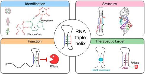
Major-groove RNA triple helices are conserved tertiary structures formed when a third strand inserts into the major groove of a classic double helix either via Hoogsteen base pairing or reverse Hoogsteen base pairing. These structures are widely distributed in eukaryotic, prokaryotic, and viral RNAs. This paper systematically summarizes the structural classification, stability-influencing factors, and identification methods of RNA triple helices. A growing body of evidence indicates that RNA triple helices are extensively involved in diverse biological processes, including RNA stability regulation, translation regulation, riboswitch ligand recognition, transposition regulation, telomerase activity, and the assembly of the spliceosome catalytic core, as well as serving as scaffolds for molecular recruitment. Notably, dysregulation of RNA triple helices is closely associated with tumorigenesis, viral infections, and genetic diseases. Based on their structural and functional characteristics, multiple therapeutic strategies targeting RNA triple helices have been explored, such as small molecules and antisense oligonucleotides. Collectively, RNA triple helices represent a pivotal link between RNA structural biology and precision medicine, with promising potential as an important candidate target for future RNA structural drug development.
Keywords: RNA triple helix, gene expression regulation, RNA stability, RNA-targeted therapeutics
Introduction
RNA molecules achieve their essential biological functions through hierarchical folding, progressively evolving from linear sequences into complex three-dimensional architectures. Among these, the RNA triple helix represents a unique structural motif within RNA tertiary structures. The RNA triple helix (also referred to as the RNA triplex) consists of three polynucleotide strands assembled through specific hydrogen bonding and stacking interactions [1]. The most common structure is the intramolecular triplex, in which the third strand originates from another single-stranded region of the same RNA molecule—such as a bulge, internal loop, or hairpin loop—and engages with the major groove of the Watson-Crick duplex. This interaction is mediated by Hoogsteen or reverse Hoogsteen hydrogen bonds, allowing the third strand to bind within the major groove of the duplex and form base triples, thereby stabilizing the local RNA architecture [2, 3]. Concurrently, intermolecular triplexes formed by three strands contributed by two or three separate RNA molecules represent another important class of RNA tertiary interactions. These intermolecular systems broaden the structural and regulatory repertoire of RNA triple helices and provide a conceptual basis for developing RNA-targeted therapeutic strategies.
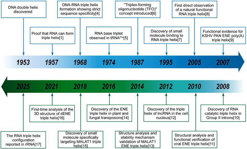
The discovery and definition of the RNA triple helix have emerged from decades of cumulative research (Figure 1) [1, 4-17]. Over the past decades, research has evolved from the DNA double helix to the DNA-RNA and RNA triple helices, expanding from telomerase RNA to spliceosomal RNA, riboswitches, and nuclear expression elements, while increasingly exploring RNA triple helices as therapeutic targets (Figure 1). In 1957, Felsenfeld, Davies, and Rich first demonstrated the existence of the triple helix in vitro [1]. Using a system of synthetic poly(U) and poly(A) homopolymers, they discovered that a third poly(U) strand could specifically associate with the poly(A)·poly(U) duplex, leading them to infer the formation of a three-stranded complex. Although the detailed arrangement of the U·A-U triple helix had not yet been established at the time, subsequent studies confirmed this structural interpretation. This pioneering work established the foundation of nucleic acid triple helix research. Over the following decades, key breakthroughs were achieved, including the observation of naturally occurring continuous U·A-U major-groove triple helices [8] and the subsequent identification of RNA triple helices with clear biological functions [11]. Advances in high-resolution structural biology—exemplified by the SAM (S-adenosylmethionine)-I riboswitch structure [18]—and increasingly precise molecular techniques have driven the continuous discovery of RNA triple helices in viral, eukaryotic and prokaryotic RNAs (Figure 1). Figure 1 illustrates key time points and significant events in the study of RNA triple helices. Collectively, these findings demonstrate that triple helices are not rare structural anomalies, but rather widespread and functionally important RNA tertiary motifs (Figure 1). Accordingly, research in this field has evolved from the initial phase of structural identification to a new phase focused on functional characterization and the development of intervention strategies targeting triple helices.
Timeline of the discovery and development of RNA triple helices. Over the past several decades, scientific research has progressed from the DNA double helix to the DNA-RNA triple helix and then to the RNA triple helix. The scope of research on the RNA triple helix has also expanded from telomerase RNA to eukaryotic spliceosome RNA, riboswitches, and nuclear expression elements. This field has advanced to exploring approaches such as targeting the RNA triple helix as a therapeutic target and other research directions.
This article reviews the structural and mechanistic basis of RNA triple helices, highlights their major biological functions, and discusses recent advances in therapeutic strategies targeting these structures. Furthermore, this paper outlines the key challenges and future directions in this rapidly developing field. Although several excellent reviews have summarized key advances in the biology of RNA triple helices [2, 3, 19], this paper places particular emphasis on the emerging field of extending structural and functional insights to disease intervention and therapeutic applications.
Structure and Classification of RNA Triple Helix
Identification and characterization of RNA triple helix
Techniques for identifying and characterizing RNA triple helices are continually advancing (Figure 1). The most definitive evidence comes from high-resolution three-dimensional structures, which can be resolved through nuclear magnetic resonance (NMR), X-ray crystallography, or single-particle cryo-electron microscopy (cryo-EM) [20-22]. The ultraviolet (UV) thermal denaturation method monitors UV absorbance under a temperature gradient; RNAs containing triple helices often exhibit a biphasic melting profile in which the first transition reflects triple-helix dissociation and the second duplex unwinding, thereby enabling the analysis of thermodynamic parameters governing triple-helix formation and stability [23]. Isothermal titration calorimetry (ITC) is also widely used to determine the thermodynamic characteristics of RNA triple helices, as heat changes can reveal binding stoichiometry and affinity constants, thereby obtaining data such as heat capacity [24]. In a competition/indicator displacement assay, a signal-reporting probe is first bound to the RNA triple helix and test compounds are then added to monitor probe displacement as a readout of ligand binding [24]. Currently, analytical methods for triple helices are becoming increasingly mature and diverse.
Since experimental discovery alone is insufficient to fully elucidate RNA triple-helical structures—particularly given that only a limited number of such structures have been identified in the human transcriptome—various software tools have been developed to search for and predict RNA triple helices. Because covariance models can capture conserved sequence and secondary-structure features, Infernal 1.1 can be used to detect homologous RNA regions that may preserve triple-helix-associated structural modules, even though it does not explicitly model the base triples themselves [25]. More recently, several new computational frameworks have emerged to characterize long non-coding RNA (lncRNA)-associated RNA triple helices and their potential functions. For example, TRIPinRNA identifies RNA triple helices by scanning transcript sequences for patterns capable of forming canonical triples, as well as non-canonical hetero-purine/hetero-pyrimidine motifs, and predicts continuous triple-helix-forming regions across the full length of human transcripts, providing structural details including the Hoogsteen strand and potential gap positions [26]; using this approach, the study identified plausible triplex-forming sequences in lncRNAs associated with X-chromosome inactivation, with selected candidates subsequently validated by biophysical assays. Researchers have developed an end-to-end computational process specifically for RNA-RNA-RNA triple helices. At the core of TripleMatcher is a dynamic-programming Matcher that searches candidates for unpaired third-strand segments aligned with continuous Watson-Crick-Franklin base-paired helices. When 3D structural information is available, these 2D candidates undergo further screening via 3DFilter, which uses C1′-C1′ distance thresholds to remove geometrically implausible cases. Finally, the ZoneCombiner integrates overlapping or nearby matches into non-overlapping candidate regions (“zones”), thereby highlighting broader RNA segments that may contain triple helices [27]. TripleMatcher successfully localized all annotated triple-helix regions in eight experimentally validated RNAs, including telomerase RNAs and the lncRNAs metastasis-associated lung adenocarcinoma transcript 1 (MALAT1) and Kaposi's sarcoma-associated herpesvirus (KSHV) polyadenylated nuclear (PAN) RNA [27]. Collectively, these advances are progressively establishing a comprehensive computational framework for discovering, annotating, and functionally interpreting RNA triple-helical structures across the human transcriptome. Nevertheless, these tools still share important limitations, including dependence on predefined sequence or secondary-structure patterns, incomplete representation of noncanonical and context-dependent triple interactions, sensitivity to the accuracy of input secondary or 3D structural models, and the need for experimental validation to distinguish true triple helices from computational candidates.
Basic structure and classification of RNA triple helix
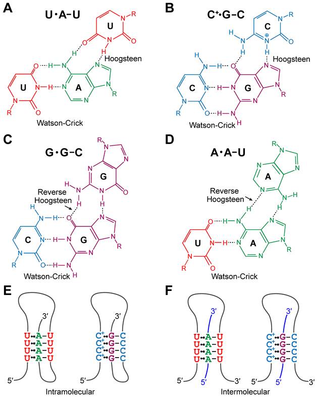
An RNA triple helix forms when a single-stranded oligonucleotide inserts itself into the major groove of a classical Watson-Crick duplex via sequence-specific hydrogen bonding, resulting in a characteristic triple-helical structure. On the basis of the composition and orientation of the third strand, as well as its mode of interaction with the underlying double helix, RNA triple helices can be classified into pyrimidine-type and purine-type triple helices. In a pyrimidine-type triple helix, the pyrimidine-rich third strand (U, C) binds parallel to the homopurine strand of the duplex through Hoogsteen interactions, forming stable U·A-U (Figure 2A) and C+·G-C triples (Figure 2B) (Where “·” represents Hoogsteen hydrogen bonds and “-” denotes Watson-Crick hydrogen bonds). Notably, this structure relies on the protonation of the N3 site in cytosine and its interaction with the N7 site in guanine, thereby forming the same number of hydrogen bonds as the Hoogsteen pair in the U·A-U triple [11, 12]. In contrast, the third strand of purine-type triple helices is rich in purines (G, A), which bind to the purine strand of the duplex in an antiparallel orientation via reverse Hoogsteen interactions, predominantly forming triples such as G·G-C (Figure 2C) and A·A-U (Figure 2D) [28, 29]. This classification highlights the decisive role of the third-strand sequence and orientation in determining the specificity and stability of triple-helix binding, providing a conceptual framework for understanding the rules governing triple-helix formation. RNA triple helices can also be categorized as intramolecular (Figure 2E) or intermolecular (Figure 2F) based on whether all three strands originate from the same transcript or from two or three separate RNA molecules. In addition, non-canonical triple-helix-like variants exist, in which the third strand inserts into the minor groove of a Watson-Crick duplex; and one of the most typical forms is the A-minor motif, in which adenine recognizes standard base pairs from the minor groove side, such as A·G-C base triples [9, 11, 13]. In certain newly characterized riboswitches, the third RNA strand can insert into the minor groove and interact specifically with the duplex, thereby generating a local triple-helical structure [30]. However, these triple helices are generally thermodynamically unstable. Therefore, this review will not elaborate further on the relevant content.
Classification of RNA triple helix. (A, B) The figures show U·A-U and C+·G-C base triples with Hoogsteen and Watson-Crick interactions, respectively. Hydrogen bonds are indicated by broken lines. Schematic diagrams of U·A-U and C+·G-C base triples are also shown on the right. Dashed lines represent hydrogen bonds. (C, D) The figure shows G·G-C and A·A-U base triples with the reverse Hoogsteen and Watson-Crick interactions. (E, F) RNA triple helices can be classified into intermolecular and intramolecular triple helices based on their molecular properties; if the three strands originate from the same transcript, they belong to intramolecular triple helices, whereas those originating from multiple transcripts (additional transcripts are indicated by blue lines) belong to intermolecular transcripts.
A growing number of RNA triple helices incorporate noncanonical base triples. For example, the group II intron in Oceanobacillus iheyensis contains a triple-helical structure composed of G·G-U and C·C-G base triples [10]. Similarly, the bacterial SAM-II riboswitch has a U·U·ASAM ligand-dependent base triple, in which the adenine moiety of SAM, rather than a regular RNA base, serves as the third base-like component in the interaction [31]. In spliceosomal U2/U6 small nuclear RNAs (snRNAs) in Schizosaccharomyces pombe triple-helical formation involves base triples such as A·A-U, U·C-G, and G·G-C [29]. The 7SK stem-loop 1, in a complex formed with a peptide fragment representing the RNA-binding domain of the human immunodeficiency virus trans-activator of transcription (HIV Tat) protein, possesses two consecutive U·A-U base triples; however, the protein recognizes its flanking C-G base pair [32], which differs from classical triples that can directly serve as protein-binding ligands. Moreover, researchers used the TRIPinRNA system to search for hetero-purine-pyrimidine intramolecular triple helices and discovered atypical A·U-A triples as well as transient motifs, such as UAA and AUU. These are less stable than classical triples and display higher structural plasticity and dynamic behavior [33]. Together with high-resolution structural observations of other noncanonical triples in natural RNAs, including C·C-G and G·G-U in the group II intron [10, 34], U·U·ASAM in the SAM-II riboswitch [31], and A·A-U, U·C-G, and G·G-C in spliceosomal U2/U6 snRNAs [29]. Therefore, the design and engineering of RNA triple-helical structures should not be restricted to classical combinations such as U·A-U. Currently resolved RNA triple-helical structures are summarized in Table 1.
Information on the resolved triple-helix structures (ordered numerically and alphabetically).
| Structure | Method and resolution | PDB ID | References |
|---|---|---|---|
| P2b-P3 pseudoknot from human telomerase RNA | Solution NMR | 1YMO | [8] |
| P2B-P3 pseudoknot of human telomerase RNA | Solution NMR | 2K95, 2K96 | [22] |
| Pyrimidine motif triple helix in the Kluyveromyces lactis telomerase RNA pseudoknot | Solution NMR | 2M8K | [82] |
| PreQ1 Class II riboswitch from Streptococcus pneumoniae | Solution NMR | 2MIY | [67] |
| SAM-II riboswitch bound to S-adenosylmethionine | X-ray diffraction, 2.80 Å | 2QWY | [31] |
| Self-spliced group II intron | X-ray diffraction, 3.10 Å | 3BWP | [10] |
| Yeast spliceosome | Electron microscopy, 3.60 Å | 3JB9 | [169] |
| ENE, a viral RNA stability element, in complex with A9 RNA | X-ray diffraction, 2.50 Å | 3P22 | [11] |
| C-di-GMP-II riboswitch from Clostridium acetobutylicum bound to c-di-GMP | X-ray diffraction, 2.51 Å | 3Q3Z | [170] |
| Class II preQ1 riboswitch | X-ray diffraction, 2.28 Å | 4JF2 | [66] |
| Triple-helical stability element at the 3′ end of MALAT1 | X-ray diffraction, 3.10 Å | 4PLX | [13] |
| PreQ1 Riboswitch | X-ray diffraction, 2.75 Å | 4RZD | [62] |
| Group II intron complexed with its reverse transcriptase | Electron microscopy, 4.50 Å | 5G2Y | [171] |
| Catalytic Step I spliceosome | Electron microscopy, 3.40 Å | 5GMK | [102] |
| Tetrahymena telomerase RNA pseudoknot | Solution NMR | 5KMZ | [172] |
| Human spliceosome activated for step 2 of splicing | Electron microscopy, 5.90 Å | 5MQF | [28] |
| Thermobifida fusca guanidine III riboswitch with guanidine | X-ray diffraction, 1.91 Å | 5NWQ | [63] |
| Group II intron lariat with an intact 3′ splice site (pre-2s state) | X-ray diffraction, 3.70 Å | 6CHR | [94] |
| MetY SAM V riboswitch | X-ray diffraction, 2.50 Å | 6FZ0 | [64] |
| Structure of a group II intron retroelement after DNA integration | Electron microscopy, 3.60 Å | 6MEC | [95] |
| U·A-U-rich RNA triple helix with 11 consecutive base triples | X-ray diffraction, 2.50 Å | 6SVS | [20] |
| Stabilized PAN ENE bimolecular triplex with a GC-clamped polyA tail, in complex with Fab-BL-3,6 | X-ray diffraction, 3.30 Å | 6X5N | [124] |
| Double-ENE RNA stability element in complex with a 28-mer poly(A) RNA | X-ray diffraction, 2.89 Å | 7JNH | [16] |
| B dENE construct complexed with a 28-mer poly(A) | Electron microscopy, 5.60 Å | 7LJY | [46] |
| NAD+-II riboswitch | X-ray diffraction, 2.15 Å/2.50 Å | 8GXB, 8GXC | [65] |
| NAD-II riboswitch (two strands) | X-ray diffraction, 1.67 Å | 8I3Z | [30] |
| Ribosomal large subunit from Entamoeba histolytica | Electron microscopy, 2.80 Å | 9V1I | [17] |
Structural stability of the RNA triple helix
Having defined the structural features of RNA triple helices, it is essential to consider the factors that govern their formation and stability, as these ultimately reflect their thermodynamic and kinetic properties. A common method for measuring the stability of RNA triple helices is ultraviolet (UV) thermal denaturation, which works by monitoring the UV absorption spectrum of the sample under a temperature gradient to track its structural disruption process [23]. Nucleotide composition is one of the primary determinants, particularly at Hoogsteen-interacting positions. Studies have shown that the identity of bases engaged in Hoogsteen (or reverse-Hoogsteen) pairing strongly influences thermodynamic stability, with pyrimidines generally providing greater stability than purines [35]. Studies on the MALAT1 3′ end protective triple helix further demonstrate that pyrimidine·purine-pyrimidine arrangements exhibit superior stability [13]. Moreover, C·G-C and U·G-C can also support highly stable RNA triple-helical formation [35]. Importantly, sufficient thermodynamic stabilization is often required to sustain RNA function under physiological conditions, which provides useful guidance for the design of structured RNAs, including mRNA therapeutics. Specifically, RNA secondary structure can regulate antigen expression and the immunogenicity of mRNA vaccines through modulating RNA stability and translation efficiency [36, 37]. In addition, RNA triple helices are dynamic rather than rigid structures, and their folding, assembly, and disassembly are influenced by multiple factors, which will be mentioned below. Accordingly, systems in which the third strand is released are also highly important.
In RNA triple helices, Hoogsteen base pairing often requires protonation of cytidine at the N3 position to form a hydrogen bond with guanine at the N7 position [12]. Therefore, environmental pH is a critical determinant of triple-helical formation and stability. Melting temperature of the RNA triple helix decreases sharply with increasing pH, which is consistent with theoretical expectations [38]. When the NaCl concentration was increased from 100 mM to 1 M, the melting temperature of the RNA trimer also decreased; this is presumed to be due to changes in salt concentration affecting the pKa value of N3 in cytosine, which in turn influences the formation of Hoogsteen hydrogen bonds in the RNA triple-helical structure [38]. These factors also provide an important basis for assessing how RNA chemical modifications influence the stability of RNA triple-helical structures.
The peripheral structure of the RNA triple helix also plays an essential role in its stability. While studying the long non-coding MALAT1 RNA, which relies on the triple helix structure to maintain stability, researchers found that the peripheral double-stranded elements of its triple-helical structure aid in anchoring and stabilizing the RNA triple helix [39].
Some chemical ligands can significantly enhance the stability of RNA triple helices upon binding. For example, various isoquinoline alkaloids [40], the cationic dye methylene blue [41], and a novel ruthenium(II) polypyridine complex [Ru(dmb)2dppz-idzo]2+ [42] can all act as ligands to recognize and bind to RNA triples, enhancing their stability with strong affinity. Importantly, in addition to enhancing triplex stability, certain small molecules can also regulate and shift the equilibrium between RNA duplex and triplex conformations; for example, coralyne has been shown to induce both duplex-to-triplex and triplex-to-duplex conversion in poly(A)·poly(U)/poly(A)2·poly(U) systems in a temperature- and ligand/RNA ratio-dependent manner [43]. Further details will be provided in the section of screening of small molecule drugs based on the RNA triple helix. The mechanism by which these ligand molecules recognize and bind to RNA triple helices provides valuable references and ideas for the future development of small molecules targeting RNA triple helices and the consequent expansion of intervention strategies for related diseases.
Biological Functions of the RNA Triple Helix
Enhancing RNA stability
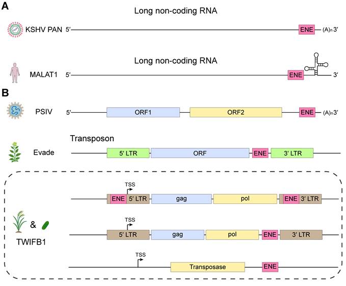
As an important conserved motif in RNA, the RNA triple helix plays a fundamental role in stabilizing the host RNA molecule and provides a platform for the establishment of diverse regulatory functions. The most representative example is the cis-acting element, expression and nuclear retention element (ENE), which contains characteristic RNA triple-helical structures. ENEs are found in diverse locations within genes and genomes, with the majority located near the 3′ end of transcripts (Figure 3). ENE was first discovered in KSHV PAN RNA [44]. ENE typically consists of a U-rich internal loop flanked by short helical segments. This architecture enables the U-rich region to form a triple helix with the 3′-poly(A) tail, thereby spatially sequestering the poly(A) tail and preventing access by deadenylases. In this way, ENEs markedly enhance RNA stability [16]. Since their initial discovery, ENEs have been identified in a wide range of cellular and viral RNAs [12, 14, 45]. The most extensively studied ENE triple-helical structures include ENE from viral RNAs, ENE from mammalian oncogenic lncRNAs, and double-domain ENEs (dENEs) found in plants and fungi [12, 14, 44].
Location of ENEs in genes and genomes. (A) The most representative ENEs found in long non-coding RNAs are the ENE of viral PAN RNA and that of mammalian MALAT1. They are located near the 3′ end of their respective transcripts, used to protect the RNA from degradation by forming RNA triple helices, and the latter does not have the classic poly(A) tail [12, 13, 44, 119]. (B) There are other ENEs that are located at the 3′ end immediately behind the ORF, such as the ENE of Plautia stali intestine virus (PSIV) and the ENE of TE in the Arabidopsis genome called Evade [23, 45]. In the common transposable elements in plants and fungi, there are not only ENEs behind the 3′ LTR, but also ENEs embedded in the 5′ and 3′ LTR and ENEs behind the transposase gene, such as the ENE of the hAT DNA transposon TWIFB1 [14]. The genes (RNAs) and ENEs are not drawn to scale.
KSHV, the etiological agent of Kaposi's sarcoma, produces an abundant 1,077-nucleotide nuclear noncoding RNA known as PAN RNA. This transcript contains an approximately 79nt ENE that prevents degradation at its 3′ end, thereby stabilizing PAN RNA. The ENE folds into a U-rich internal loop flanked by short 5′ and 3′ helical stems. The U-rich loop engages with the 3′-poly(A) tail via simultaneous Watson-Crick-Franklin and Hoogsteen hydrogen bonds, forming canonical U·A-U base triples that constitute the core of the triple helix (Figure 4A) [46]. This RNA triple-helix structure isolates the poly(A) tail to prevent RNA degradation by exonucleases such as deadenylases [11]. In addition, ENE causes intron-less transcripts to be retained in the nucleus; however, its triple-helical structure only protects and maintains the transcript and does not necessarily enhance protein expression [47]. These features establish the PAN ENE-mediated triple helix as a canonical model for poly(A) tail sequestration and viral nuclear RNA stabilization.
The well-studied triple-helix of ENE elements primarily includes viral single-domain ENEs, blunt-ended ENEs of mammalian oncogenic lncRNAs, and dENEs from plants and fungi. (A) Schematic diagram of the ENE element structure of KSHV PAN RNA, which belongs to the single-domain ENE category. The U-rich loop interacts with the poly(A) tail via hydrogen bonds, forming a U·A-U major-groove triple helix [11]. (B) Schematic diagram of the 3′ end ENE element structure of MALAT1, which belongs to the blunt-ended ENE category. It forms a blunt end through the U-rich internal stem-loop and the downstream A-rich region, rather than terminating with a 3′-poly(A) tail [13]. (C) Schematic diagram of the dENE structure in the rice hAT DNA transposon TWIFB1, which comprises the dENE, separated into two ENE motifs by a short double helix region. The right panel shows a schematic of the ENE triple helix structure [16]. Structural models of RNA secondary and tertiary structures are shown in the supplementary files.
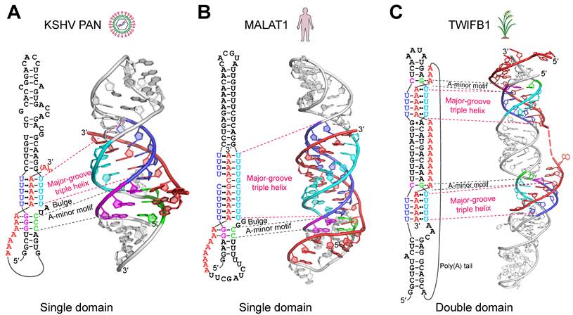
In human lncRNAs, the highly abundant and oncogenic MALAT1 and multiple endocrine neoplasia β (MENβ, also known as nuclear enriched abundant transcript 1 transcript variant 2, NEAT1_2) RNA contain triple helix elements analogous to the ENE found in PAN RNA. Unlike the PAN ENE, which interacts with the 3′-poly(A) tail, these lncRNA triple helices are formed by pairing a U-rich internal stem-loop with a downstream A-rich region, creating a triple helix possessing blunt ends. A canonical structural model, which is best characterized for MALAT1 and closely related in MENβ, comprises nine U·A-U base triples interrupted by a single C·G-C base triple and a C-G Watson-Crick base pair (Figure 4B) [12]. Its mechanism of action also prevents degradation of the 3′ end by relevant enzymes [48]. Functional dissection has been particularly well established for the MALAT1 3′-end triple-helix element, although much of this evidence has been obtained using exogenous reporter mRNAs or MALAT1-derived minimal constructs rather than full-length endogenous MALAT1. The same is true for studies on other types of RNA triples. Mutation-based β-globin reporter assays in MALAT1 further demonstrate that the precise arrangement of the internal U-rich loop and the downstream A-rich segment is critical for triplex stability and lncRNA activity [49]. In summary, the MALAT1 and MENβ triple helices extend the ENE-mediated stabilization strategy to intron-less oncogenic lncRNAs. It is worth noting that through mass spectrometry identification, Clustered Regularly Interspaced Short Palindromic Repeats (CRISPR)-mediated deletion of the NEAT1_2 triple helix domain, and in situ hybridization, a recent study revealed that the triple helix maintains the integrity of nuclear paraspeckles, thereby mediating the nuclear localization and radiation-induced rapid activation of unphosphorylated microRNA 34a [50].
ENE-like cis-acting elements have also been identified in transcripts derived from plant and fungal transposable elements (TEs), termed dENEs [14]. These elements are characterized by two ENE-like motifs separated by a short duplex region (Figure 4C). This structure, similar to the classic ENE domain, forms a protective triple helix or isolation structure with the 3′ end poly(A) tail, thereby blocking exonucleolytic attacks—including deadenylation—and promoting transcript stability [14]. All structurally characterized dENEs harbor a highly conserved adenosine triad, and their flanking stem regions display strong sequence preferences [16]. Unlike previously described ENEs, dENEs also possess a pocket motif at the downstream end of the triple helix. This pocket terminates the triple helix in a near blunt-end manner through steric hindrance, while simultaneously locking the terminal poly(A) adenosine within the major groove via hydrogen bonding, thereby shielding the 3′ end. By restricting accessibility to the 3′ end, this mechanism effectively inhibits degradation pathways such as deadenylation and reinforces RNA stability [46]. Collectively, dENEs represent an evolutionarily adapted ENE variant that combines a conserved adenosine triad with a terminal pocket motif to maximize 3′ end protection and sustain RNA stability across diverse eukaryotic transcripts.
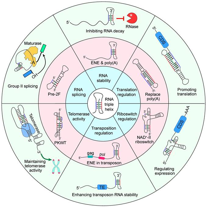
In addition to forming a triple helix with the 3′-poly(A) tail or an A-rich stretch to protect transcripts from exonuclease degradation, ENEs have also been shown to enhance the stability of nuclear transcripts, thereby increasing the mRNA levels of the corresponding genes; this phenomenon is particularly evident in intron-less mRNAs [14, 39]. This is consistent with the mechanism by which ENE maintains viral RNA stability. These transposon-derived transcripts, having lost their introns (an adaptation to evade gene silencing), tend to reside longer in the nucleus and are thus more susceptible to nuclear RNA decay pathways [51]. In this context, the presence of an ENE becomes especially important, as it confers nuclear stability and promotes the preferential accumulation of ENE-containing transcripts. It is worth noting that, besides RNA triple helices, evolutionarily conserved long-range RNA structures can also orchestrate complex gene regulation, as exemplified by the Dscam1 exon 6 cluster, where multiplexed RNA secondary structures regulate the stochastic selection of variable exons in both temporal and spatial dimensions [52]. Overall, RNA triple helices enhance host RNA stability by sequestering the 3′-poly(A) tail or A-rich region, thereby blocking deadenylases and other exonucleases from accessing the RNA end (Figure 5).
Overview of the function of RNA triple helices. The inner ring describes the biological functions of the RNA triple helix. The middle ring displays the structural features corresponding to the RNA triple helix performing different functions. The outer ring introduces the corresponding biological effects produced. Among these triple helices, group II introns, the eukaryotic spliceosome, and some types of riboswitches all possess antiparallel major-groove triple helices.
Translation regulation
RNA triple helices can promote translation and, in some contexts, functionally replace the poly(A) tail (Figure 5). As early as in replication-dependent histone mRNAs, it was observed that other structures could replace the poly(A) tail [53]. Researchers generated reporter constructs placing the 3′ region of MALAT1, which contains an ENE, downstream of the reporter gene to replace the poly(A) tail. They found that transcripts produced from these ENE-containing constructs exhibited protein expression levels comparable to those of transcripts bearing a normal poly(A) tail, suggesting that the ENE-mediated triple-helical formation stabilizes transcripts and sustains productive expression even without canonical polyadenylation [48]. Transcripts with the poly(A) tail replaced by an ENE-mediated RNA triple helix can still be repressed by microRNAs (miRNAs), suggesting that triple-helix-based stabilization does not preclude interaction with standard post-transcriptional regulatory pathways. This supports the notion that ENE-driven triple helix formation can serve as an alternative 3′ end stabilization strategy without sacrificing normal regulatory inputs [48]. Overexpression of PABPC1 in frog oocytes significantly enhanced the translation of short-tailed mRNAs, yet did not significantly promote the translation of mRNA at the 3′ end of the histone stem-loop or the MALAT1 triple helix, demonstrating that triple-helix-containing mRNAs can still maintain a certain level of translational output even in the absence of poly(A) tails, but are unable to bind to the poly(A)-binding protein in the cytoplasm to form closed-loop mRNAs [54]. In most cases, enhanced protein yield arises indirectly through increased RNA stability rather than direct stimulation of the translational machinery. RNA triple helices restrict access of exonucleases and deadenylation complexes by forming a locally stable triple helix at the 3′ end, thereby prolonging mRNA half-life and expanding the pool of transcripts available for ribosome engagement, which in turn elevates translational output [46, 55]. A recent study replaced the poly(A) tail of synthetic mRNA with the Malat1 RNA triple helix and found that while it can support translation, its efficiency is far lower than that of the canonical poly(A) tail, suggesting that future optimization of triple helix sequence and folding is needed to enhance translational capacity [56]. These findings establish the 3′ end RNA triple helix as a versatile, context-dependent regulatory module that, under certain conditions, can substitute for the poly(A) tail to maintain mRNA stability and translational competence, while still integrating into conventional post-transcriptional regulatory networks.
Major-groove base-triple interactions can also directly influence translation. A representative study showed that a major-groove triple helix within an mRNA stimulatory element enhances programmed ribosomal frameshifting by stabilizing an RNA structure that impedes ribosomal unwinding and promotes pausing at the slippery sequence [57]. Disruption of the triplex reduced frameshifting efficiency, supporting the view that RNA triple-helical motifs play a direct role in translational recoding [57].
In the 25S rRNA of the large ribosomal subunit (LSU) from Entamoeba histolytica, a major-groove RNA triple helix has been identified, consisting of three U·A-U and one U·G-C base triples. This triple helix is located in domain VI (DVI), on the solvent-exposed side near the peptide exit tunnel (PET), marking the first reported instance of such a triplex conformation in rRNA [17]. It is plausible that this triple helix, like its ENE counterpart, provides local protection against 3′ exonucleolytic or other rRNA processing or degradation activities, although this protective role remains to be experimentally confirmed. Furthermore, studies have found that ribosomal proteins and their extensions interact with the rRNA surface surrounding this triple helix, suggesting that the triple helix and its protein interactions help reinforce the outer surface of the LSU, thereby facilitating ribosome stability and subunit assembly, which in turn may contribute to more efficient translation.
Riboswitch regulation
Riboswitches are a class of cis-regulatory RNA elements located in the non-coding region of mRNAs (usually the 5′ Untranslated Region, UTR) that can regulate gene expression by directly binding to small-molecule ligands (e.g., metabolites, ions, or coenzymes) [58, 59]. Since the existence of riboswitches was first experimentally confirmed in 2002, over 50 different types have been identified, with widespread occurrence in bacteria and a small number found in archaea and eukaryotes [60, 61]. Riboswitches act as receptors, capable of forming ligand-binding structures and executing regulatory commands, and they require no protein involvement to perform translationally regulated switch transitions [59].
A variety of riboswitches can form RNA triple helices in their ligand-bound states, including prequeuosine1 (preQ1)-I/preQ1-II/preQ1-III, guanidine III, SAM-V and SAM-II, cyclic-dimeric-guanosine monophosphate (c-di-GMP)-II, nicotinamide adenine dinucleotide (NAD+)-II riboswitch, Mn²+ riboswitch, adenine or purine riboswitch, and so on (Figure 5). Riboswitch triple helices are typically organized around a Watson-Crick duplex, with a third strand docking into either the major or minor groove. Such triple-helical motifs frequently occur in the principal closing helix of the aptamer domain (P1), the short local sub-helix adjacent to the ligand-binding core (P1.1), or in pseudoknot regions formed by long-range intramolecular pairing [30, 31, 62-65]. Typically, the triple-helix structure of a riboswitch is directly formed by two U·A-U or A·G-C triple base pairs along with several nearby non-classical triple base pairs, which define the binding pocket for small molecules, thereby achieving highly selective recognition. Among the early identified riboswitch classes, SAM-V and SAM-II form major-groove triple helices, in which the third RNA strand docks into the major groove, with the SAM ligand positioned along the helical axis and stabilized by hydrogen-bonding interactions with surrounding bases at its ends [31, 64]. One side of the guanidine III binding site has an opening that can accommodate ligand derivatives carrying small side chains or substituent groups, indicating both binding selectivity and a certain degree of structural plasticity [63]. The preQ1-I/preQ1-II/preQ1-III classes adopt different folding patterns for the same ligand (preQ1), but all classes stabilize the RNA upon ligand binding, thereby altering expression platform accessibility [62, 66-68]. C-di-GMP-II incorporates ligands as part of its three-stranded structure, suggesting that proteins may be involved in shaping the riboswitch conformation [68]. These examples illustrate that riboswitch-associated RNA triple helices employ diverse architectural strategies to embed their ligands within three-stranded cores, thereby coupling highly selective small-molecule recognition with precise, structure-driven control of gene expression.
The NAD+-II riboswitch is a recently characterized RNA sensor capable of recognizing NAD+-related metabolites, with its core architecture stabilized by layered noncanonical interactions, including A·G-C base triples. This architecture broadens the structural repertoire of RNA triple interactions and illustrates that base triples can function not only as stabilizing elements of RNA folding, but also as direct contributors to metabolite recognition and riboswitch-mediated gene regulation [30, 65].
Furthermore, different riboswitch families employ distinct approaches to regulate RNA triple helices and are not confined to a single gene regulatory model. Some of the aforementioned riboswitch types can activate translation [69, 70], while others inhibit the translational process [30, 31, 62, 65, 69, 71-73]. Still others can extend, pause, or terminate transcription [71, 72, 74]. The ligand-binding triple helix regulatory mechanism of riboswitches exhibits remarkable flexibility and diversity. Future studies should employ high-resolution structural and kinetic analyses to determine the sequence of interactions between ligands and triple-helical structures. Characterizing these ligand interactions could serve as a reference for the design of RNA-targeting small-molecule drug design. Additionally, a systematic analysis should be conducted to investigate how RBPs influence the function of riboswitches under different strain conditions.
Transposition regulation
Transposable elements, also known as transposons, are mobile DNA sequences found in nearly all eukaryotic genomes that can replicate and relocate within the host genome [75, 76]. In eukaryotes, they are generally suppressed by transcriptional or post-transcriptional gene silencing mediated by small RNAs such as small interfering RNAs (siRNAs) and Piwi-interacting RNAs (piRNAs) [77, 78]. In addition, RNA deadenylation removes the poly(A) tails and selectively degrades certain transposon transcripts that escape siRNA regulation, thereby suppressing new transposition events [79]. However, some retrotransposons contain ENE elements, which form RNA triple helices to protect their transcripts from host-mediated decay, thereby modulating transposition (Figure 5) [13]. Typically, ENE elements in transposons contain a U-rich internal loop (URIL) that binds to the 3′ poly(A) tail via concurrent Hoogsteen and Watson-Crick-Franklin interactions, while short flanking stems assemble the structure into a pocket-like architecture [16].
A notable example is the Arabidopsis Evade retrotransposon, which displays unusually strong transpositional activity. Sequence analysis revealed four U-rich repeat segments between its open reading frame (ORF) and the downstream long terminal repeat (LTR), capable of forming ENE-like stem-loop structures and supporting triple-helix formation [23]. In vitro thermal denaturation experiments produced a characteristic biphasic melting profile, providing strong evidence for the presence of an RNA triple helix. Functional assays using a luciferase-based transient expression system with inducible mutagenesis further demonstrated that the ENE motif stabilizes the transcript and enhances gene expression. Finally, mutation experiments targeting the deadenylase CCR4a (an enzyme that shortens the 3′ end of retrotransposon RNAs) confirmed that the triple-helix-mediated protection is achieved by blocking CCR4a-dependent degradation [23]. This indicates that the RNA triple helix structure serves as a vital survival strategy for transposable elements to maintain their activity within the host genome. These findings highlight that ENE-mediated RNA triple helices serve as a specialized adaptive mechanism through which retrotransposons evade host RNA decay pathways, thereby sustaining their transcript levels and ensuring their continued transpositional competence within the genome.
Telomerase activity and telomere maintenance
Telomerase is a specialized ribonucleoprotein (RNP) that uses its RNA template to replicate chromosome ends, thereby solving the end-replication problem in eukaryotes [80]. Its core components include telomerase reverse transcriptase (TERT), which catalyzes the synthesis of telomeric DNA repeats, and telomerase RNA (TR), which contains the template for telomeric DNA synthesis (referred to as hTR or TERC in humans, TER in ciliates, and TLC1 in yeast); in addition, several accessory factors are involved in its assembly, maturation, and localization [81]. TR also contains several conserved sequences, among which the H-type pseudoknot structure is one of the most critical functional regions. A recent structural biology study has shown that the RNA triple helix found within this pseudoknot is a conserved structural element essential for telomerase catalytic activity (Figure 5) [82]. Thus, the conserved triple helix embedded in the TR pseudoknot serves as a critical architectural hub that couples proper RNA folding with telomerase assembly and catalysis, thereby safeguarding telomere maintenance in eukaryotic cells.
By successively resolving the structures of human TR, Kluyveromyces lactis TR, several ciliate TERs, and Tetrahymena thermophila TER, it was found that this RNA triple helix is located in the stem of the pseudoknot forming a major-groove triplex composed of several consecutive U·A-U base triples; moreover, C+·G-C or U·G-C triples are present in yeast, whereas A+·G-C triples are present in T. thermophila [8, 22, 82, 83]. C+ and A+ denote protonated cytosine and protonated adenine, respectively. Mutational studies of human TR show that disrupting these triple interactions markedly reduces the thermal stability of the pseudoknot and leads to loss of telomerase activity; conversely, compensatory mutations that allow the formation of C+·G-C triples at low pH partially restore structural stability and enzymatic activity, demonstrating the critical role of the triple helix in telomerase catalysis [8]. Similar experiments conducted in K. lactis, T. thermophila, Saccharomyces cerevisiae, and other species have yielded consistent results [82-85], indicating that precise structural arrangement is essential for telomerase. Comparison with archaeal H/ACA ribonucleoprotein complexes, which normally guide the pseudouridylation of target RNAs, suggest that intramolecular pairing and fold-back extension of an opposing RNA strand in telomerase RNA generates a triple helix similar to the substrate-bound state, thereby blocking pseudouridylation and protecting telomerase RNA from nonspecific modification [21]. These experiments demonstrate that RNA triple helices are necessary and widely conserved for maintaining telomerase activity.
Single-molecule experiments, NMR, and cryo-EM studies have shown that in full-length TR without protein, the pseudoknot triple helix does not usually represent the dominant conformation due to the presence of competing folding pathways; only when protein subunits (such as TERT and H/ACA-related proteins) participate in assembly does the triple helix form and stabilize the active state, suggesting that the triple helix may facilitate correct pseudoknot folding and may directly contribute to the positioning and assembly of groups near the catalytic site [8, 82-87].
The function of the triple helix is not isolated; together with surrounding domains such as the CR4/5 domain of Japanese medaka telomerase and the H/ACA domain and auxiliary proteins, it forms an RNP network that jointly determines telomerase activity and its biogenesis pathway [8, 88, 89]. Recent studies have revealed that an RNA chaperone, La-related protein 3 (LARP3), competes with triple helix formation. This protein recognizes long 3′-extended transcripts that fail to fold correctly into the triplex and mediates their degradation; conversely, the formation of the triplex prevents LARP3 binding, blocks its degradation pathway, and promotes hTR maturation, thus maintaining a dynamic balance between the two [90]. When this balance is disrupted—for example, by LARP3 overexpression—triple helix formation is inhibited, telomerase activity is reduced, and telomeres become shorter; in contrast, LARP3 knockdown promotes the production of mature hTR and leads to telomere elongation [90], both of which highlight the central regulatory role of the triple helix in quality control and RNA maturation.
In addition, some special cases have been observed in telomerases from different organisms. In K. lactis, mutations in the triple helix led to truncated telomeric repeat synthesis, suggesting that the triple helix may affect template usage or the translocation process [84]. However, in S. cerevisiae, TERT does not require a triple helix to associate with TR [85]. Furthermore, studies on TR biogenesis indicate that the RNA triple helix participates in determining the fate of precursor TR transcripts, particularly 3′-extended immature TR molecules, as the extension of TR transcripts can form an RNA triple-helical structure and recruit processing-related proteins [89]. These organism-specific observations suggest that the telomerase RNA triple helix not only exerts flexible, species-dependent control over template utilization and enzyme translocation but also contributes to TR biogenesis by shaping the precursor processing and maturation pathways.
RNA splicing regulation
Pre-mRNAs in eukaryotic cells contain introns, which are noncoding RNA sequences. Eukaryotic pre-mRNA splicing is a two-step reaction involving the removal of introns and the ligation of exons. The chemical steps of this mechanism are highly similar to those of self-splicing group II introns, as both proceed through two sequential transesterification reactions [91]. Group II introns are usually 400-800 nt in length and fold into a characteristic six-domain (DI-DVI) spoke-like architecture around a central hub [92]. Both systems exhibit the formation of a catalytic triplex to perform catalysis: in group II introns, the catalytic triplex is located in Domain V (DV), whereas in the spliceosome, the catalytic core is an RNA-based catalytic center analogous to that of group II introns (Figure 5). At the evolutionary and structural levels, the RNA scaffold of group II introns is considered an ancestral model for the spliceosome RNA-protein complex. Although many of the extensive scaffolding functions once performed by intron RNA have been replaced by proteins, the strategy involving the catalytic triplex has been retained [91, 92]. The shared reliance on a conserved catalytic triplex in both group II introns and the spliceosome underscores a deep evolutionary continuity; even as much of the surrounding structural framework has gradually been handed over to proteins, an ancient RNA-based catalytic strategy has been preserved.
As mentioned earlier, the core catalytic structure of group II introns is located in the DV region. This region contains a highly conserved A-G-C catalytic triad and a C-G-C motif, which, together with the J2/3 joining strand—the RNA segment connecting domains II and III—form a catalytic triple helix. In conjunction with the dinucleotide bulge of DV, this triple helix coordinates two catalytic Mg²+ ions (M1 and M2) and participates directly in catalysis [10, 93-96]. Among these, M1 accurately locates 2′-OH and stabilizes the departing group. M2 assists the departing group and coordinates an oxygen atom at the breaking site, thereby activating the nucleophile [97, 98]. Cryo-EM and X-ray analyses at 3.0-3.5 Å resolution reveal that the metal-triplex catalytic platform remains unchanged during both transesterification reactions, while the substrate is exchanged through oscillation of the D6 branch helix. D6, the branch-site-containing domain VI of the group II intron, is anchored in the first step by contact with the thumb subdomain and DNA-binding (DBD) domain of the maturase; in the second step, it contacts intron domain II (D2) via downward oscillation [99]. Group II intron uses this mechanism of solely exchanging substrates while maintaining the catalytic platform to fix magnesium ions, thereby maintaining the catalytic geometric configuration and stability.
The spliceosome in eukaryotes is a highly dynamic molecular machine composed of small nuclear ribonucleoproteins (snRNPs; U1, U2, U4, U5, and U6) and their associated auxiliary factors. It catalyzes intron removal and exon ligation through two sequential transesterification reactions [100]. The core catalytic triplex of the eukaryotic spliceosome resembles the active-site architecture of self-splicing group II introns. In particular, the terminal GA of the U6 snRNA ACAGAGA sequence forms a major-groove triple helix interaction with the U2/U6 helix Ib, a segment of the U2-U6 complementary duplex. Concurrently, the apex of the adjacent U6 internal stem-loop (ISL) contains an AGC triad, which establishes three consecutive base triples with the U2/U6 helix Ib in a one-to-one fashion [96, 99, 101]. Using cryo-EM, crosslinking mass spectrometry, genetic experiments, and metal-rescue approaches, researchers have demonstrated that these RNA elements enable U6 snRNA to coordinate two catalytic Mg2+ ions, thereby stabilizing the spliceosome conformation and promoting cooperative metal-dependent catalysis during splicing [28, 29, 102-104]. Unlike group II introns, the eukaryotic spliceosome relies on extensive protein interactions with the U2/U6 RNA core to maintain structural stability and enhance catalytic efficiency [102, 103]. Among these proteins, Prp8 is the largest and most conserved component of the spliceosome. Its core structure, together with surrounding subunits, forms a positively charged catalytic cavity that encloses the triplex active center. This surface neutralizes the negatively charged RNA phosphate backbone, facilitates substrate positioning, and is highly conserved from Stentor coeruleus to humans [101, 105]. Moreover, the N⁶-methyladenosine (m⁶A) modification of the ACAGAGA sequence in U6 snRNA stabilizes the docking of U6 to the 5′ splice site (5′ SS) and promotes substrate entry into the catalytic triplex active center [106]. These studies elucidate how proteins enhance spliceosome stability through precise contacts with the RNA triple helix and propose new mechanisms for the recognition and regulation of RNA base triples.
In addition, cryo-EM analysis revealed that the U12/U6atac helix Ib in the U12 small spliceosome forms a catalytic triple helix together with G19, A20, and U46 of U6atac, which also coordinates two metal ions, thereby inheriting the core geometrical conformation of the catalytic triplex of the Group II intron [104]. Compared to the main spliceosome, it lacks the 5′ stem-loop but possesses a unique 3′ stem-loop with tighter base pairing and a higher number of pairs, compensating for the missing helix II structure and reflecting the conservation of U12 introns. These features indicate that the U12-dependent minor spliceosome preserves the ancestral triplex-based catalytic core of group II introns while maintaining splice site recognition and catalytic functions through distinct architectural adjustments.
Structural scaffold
RNA triple helices function as dynamic, structure-dependent protein-binding scaffolds that mediate selective protein recruitment and regulate RNA maturation and activity. RNA triple helices can bind to proteins. The RNA methyltransferase METTL16 mentioned above interacts with RNA triple helices the need for methylation, whereas chemical modification of the triple-helical structure markedly reduces its binding affinity for METTL16 [107]. This suggests that RNA triple helices act as structural scaffolds that provide specific platforms for the binding of certain functional proteins and recruit effector molecules, with protein recognition being determined jointly by the triple helix architecture and the local chemical environment [107, 108]. The triple helix arranges specific base triples into a continuous groove that forms a hydrogen-bonding network and hydrophobic stacking steps complementary to the side strands of α-helices or β-strands, which explains why some RBPs (such as METTL16) are highly sensitive to the in situ chemical environment of the triple helix [55]. As modular, structure-specific docking platforms, RNA triple helices translate local chemical signatures into selective protein recruitment and downstream regulatory outcomes.
In addition, in living cells, the triple helix within the hTR pseudoknot undergoes conformational changes before and after assembly, thereby conferring the structural flexibility required for it to function as a protein-recruiting scaffold. As mentioned earlier, LARP3, LARP7, and MePCE dynamically bind to and exchange positions on different conformations of the hTR triple helix during its early maturation, thereby influencing hTR localization and activity and highlighting the continuous role of hTR as protein recruitment platform throughout its maturation process [17, 90, 109]. The conformational plasticity of the hTR triple helix endows the telomerase RNA with a dynamic scaffold function, orchestrating the stepwise recruitment and exchange of RNP assembly factors during telomerase biogenesis.
Diseases and Therapeutic Applications
Mechanisms of RNA triple-helix-related diseases
Cancer
LncRNAs such as MALAT1 and MENβ promote cancer initiation and progression through multiple molecular mechanisms. The RNA triple helix structures at their 3′ ends can sequester the poly(A) tail, thereby preventing the activation of cellular RNA surveillance and decay pathways; moreover, in the absence of a canonical poly(A) tail, these triple helices can functionally substitute for the poly(A) tail to maintain transcript stability, enhance intracellular accumulation, and, to some extent, promote translation [13, 46, 110].
Although MALAT1 and MENβ are strongly implicated in cancer, there is a lack of direct evidence demonstrating that their 3′-end triple helices play a functional role. The best-established function of these triple helices is to stabilize transcripts and promote their nuclear accumulation [13, 46, 48, 110]. This structural support may facilitate the oncogenic activities of MALAT1 and MENβ by maintaining sufficient RNA abundance. However, most of the cancer-related phenotypes reported for MALAT1 or MENβ have been observed in the context of the full-length lncRNAs and their associated RNP networks, rather than being directly attributed to the triple-helix structure itself [111-114]. Thus, the triple helix is more accurately viewed as a supporting structural element in the functional activity of oncogenic lncRNAs.
In human telomerase RNA (hTR/TERC), the triple-helix-containing pseudoknot domain is essential for proper RNA folding, ribonucleoprotein assembly, and telomerase catalytic activity [8, 83, 85]. Dysfunction of this structural element can lead to profound pathological effects. On the one hand, the maintenance of the hTR triple helix supports telomerase activity, which is frequently reactivated in cancer cells and contributes to their ability for unlimited proliferative capacity by sustaining telomere length [115, 116]. On the other hand, destabilizing mutations or structural defects in hTR impair telomerase assembly or function, leading to telomere shortening, stem cell exhaustion, premature aging phenotypes, and telomere biology disorders such as dyskeratosis congenita [117, 118]. Thus, the hTR triple helix links RNA structural integrity to both tumorigenesis and aging through its central role in telomere homeostasis.
Viral infection
As mentioned earlier, human herpesvirus KSHV PAN RNA contains a poly(A) tail or an A-rich region at its 3′ end, which folds back into the major groove to form a stable triple helix domain known as the ENE. This structure seals the 3′ end, preventing exonuclease-mediated deadenylation and degradation. As a result, PAN RNA accumulates to very high levels within the cell nucleus, accelerating the transition from viral latency to the lytic phase [9, 44, 119, 120]. The presence of the triple helix enhances lytic replication efficiency, promotes virion production, and thereby increases the likelihood of reinfection of target cells [119, 121, 122]. In addition, viral RNAs such as PAN RNA can directly modulate host transcriptional programs and the expression of cytokines and chemokines, contributing to chronic local inflammation or immunosuppression [123]. Importantly, KSHV tunes triplex stability by structurally modifying its RNA across its life cycle, with reduced stability during latency when shorter RNA persistence aids immune evasion [124]. Thus, the ENE-mediated triple helix in PAN RNA functions as a structural rheostat for viral RNA stability, enabling KSHV to amplify lytic replication, reshape local immune and inflammatory responses, and flexibly toggle between latent persistence and productive infection.
Another important function of this class of ENE triple helix is to promote nuclear retention of PAN RNA and recruit host chromatin-modifying complexes, thereby establishing an epigenetic environment favorable for viral transcription and activating and enhancing the transcription of lytic genes [122]. At the same time, several protruding bases within the triple helix confer conformational flexibility, enabling it to stabilize PAN RNA while maintaining sufficient adaptability for protein binding and recruitment [124]. Furthermore, some long-term latent human viruses rely on triple-helical structures to maintain high levels of transcription, thereby contributing to chronic inflammation, immune evasion, and even tumor formation [11, 12, 120, 121], highlighting the convergent evolutionary significance of this structure in the regulation of viral RNA homeostasis. These findings indicate that ENE-like triple helices function as conserved viral RNA platforms that couple nuclear retention, chromatin remodeling, and RNA processing to sustain high-level viral gene expression; in long-term infections, they serve as a co-evolutionary strategy enabling immune evasion, chronic inflammation, and even oncogenic transformation, thereby controlling viral RNA homeostasis.
Genetic diseases
Mutations in genes that control the formation of RNA triple helices can disrupt or fail to form these triple helices, resulting in loss or gain of function and consequently triggering disease; such disorders are generally hereditary. The triple-helical structures present in telomerase are crucial for its catalytic activity and play an important role in the correct folding of specific regions of hTR and the maturation of precursor mRNAs. Therefore, if the genes that govern the formation of these triple helices are mutated, leading to mutations in the template/pseudoknot (t/PK) core region of hTR (such as the hTR n.96_97delCT mutation and mutations at the hTR 107/108 sites), the resulting disruption of the triple-helical structure decreases hTR stability, reduces telomerase catalytic activity, aggravates oxidative damage to telomeric DNA, accelerates telomere shortening and premature cellular aging, and triggers genetic diseases such as dyskeratosis congenita (DC), idiopathic pulmonary fibrosis (IPF), and aplastic anemia (AA) [8, 125].
Furthermore, the pathological consequences of hTR triple-helix disruption extend beyond the loss of catalytic efficiency alone. Studies have shown that the H/ACA complex can disrupt the precursor hTR triple helix, thereby permitting processing by RRP6 and PARN; meanwhile, LARP3 preferentially recognizes misfolded 3′-extended hTR precursors and promotes their turnover [89, 90]. Notably, there is currently a lack of a sufficiently validated spectrum of recurrent human hereditary disease-causing mutations.
Screening of small molecule drugs based on the RNA triple helix
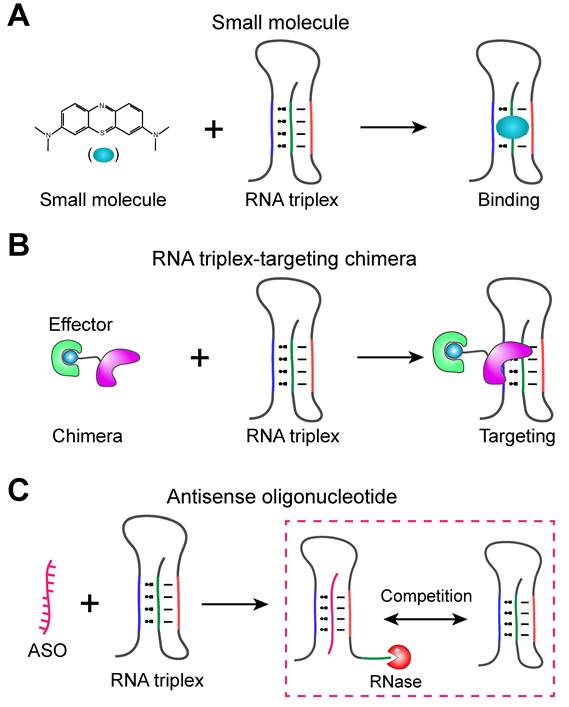
The application of RNA triple helices as therapeutic targets for related diseases is still in its infancy, with several targeting strategies currently in development, among which research on small-molecule drugs based on RNA triple-helical structures has been relatively well-established (Figure 6A). A key challenge in designing small molecules that target RNA triple helices is ensuring their specific binding to RNA triple helices [126].
RNA triple helix acts as therapeutic target. (A) Small molecules directly target RNA triplexes and produce corresponding effects. (B) RNA triplex-targeting chimeras employ molecules that target RNA triplexes as probes and are coupled with effectors that selectively reduce harmful transcripts. (C) ASOs target regions of the U-rich main strand or A-rich complementary strand required for triplex formation, competitively displacing the natural third strand. Among these, small molecules (A) and ASOs (C) have been experimentally verified in RNA triple helices, while RNA triplex-targeting chimeras (B) are mainly derived from other RNA-targeted therapeutic strategies. A strategy similar to it, PINADs, has been experimentally verified in RNA triple helices.
Cancer therapeutic targets
In cancers driven by lncRNAs such as MALAT1 and MENβ that harbor RNA triple helices, these triple-helix motifs enhance transcript stability by shielding the RNA from nuclease-mediated decay, making them highly attractive therapeutic targets. Notably, these lncRNAs do not acquire the canonical 3′-poly(A) tail. Instead, their precursor transcripts are processed by RNase P to generate a mature 3′ end, followed by subsequent processing to produce MALAT1-associated small cytoplasmic RNA (mascRNA) [127]. During maturation, the ENE element containing a U·U base pair folds into an extended and conformationally restricted structural unit. However, the RNA remains vulnerable to exonucleolytic degradation at this stage, indicating that the triple helix is in a metastable state [110]. This U·U base pair thus represents a pharmacological hotspot: rationally designed small molecules can recognize and disrupt this motif [128, 129], thereby impairing triple-helical formation and stability, and consequently reducing steady-state lncRNA accumulation and oncogenic activity. Wilusz et al. demonstrated by constructing expression plasmids that recapitulate the 3′ end processing of MALAT1 that the internal U-loop of the ENE and the short A-rich tract are essential for transcript stability [48]. Accordingly, therapeutic strategies may aim to perturb this region through structural engineering, chemical modification, or small-molecule targeting, thereby disrupting triplex integrity and reducing RNA stability. For instance, incorporating 2′-O-methyl sugar modifications can destabilize the triple helix [130], rendering the RNA susceptible to nuclease-mediated degradation and thereby eliminating oncogenic lncRNAs. Another small-molecule-based approach involves inhibiting other cofactors that associate with the triple helix to block lncRNA function, such as METTL16, which interacts with the aforementioned ENE triple helix and represents a potential therapeutic target [107]. A recent study integrated a structure-based drug discovery (SBDD) pipeline comprising molecular dynamics simulations, pocket dynamics analysis, ensemble docking, and multiple scoring functions to evaluate diminazene ligands targeting the MALAT1 triple helix, identified two potential binding sites, revealed that binding affinity is primarily driven by hydrogen-bonding and electrostatic interactions, yet highlighted the limited predictive accuracy of current computational workflows [131].
Multiple small molecules have emerged as potential therapeutics targeting RNA triple helices (Table 2). Since discovering ligands with sufficient specificity for RNA triple helices is essential for therapeutic development, high-throughput methods, such as small-molecule microarray (SMM) screening, have been employed for initial hit identification [132]. In the case of the MALAT1 triple helix, Abulwerdi et al. identified two imidazole derivatives, compounds 5 and 16, that specifically bind this triple helix through SMM screening and validation in breast tumor organoids [133]. These compounds reduced MALAT1 RNA levels by 54% in breast tumor models without affecting homologous triple-helix-forming RNAs, indicating that they possess a certain degree of selectivity and limited off-target effects [133]. The natural flavonoid quercetin also exhibits similar activity [134]. Several diminazene derivatives targeting the MALAT1 triple helix can achieve bidirectional modulation: they can either stabilize the triplex via hydrogen bonding and electrostatic interactions, reducing nuclease accessibility, or induce conformational rearrangements to expose the poly(A) tail, thereby promoting nuclease recognition [24]. The aromatic heterocycle M5, based on a diazaindene scaffold, can disrupt the stability of the base triple [135, 136]. A recent study developed a time-resolved fluorescence resonance energy transfer (FRET)-based high-throughput screening platform and identified novel small-molecule ligands (kynuramine derivatives) that destabilize the MALAT1 RNA triple helix, providing a new strategy for discovering chemical probes targeting lncRNA triple helices [137]. In addition, clinically used small-molecule drugs can dock directly with RNA triple helices, offering the potential to accelerate translational validation due to their established safety profiles, as exemplified by chlorhexidine and kanamycin; however, further cellular and in vivo testing remains necessary [138]. In contrast, the MENβ triple helix has been less explored: compounds such as emodin, GW5074, and L-798106 bind to the triple helix but exhibit limited inhibitory or toxic effects, highlighting the need for additional screening or conjugation strategies [139]. Reverse transcription quantitative polymerase chain reaction (RT-qPCR) can be used to assess the stability of RNA triple helices, whose Cycle threshold (Ct) is determined by the reverse transcriptase extension efficiency, allowing for the simultaneous detection of both stable and unstable ligands, thereby supporting bidirectional functional interrogation [140]. Mousseau et al. recently conducted a systematic evaluation of eight triplex-targeting molecules, confirming that their primary binding site is the MALAT1 major-groove triple helix rather than adjacent duplex regions. Furthermore, different small molecules display varying sensitivities to triple-helical length, base composition, and A-minor pocket conformation. Flavonoids primarily recognize base composition and A-minor pocket conformation, whereas alkaloids and berenil show a stronger preference for recognizing triple-helical length. Moreover, with the exception of neomycin, most interactions with both precursor and mature MALAT1 conformations are rapid and reversible, suggesting their applicability in dynamic regulatory contexts [141]. Table 2 lists detailed information on small molecules targeting RNA triple helices. Most currently identified triple-helix-targeting small molecules still require further structural refinement or affinity optimization to enhance their potency.
Summary of RNA triple-helix-targeting small molecules (chemical structures, shown in the Figure S1, are ordered alphabetically).
| Small molecule | Targeted triple helix | Effect on triple helix | References |
|---|---|---|---|
| Aromatic heterocyclic compound M5 | MALAT1 triple helix | Disrupting the stability of triple helix | [135, 136] |
| Benzoquinoquinoxaline-neomycin conjugate | U·A-U RNA triple helix | Enhancing the thermal stability of triple helix | [173] |
| Berberine, coralyne, palmatine | U·A-U RNA triple helix | Enhancing the thermal stability of triple helix | [40] |
| Berenil (diminazene aceturate) | U·A-U RNA triple helix | Promoting the thermal stability of triple helix | [7] |
| Chelerythrine | U·A-U RNA triple helix | Increasing binding affinity and the thermal stability | [174] |
| Chlorhexidine, kanamycin | MALAT1 triple helix | Predicted to bind to triple helix | [138] |
| Compound 15 | PAN triple helix and two nucleotides bulge at the bottom | Enhancing resistance to nucleic acid exoribonucleases | [124] |
| Dibenzofuran and its derivatives | MALAT1 triple helix | Selective binding, stabilizing the structure and inhibiting degradation | [15, 175] |
| Diminazene derivatives | MALAT1 triple helix | Bidirectional regulation: Some reduce nuclease accessibility; others promote degradation | [24] |
| Emodin, GW5074, L-798106 | MENβ/NEAT1_2 triple helix or immature fragments | Inhibitory effect decreases progressively | [139] |
| Fisetin | U·A-U RNA triple helix | Enhancing the thermal stability of triple helix | [176] |
| Imidazole compounds 5, 16 | MALAT1 triple helix | Specific binding, MALAT1 RNA decreased in breast tumor models | [133] |
| Kynuramine class of derivatives compounds 7, 10, 15 | MALAT1 triple helix | Destabilizing the triple helix through non-intercalative interactions | [137] |
| Luteolin | U·A-U RNA triple helix | Enhancing the thermal stability of RNA triple helix | [141, 177] |
| Methylene blue | U·A-U RNA triple helix | Stabilizing Watson-Crick strand, destabilizing Hoogsteen strand | [41] |
| Neomycin, gentamicin, paromomycin | U·A-U RNA triple helix | Enhancing the thermal stability of triple helix with neomycin showing the best | [141, 178] |
| N-fused quinazolino-quinazoline-dione 2z | MALAT1 triple helix | Recognizing triple helix UUG pockets; exhibiting toxicity to tumor cells with high expression of MALAT1 | [179] |
| Phenosafranine | U·A-U RNA triple helix | Increasing binding affinity and the thermal stability | [180] |
| PINAD-1 | MALAT1 triple helix | Binder-induced triplex destabilization | [147] |
| Quercetin | MALAT1 triple helix | Inducing structural changes, resulting in decrease in MALAT1 expression | [134] |
| [Ru(bpy)₂(uip)]²+ (Ru1) and [Ru(phen)₂(uip)]²+ (Ru2) | U·A-U RNA triple helix | Enhancing the thermal stability of RNA triple helix | [181] |
| [Ru(dmb)₂(dppz-idzo)]²+ | U·A-U RNA triple helix | Enhancing the thermal stability of RNA triple helix | [42] |
| Sanguinarine | U·A-U RNA triple helix | Enhancing the thermal stability of triple helix | [141, 182] |
| Two types 9-amino alkyl berberine analogs BC1, BC2 | U·A-U RNA triple helix | Increasing binding affinity, improving triple helix stability | [183] |
Emerging strategies that use small molecules as probes to recruit nucleases or degradation effectors, thereby selectively reducing harmful transcripts containing triple helices, are gradually attracting attention. These approaches mainly include RNA-targeting chimeras (RNATACs) (Figure 6B) and Proximity-Induced Nucleic Acid Degraders (PINADs), which are new molecular modalities based on the proximity-induced strategy [142]. PINADs achieve catalytic cleavage by incorporating imidazole groups—which mimic the active site of RNase A—into RNA-targeting small molecules (rSMs) localized to triple helix regions or other specific three-dimensional structures, thereby selectively cleaving abnormally stable or pathogenic RNA triple helices [143-145]. The design of RNATACs is inspired by the principle of Proteolysis Targeting Chimeras (PROTAC), but their targets are RNA rather than proteins. Their basic architecture is a heterobifunctional small molecule: one end binds to a specific RNA structure (e.g., a triple helix), while the other end recruits intracellular RNA effectors (e.g., RNase L, Regnase-1, or SMG6) to mediate degradation [146]. Recently, researchers have reported PINAD-1, the first bifunctional small-molecule degrader targeting the 3′-terminal triple helices of MALAT1, which couples a triplex-binding ligand to an imidazole-based cleavage warhead to achieve proximity-induced and selective MALAT1 degradation, while demonstrating that efficient RNA degradation depends not only on proximity but also on local triplex geometry and binder-induced destabilization [147]. Further optimization of the ligandability and tissue-specific expression of RNA effectors is still required, along with the development of additional usable rSMs.
Antiviral targets
The RNA triple helix within the ENE element of KSHV PAN RNA protects the transcript from host-mediated degradation, while PAN itself drives global lytic gene activation and contributes to evasion of innate immunity, making it essential for efficient lytic replication [119]. Knockdown of the PAN gene at its source suppresses nuclear mRNA export and reduces cytoplasmic accumulation of transcripts encoding late lytic viral proteins [148]. However, deletion of ENE does not directly impair lytic replication but instead increases cytoplasmic RNA levels [47], indicating that direct engineering of the ENE triple helix is not a viable approach. Therefore, efforts should focus on exploring small molecules that target ENE to directly or indirectly modulate the activity of functional RNA, with the potential to obtain KSHV-specific antiviral agents; this strategy may also be applicable to other viral infections involving RNA triple helices or similar structures containing ENEs.
Using the SMM screening strategy, compounds capable of specifically recognizing and binding to RNA triple helices were identified and subjected to biochemical analyses. Ultimately, a small-molecule ligand was identified that specifically binds to both the PAN triple helix and the pocket formed by the AU dinucleotide bulge at its base (Figure 4A) [124]. This compound is likely to disrupt the interaction between the triple helix and the minor capsid protein ORF26, thereby impacting KSHV pathogenesis and presenting a new therapeutic strategy that targets a viral lncRNA instead of a host factor [124]. Notably, such single-stranded RNA bulges forming pocket-like structures are present in many ENE-containing triple helices [14], and they may represent important sites for future small-molecule targeting. These results indicate that the pocket-like bulges at the base of ENE-containing triple helices constitute structurally privileged small-molecule-druggable hotspots, where ligands identified via SMM screening can selectively perturb triplex-protein interactions and potentially destabilize pathogenic viral RNAs such as PAN.
Oligonucleotide therapy
Antisense oligonucleotides (ASOs) are short (typically 12-30 nt) synthetic oligonucleotides that bind to target RNAs (and occasionally DNAs) through Watson-Crick or Hoogsteen base pairing, thereby regulating the processing, stability, or translation of nucleic acids, which in turn alters gene expression or exerts therapeutic effects [149]. ASOs can disrupt RNA's secondary and tertiary structures via competitive binding or by inducing conformational changes, making transcripts more prone to degradation or functional loss [150, 151]. Thus, ASOs can be designed to disrupt or block the formation and stabilization of pathogenic RNA triple helices (Figure 6C). When ASOs are directed to the U-rich main strand or the A-rich complementary strand regions required for triplex formation, they can competitively replace the natural third strand or prevent closure of the triple helix [110]. In MALAT1, ASO-mediated disruption of the triple helix exposes the 3′ end, abolishes ENE protection, and promotes rapid exonucleolytic degradation. Competitive displacement assays further showed that locked nucleic acid (LNA) 15 and its phosphorothioate-modified derivative—phosphorothioate LNA 15 (PS-L15)—can displace A-tract and the RNA triple-helix-binding protein METTL16 from the triple helix, forming an RNA·LNA-RNA triple helix, and markedly reduce MALAT1 and MENβ expression [152], representing a practical dual-target inhibition strategy. These studies establish ASOs (particularly chemically optimized, triplex-targeting designs such as LNA and PS-LNA oligonucleotides) as versatile tools for selectively dismantling pathogenic RNA triple helices, while simultaneously destabilizing oncogenic lncRNAs (such as MALAT1/MENβ) and eliminating toxic repeat-expanded transcripts in neurodegeneration, thereby offering a broadly applicable therapeutic strategy across diverse RNA structure-driven diseases.
In addition, bifacial peptide nucleic acids (bPNAs), which are built on a peptide scaffold attached with bases every two amino acids, can be designed to target RNA triple helices and induce structural displacement, thereby modulating the activity of the parent RNA. This approach exploits the ability of the artificial base melamine to form an UMU base triple with two uridines in the URIL of an RNA triplex [153-155] and competitively displace the natural poly(A) tail [128, 156-158]. Since the melamine base in bPNAs can recognize uridine-rich RNA motifs, this strategy may be applicable to a variety of RNA targets. On this basis, several bPNA scaffolds, including normal peptide, diketopiperazine, and isodipeptide backbones, have been developed to target the MALAT1 triple helix. Among these, the K2M-A-K2M tripeptide (a tripeptide-based bPNA containing four melamine groups, hence also termed 4M bPNA) exhibits the best activity, reducing MALAT1 RNA half-life to 0.5 h and suppressing MALAT1 expression by approximately 50% in PANC-1 pancreatic cancer cells [159]. This strategy provides a new avenue for lncRNA-targeted cancer therapy, particularly for oncogenic transcripts whose stability depends on a triple helix.
Challenges and Strategies
Improving target specificity and avoiding off-target effects
RNA triple helices often contain conserved or repeated nucleoside patterns and interact with other RNAs or proteins within the cell; small molecules or oligonucleotides tend to bind non-specifically to similar sequences or structures, leading to off-target effects [48]. To overcome the low selectivity caused by sequence or structural similarity, a structural recognition approach can be considered instead of relying solely on sequence recognition. This involves utilizing high-resolution structural information to locate specific triple interfaces or peripheral anchoring secondary structures. In drug design, one can consider introducing ligand features capable of recognizing specific sites on triple helices (e.g., ligands that recognize the U·A-U geometric feature unique to triples) [107]. These high-quality structural and dynamic data can significantly enhance the efficiency of selective screening. To address potential hybridization issues between oligonucleotides and non-target transcripts, ASOs can undergo chemical modification and length optimization to improve affinity and reduce non-specific binding. Additionally, in vivo off-target prediction at the sequence level and RNA sequencing (RNA-seq) can be employed to screen candidates [149, 160]. For the screening of highly specific small-molecule ligands, a multimodal screening workflow (high-throughput binding screening → structural validation → intracellular functional validation) can be employed, incorporating bispecific molecules to enhance functional selectivity and direct therapeutic efficacy [142, 161, 162]. Additionally, telomerase and spliceosomal RNA triple helices represent mechanistically compelling but still underexploited therapeutic targets. Although current telomerase-directed and spliceosome-modulating agents mainly act on the hTR template region, telomerase-associated pathways, or spliceosomal protein components rather than the triple helices themselves, these systems provide important precedents for future structure-guided intervention against disease-relevant RNA triplex motifs [163, 164].
Overcoming the dynamics and complexity of RNA structure
The RNA triple helix is not a rigid structure; its folding, formation and disassembly are influenced by post-transcriptional processing, protein binding, modifications, and the ionic environment, leading to variations in the accessibility of target sites under different conditions. If kinetic factors are not taken into account, designed ligands may be ineffective in vivo [110]. Before the RNA triple helix is fully processed, ligand molecules or ASOs can be designed to bind to its folded intermediate states, thereby preventing the triple helix from fully forming and stabilizing, which relies heavily on relevant structural and kinetic data [165]. Establishing drug-binding sites or serving as co-targeting sites through chemical modification is another strategy [55], which can ignore the dynamics of RNA structure to the greatest extent possible. In addition, given that many RNA triple helices and their corresponding RBPs constitute functional units, it is possible to target the interacting RBPs, such as by connecting small molecules to protein degradation modules [142], thereby bypassing the RNA itself and directly intervening in the overall function of the triple helix. Although RNA triple-helix-based drug treatment strategies are diverse, they still face many challenges and require refinement in numerous aspects before they can ultimately be applied clinically to benefit mankind. Moreover, since many RNA triple helices reside within the cell nucleus, current delivery systems face challenges in efficiently and selectively penetrating the nucleus, which further limits the development of therapeutics against these targets [166-168]. Finally, RNA secondary structures play a crucial role in regulating antigen expression and the immunogenic efficacy of mRNA vaccines by modulating translation initiation, transcript stability, and innate immune sensing [36, 37]. Given the reported roles of RNA triple helices in inhibiting RNA decay [16] and promoting both transcription [14] and translation [48], these structures may offer a promising avenue for enhancing RNA stability and translational efficiency in mRNA-based therapeutics. Rosemann et al. showed that Malat1-derived 3′-end motifs, including the triple-helix element, could support translation of synthetic reporter mRNAs in human cells, although they consistently underperformed canonical poly(A) tails, suggesting that triple helices may serve as partial poly(A)-replacement elements but still require further optimization [56].
Conclusions
As an important motif in higher-order nucleic acid structures, research on RNA triple helix is gradually shifting from basic structural studies to interdisciplinary frontier areas such as functional dissection and disease therapy. Over the past several decades, researchers have progressed from the initial discovery of RNA triple helices in artificial systems to the continuous identification of natural examples in viral, prokaryotic, and eukaryotic RNAs. This has gradually revealed the central role of RNA triple helices in key biological processes, including RNA stabilization, transcriptional and translational regulation, maintenance of telomerase activity, ligand recognition and regulation mediated by riboswitches, control of transposition, and RNA splicing. With the rapid development of structural biology and high-throughput techniques, such as cryo-EM, single-molecule FRET, and selective 2′-hydroxyl acylation analyzed by primer extension and mutational profiling (SHAPE-MaP), our understanding of the structural diversity, dynamic equilibria, and biological significance of RNA triple helices has continued to deepen.
At disease level, accumulating evidence indicates that the formation, disruption, or aberrant stabilization of RNA triple helices is closely associated with a variety of pathological processes, including cancer, viral infections, neurodegenerative disorders, and genetic diseases. Therapeutic strategies targeting these structural abnormalities are gradually shifting from functional validation to targeted intervention. Also, small-molecule ligands, antisense oligonucleotides, siRNAs, CRISPR, and emerging RNA degradation technologies are opening new directions for structured RNA-targeting drugs. This marks the transition of RNA triple helices from a concept in structural biology to a potential druggable structural motif.
Overall, RNA triple helices are essential and highly diverse functional elements within gene regulatory networks. Positioned at the intersection of RNA structure and function, they directly encode structural information into RNA folding through processes such as stability regulation, catalytic activity modulation, ligand recognition, and protein recruitment, thereby bridging the gap between basic RNA biology and clinical applications.
Supplementary Material
Supplementary figures and tables.

Acknowledgements
The authors apologize to the many colleagues whose relevant publications we could not cite for reasons of focus and space limitations. We thank the graduate students from Jin Laboratory for their assistance. This work was supported by research grants from the National Key Research and Development Program of China (2023YFC2604300 to Y.J.), the National Natural Science Foundation of China (92581117 to Y.J.), the National Natural Science Foundation of China (32201054 and 32471345 to H.D.), the National Natural Science Foundation of China (32301075 to Z.W.), the Fundamental Research Funds for the Central Universities (No. K20220228 to Y.J.) and Zhejiang Chinese Medical University Scientific Research Project (No. 2024RCZXZK46 to Z.W.).
Author Contributions
Yiqi Ding: Conceptualization; visualization; writing-original draft; writing-review & editing. Zhechao Wang: writing-original draft; writing-review & editing. Zimo Wu: writing-original draft; writing-review & editing. Benyuan Cao: writing-review & editing. Haiyang Dong: writing-original draft; writing-review & editing. Yongfeng Jin: Conceptualization; funding acquisition; supervision; visualization; writing-review.
Competing Interests
The authors have declared that no competing interest exists.
References
1. Felsenfeld G, Davies DR, Rich A. FORMATION OF A THREE-STRANDED POLYNUCLEOTIDE MOLECULE. Journal of the American Chemical Society. 1957;79:2023-4
2. Brown JA. Unraveling the structure and biological functions of RNA triple helices. WIREs RNA. 2020 11
3. Devi G, Zhou Y, Zhong Z, Toh D-FK, Chen G. RNA triplexes: from structural principles to biological and biotech applications. Wiley Interdisciplinary Reviews: RNA. 2015;6:111-28
4. Morgan AR, Wells RD. Specificity of the three-stranded complex formation between double-stranded DNA and single-stranded RNA containing repeating nucleotide sequences. Journal of molecular biology. 1968;37:63-80
5. Kim SH, Suddath FL, Quigley GJ, McPherson A, Sussman JL, Wang AHJ. et al. Three-dimensional tertiary structure of yeast phenylalanine transfer RNA. Science (American Association for the Advancement of Science). 1974;185:435-40
6. Moser HE, Dervan PB. Sequence-Specific Cleavage of Double Helical DNA by Triple Helix Formation. Science (American Association for the Advancement of Science). 1987;238:645-50
7. Pilch DS, Kirolos MA, Breslauer KJ. Berenil binding to higher ordered nucleic acid structures: complexation with a DNA and RNA triple helix. Biochemistry (Easton). 1995;34:16107-24
8. Theimer CA, Blois CA, Feigon J. Structure of the Human Telomerase RNA Pseudoknot Reveals Conserved Tertiary Interactions Essential for Function. Molecular cell. 2005;17:671-82
9. Conrad NK, Shu M-D, Uyhazi KE, Steitz JA. Mutational analysis of a viral RNA element that counteracts rapid RNA decay by interaction with the polyadenylate tail. Proceedings of the National Academy of Sciences - PNAS. 2007;104:10412-7
10. Toor N, Keating KS, Taylor SD, Pyle AM. Crystal Structure of a Self-Spliced Group II Intron. Science (American Association for the Advancement of Science). 2008;320:77-82
11. Mitton-Fry RM, DeGregorio SJ, Wang J, Steitz TA, Steitz JA. Poly(A) Tail Recognition by a Viral RNA Element Through Assembly of a Triple Helix. Science (American Association for the Advancement of Science). 2010;330:1244-7
12. Brown JA, Valenstein ML, Yario TA, Tycowski KT, Steitz JA. Formation of triple-helical structures by the 3′-end sequences of MALAT1 and MENβ noncoding RNAs. Proceedings of the National Academy of Sciences - PNAS. 2012;109:19202-7
13. Brown JA, Bulkley D, Wang J, Valenstein ML, Yario TA, Steitz TA. et al. Structural insights into the stabilization of MALAT1 noncoding RNA by a bipartite triple helix. Nature Structural and Molecular Biology. 2014;21:633-40
14. Tycowski Kazimierz T, Shu M-D, Steitz Joan A. Myriad Triple-Helix-Forming Structures in the Transposable Element RNAs of Plants and Fungi. Cell reports (Cambridge). 2016;15:1266-76
15. Donlic A, Morgan BS, Xu JL, Liu A, Roble C, Hargrove AE. Discovery of Small Molecule Ligands for MALAT1 by Tuning an RNA-Binding Scaffold. Angewandte Chemie International Edition. 2018;57:13242-7
16. Torabi S-F, Vaidya AT, Tycowski KT, DeGregorio SJ, Wang J, Shu M-D. et al. RNA stabilization by a poly(A) tail 3′-end binding pocket and other modes of poly(A)-RNA interaction. Science (American Association for the Advancement of Science). 2021;371:584
17. Sharma S, Mishra S, Gourinath S, Kaushal PS. Cryo-EM structure of ribosome from pathogenic protozoa Entamoeba histolytica reveals unique features of its architecture. Nat Commun. 2025;16:7758
18. Montange RK, Batey RT. Structure of the S-adenosylmethionine riboswitch regulatory mRNA element. Nature. 2006;441:1172-5
19. Conrad NK. The emerging role of triple helices in RNA biology. Wiley Interdiscip Rev RNA. 2014;5:15-29
20. Ruszkowska A, Ruszkowski M, Hulewicz JP, Dauter Z, Brown JA. Molecular structure of a U•A-U-rich RNA triple helix with 11 consecutive base triples. Nucleic Acids Res. 2020;48:3304-14
21. Ghanim GE, Sekne Z, Balch S, van Roon AM, Nguyen THD. 2.7 Å cryo-EM structure of human telomerase H/ACA ribonucleoprotein. Nat Commun. 2024;15:746
22. Kim N-K, Zhang Q, Zhou J, Theimer CA, Peterson RD, Feigon J. Solution Structure and Dynamics of the Wild-type Pseudoknot of Human Telomerase RNA. Journal of molecular biology. 2008;384:1249-61
23. Li H, Wang L, Lei Z, Biacsi AS, Jeong DH, Cho J. RNA triple helix assembled by the poly(A) tail enhances retrotransposon mobilization by preventing RNA deadenylation. Proceedings of the National Academy of Sciences - PNAS. 2025;122:e2510774122
24. Zafferani M, Martyr JG, Muralidharan D, Montalvan NI, Cai Z, Hargrove AE. Multiassay Profiling of a Focused Small Molecule Library Reveals Predictive Bidirectional Modulation of the lncRNA MALAT1 Triplex Stability In Vitro. ACS chemical biology. 2022;17:2437-47
25. Nawrocki EP, Eddy SR. Infernal 1.1: 100-fold faster RNA homology searches. BIOINFORMATICS. 2013;29:2933-5
26. Rakheja I, Bharti V, Sahana S, Das PK, Ranjan G, Kumar A. et al. Development of an In Silico Platform (TRIPinRNA) for the Identification of Novel RNA Intramolecular Triple Helices and Their Validation Using Biophysical Techniques. Biochemistry (Easton). 2025;64:250-65
27. Matarrese MAG, Quadrini M, Luchetti N, Di Petta F, Durante D, Ballarino M. et al. Decoding RNA triple helices: identification from sequence and secondary structure. Brief Bioinform. 2026;27:bbag009
28. Bertram K, Agafonov DE, Liu W-T, Dybkov O, Will CL, Hartmuth K. et al. Cryo-EM structure of a human spliceosome activated for step 2 of splicing. Nature (London). 2017;542:318-23
29. Hang J, Wan R, Yan C, Shi Y. Structural basis of pre-mRNA splicing. Science (American Association for the Advancement of Science). 2015;349:1191-8
30. Peng X, Liao W, Lin X, Lilley DMJ, Huang L. Crystal structures of the NAD+-II riboswitch reveal two distinct ligand-binding pockets. Nucleic Acids Res. 2023;51:2904-14
31. Gilbert SD, Rambo RP, Van Tyne D, Batey RT. Structure of the SAM-II riboswitch bound to S-adenosylmethionine. Nature structural & molecular biology. 2008;15:177-82
32. Pham VV, Salguero C, Khan SN, Meagher JL, Brown WC, Humbert N. et al. HIV-1 Tat interactions with cellular 7SK and viral TAR RNAs identifies dual structural mimicry. Nature communications. 2018;9:4266-12
33. Rakheja I, Panda G, Maiti S, Ray A. Molecular Modeling of Non-Canonical Intramolecular RNA Triple Helix Structures Predicted from TRIPinRNA and Their In Vitro Biophysical Structure Validation. J Phys Chem B. 2025;129:4298-308
34. Fica SM, Mefford MA, Piccirilli JA, Staley JP. Evidence for a group II intron-like catalytic triplex in the spliceosome. Nature structural & molecular biology. 2014;21:464-71
35. Brown JA, Kinzig CG, DeGregorio SJ, Steitz JA. Hoogsteen-position pyrimidines promote the stability and function of the MALAT1 RNA triple helix. RNA (Cambridge). 2016;22:743-9
36. Pardi N, Hogan MJ, Porter FW, Weissman D. mRNA vaccines - a new era in vaccinology. Nature Reviews Drug Discovery. 2018;17:261-79
37. Wayment-Steele HK, Kim DS, Choe CA, Nicol JJ, Wellington-Oguri R, Watkins AM. et al. Theoretical basis for stabilizing messenger RNA through secondary structure design. Nucleic Acids Res. 2021;49:10604-17
38. Zhou Y, Kierzek E, Loo ZP, Antonio M, Yau YH, Chuah YW. et al. Recognition of RNA duplexes by chemically modified triplex-forming oligonucleotides. Nucleic Acids Res. 2013;41:6664-73
39. Mwangi MN, Yonkunas MJ, Ageeli AA, McGovern-Gooch KR, Yilmaz S, Baird NJ. et al. A Newly Identified Peripheral Duplex Anchors and Stabilizes the MALAT1 Triplex. Biochemistry (Easton). 2024;63:2280-92
40. Sinha R, Kumar GS. Interaction of Isoquinoline Alkaloids with an RNA Triplex: Structural and Thermodynamic Studies of Berberine, Palmatine, and Coralyne Binding to Poly(U).Poly(A)Poly(U). The journal of physical chemistry B. 2009;113:13410-20
41. Goswami R, Paul L, Das H, Ghosh A, Chowdhury S, Das S. Spectroscopic and Theoretical Insights into the Binding of Phenothiazinium Dye Methylene Blue with Triple- and Double-Helical Forms of RNA: A Comparative Study. The journal of physical chemistry B. 2025;129:9068-82
42. Wang H, Liu X, Tan L. Binding properties of a molecular “light switch” ruthenium(II) polypyridyl complex toward double- and triple-helical forms of RNA. International journal of biological macromolecules. 2023;242:124710
43. Hoyuelos FJ, García B, Leal JM, Busto N, Biver T, Secco F. et al. RNA triplex-to-duplex and duplex-to-triplex conversion induced by coralyne. Phys Chem Chem Phys. 2014;16:6012-8
44. Conrad NK, Steitz JA. A Kaposi's sarcoma virus RNA element that increases the nuclear abundance of intronless transcripts. The EMBO journal. 2005;24:1831-41
45. Tycowski KT, Shu MD, Borah S, Shi M, Steitz JA. Conservation of a triple-helix-forming RNA stability element in noncoding and genomic RNAs of diverse viruses. Cell Rep. 2012;2:26-32
46. Torabi S-F, Chen Y-L, Zhang K, Wang J, DeGregorio SJ, Vaidya AT. et al. Structural analyses of an RNA stability element interacting with poly(A). Proceedings of the National Academy of Sciences - PNAS. 2021;118:1-11
47. Gutierrez IV, Dayton J, Harger S, Rossetto CC. The Expression and Nuclear Retention Element of Polyadenylated Nuclear RNA Is Not Required for Productive Lytic Replication of Kaposi's Sarcoma-Associated Herpesvirus. Journal of virology. 2021;95:e0009621
48. Wilusz JE, JnBaptiste CK, Lu LY, Kuhn C-D, Joshua-Tor L, Sharp PA. A triple helix stabilizes the 3′ ends of long noncoding RNAs that lack poly(A) tails. Genes & development. 2012;26:2392-407
49. Piórkowska K, Zygmunt K, Hunter W, Wróblewska K. MALAT1: A Long Non-Coding RNA with Multiple Functions and Its Role in Processes Associated with Fat Deposition. Genes (Basel). 2024 15
50. Read GH, McGreevy K, Issawi H, Yang T, Tsang C, Turk IA. et al. Paraspeckles Are Associated with the Activation and Nuclear Localization of Unphosphorylated miR-34a. Noncoding RNA. 2026 12
51. Ilık İ A, Glažar P, Tse K, Brändl B, Meierhofer D, Müller FJ. et al. Autonomous transposons tune their sequences to ensure somatic suppression. Nature. 2024;626:1116-24
52. Dong H, Xu B, Wu L, Wang Z, Li Z, Xie J. et al. Regulatory paradigm of Dscam1 stochastic alternative splicing through conserved long-range RNA structures. Nucleic Acids Res. 2026 54
53. Marzluff WF, Wagner EJ, Duronio RJ. Metabolism and regulation of canonical histone mRNAs: life without a poly(A) tail. Nature reviews Genetics. 2008;9:843-54
54. Xiang K, Bartel DP. The molecular basis of coupling between poly(A)-tail length and translational efficiency. Elife. 2021;10:e66493
55. Schievelbein MJ, Resende C, Glennon MM, Kerosky M, Brown JA. Global RNA modifications to the MALAT1 triple helix differentially affect thermostability and weaken binding to METTL16. The Journal of biological chemistry. 2024;300:105548
56. Rosemann J, Jahr GT, Macho J, Haemmerle M, Gutschner T. Evaluating Malat1-derived 3'end sequences for functional transgene expression in human cells using synthetic mRNA delivery. RNA Biol. 2026;23:1-12
57. Chen G, Chang KY, Chou MY, Bustamante C, Tinoco I Jr. Triplex structures in an RNA pseudoknot enhance mechanical stability and increase efficiency of -1 ribosomal frameshifting. Proc Natl Acad Sci U S A. 2009;106:12706-11
58. McCown PJ, Corbino KA, Stav S, Sherlock ME, Breaker RR. Riboswitch diversity and distribution. RNA (Cambridge). 2017;23:995-1011
59. Breaker RR. Riboswitches and Translation Control. Cold Spring Harbor perspectives in biology. 2018;10:a032797
60. Breaker RR. The Biochemical Landscape of Riboswitch Ligands. Biochemistry (Easton). 2022;61:137-49
61. Salvail H, Breaker RR. Riboswitches. Current biology. 2023;33:R343-R8
62. Liberman JA, Suddala KC, Aytenfisu A, Chan D, Belashov IA, Salim M. et al. Structural analysis of a class III preQ₁ riboswitch reveals an aptamer distant from a ribosome-binding site regulated by fast dynamics. Proceedings of the National Academy of Sciences - PNAS. 2015;112:E3485-E94
63. Huang L, Wang J, Wilson TJ, Lilley DMJ. Structure of the Guanidine III Riboswitch. CELL CHEMICAL BIOLOGY. 2017;24:1407-15.e2
64. Huang L, Lilley DMJ. Structure and ligand binding of the SAM-V riboswitch. Nucleic acids research. 2018;46:6869-79
65. Xu X, Egger M, Li C, Chen H, Micura R, Ren A. Structure-based investigations of the NAD+-II riboswitch. Nucleic Acids Res. 2023;51:54-67
66. Liberman JA, Salim M, Krucinska J, Wedekind JE. Structure of a class II preQ1 riboswitch reveals ligand recognition by a new fold. Nature chemical biology. 2013;9:353-5
67. Kang M, Eichhorn CD, Feigon J. Structural determinants for ligand capture by a class II preQ₁ riboswitch. Proceedings of the National Academy of Sciences - PNAS. 2014;111:E663-E71
68. Dutta D, Wedekind JE. Nucleobase mutants of a bacterial preQ1-II riboswitch that uncouple metabolite sensing from gene regulation. The Journal of biological chemistry. 2020;295:2555-67
69. Lee ER, Baker JL, Weinberg Z, Sudarsan N, Breaker RR. An Allosteric Self-Splicing Ribozyme Triggered by a Bacterial Second Messenger. Science (American Association for the Advancement of Science). 2010;329:845-8
70. Sherlock ME, Breaker RR. Biochemical Validation of a Third Guanidine Riboswitch Class in Bacteria. Biochemistry (Easton). 2017;56:359-63
71. Van Vlack ER, Topp S, Seeliger JC. Characterization of Engineered PreQ1 Riboswitches for Inducible Gene Regulation in Mycobacteria. Journal of bacteriology. 2017;199:E00656
72. Dutta D, Belashov IA, Wedekind JE. Coupling Green Fluorescent Protein Expression with Chemical Modification to Probe Functionally Relevant Riboswitch Conformations in Live Bacteria. Biochemistry (Easton). 2018;57:4620-8
73. Neuner E, Frener M, Lusser A, Micura R. Superior cellular activities of azido- over amino-functionalized ligands for engineered preQ1 riboswitches in E.coli. RNA biology. 2018;15:1376-83
74. Chauvier A, Dandpat SS, Romero R, Walter NG. A nascent riboswitch helix orchestrates robust transcriptional regulation through signal integration. Nat Commun. 2024;15:3955
75. Zhang H, Lang Z, Zhu J-K. Dynamics and function of DNA methylation in plants. Nature reviews Molecular cell biology. 2018;19:489-506
76. Wells JN, Feschotte C. A Field Guide to Eukaryotic Transposable Elements. Annual Review of Genetics. 2020;54:539-61
77. Matzke MA, Mosher RA. RNA-directed DNA methylation: an epigenetic pathway of increasing complexity. Nature reviews Genetics. 2014;15:394-408
78. Kim EY, Wang L, Lei Z, Li H, Fan W, Cho J. Ribosome stalling and SGS3 phase separation prime the epigenetic silencing of transposons. Nature plants. 2021;7:303-9
79. Wang L, Li H, Lei Z, Jeong DH, Cho J. The CARBON CATABOLITE REPRESSION 4A-mediated RNA deadenylation pathway acts on the transposon RNAs that are not regulated by small RNAs. The New phytologist. 2024;241:1636-45
80. Wu RA, Upton HE, Vogan JM, Collins K. Telomerase Mechanism of Telomere Synthesis. Annual Review of Biochemistry. 2017;86:439-60
81. Dey A, Monroy-Eklund A, Klotz K, Saha A, Davis J, Li B. et al. In vivo architecture of the telomerase RNA catalytic core in Trypanosoma brucei. Nucleic Acids Res. 2021;49:12445-66
82. Cash DD, Cohen-Zontag O, Kim N-K, Shefer K, Brown Y, Ulyanov NB. et al. Pyrimidine motif triple helix in the Kluyveromyces lactis telomerase RNA pseudoknot is essential for function in vivo. Proceedings of the National Academy of Sciences - PNAS. 2013;110:10970-5
83. Cash DD, Feigon J. Structure and folding of the Tetrahymena telomerase RNA pseudoknot. Nucleic acids research. 2017;45:482-95
84. Shefer K, Brown Y, Gorkovoy V, Nussbaum T, Ulyanov NB, Tzfati Y. A Triple Helix within a Pseudoknot Is a Conserved and Essential Element of Telomerase RNA. Molecular and Cellular Biology. 2007;27:2130-43
85. Qiao F, Cech TR. Triple-helix structure in telomerase RNA contributes to catalysis. Nature structural & molecular biology. 2008;15:634-40
86. Mihalusova M, Wu JY, Zhuang X. Functional importance of telomerase pseudoknot revealed by single-molecule analysis. Proceedings of the National Academy of Sciences - PNAS. 2011;108:20339-44
87. Nguyen THD, Tam J, Wu RA, Greber BJ, Toso D, Nogales E. et al. Cryo-EM structure of substrate-bound human telomerase holoenzyme. Nature (London). 2018;557:190-5
88. Huang J, Brown AF, Wu J, Xue J, Bley CJ, Rand DP. et al. Structural basis for protein-RNA recognition in telomerase. Nature structural & molecular biology. 2014;21:507-12
89. Tseng C-K, Wang H-F, Schroeder MR, Baumann P. The H/ACA complex disrupts triplex in hTR precursor to permit processing by RRP6 and PARN. Nature communications. 2018;9:5430-12
90. Kao TL, Huang YC, Chen YH, Baumann P, Tseng CK. LARP3, LARP7, and MePCE are involved in the early stage of human telomerase RNA biogenesis. Nat Commun. 2024;15:5955
91. Galej WP, Toor N, Newman AJ, Nagai K. Molecular Mechanism and Evolution of Nuclear Pre-mRNA and Group II Intron Splicing: Insights from Cryo-Electron Microscopy Structures. Chemical reviews. 2018;118:4156-76
92. Smathers CM, Robart AR. The mechanism of splicing as told by group II introns: Ancestors of the spliceosome. Biochimica et biophysica acta Gene regulatory mechanisms. 2019;1862:194390
93. Marcia M, Pyle AM. Visualizing Group II Intron Catalysis through the Stages of Splicing. Cell. 2012;151:497-507
94. Chan RT, Peters JK, Robart AR, Wiryaman T, Rajashankar KR, Toor N. et al. Structural basis for the second step of group II intron splicing. Nature communications. 2018;9:4676-10
95. Haack DB, Yan X, Zhang C, Hingey J, Lyumkis D, Baker TS. et al. Cryo-EM Structures of a Group II Intron Reverse Splicing into DNA. Cell. 2019;178:612-23.e12
96. Costa M. Group II Introns: Flexibility and Repurposing. Front Mol Biosci. 2022;9:916157
97. Gordon PM, Piccirilli JA. Metal ion coordination by the AGC triad in domain 5 contributes to group II intron catalysis. Nature Structural Biology. 2001;8:893-8
98. Gordon PM, Fong R, Piccirilli JA. A Second Divalent Metal Ion in the Group II Intron Reaction Center. Chemistry & biology. 2007;14:607-12
99. Xu L, Liu T, Chung K, Pyle AM. Structural insights into intron catalysis and dynamics during splicing. Nature. 2023;624:682-8
100. Wilkinson ME, Charenton C, Nagai K. RNA Splicing by the Spliceosome. Annual Review of Biochemistry. 2020;89:359-88
101. Nuadthaisong J, Phetruen T, Techawisutthinan C, Chanarat S. Insights into the Mechanism of Pre-mRNA Splicing of Tiny Introns from the Genome of a Giant Ciliate Stentor coeruleus. Int J Mol Sci. 2022 23
102. Wan R, Yan C, Bai R, Huang G, Shi Y. Structure of a yeast catalytic step I spliceosome at 3.4 Å resolution. Science (American Association for the Advancement of Science). 2016;353:895-904
103. Yan C, Wan R, Bai R, Huang G, Shi Y. Structure of a yeast activated spliceosome at 3.5 Å resolution. Science (American Association for the Advancement of Science). 2016;353:904-11
104. Bai R, Wan R, Wang L, Xu K, Zhang Q, Lei J. et al. Structure of the activated human minor spliceosome. Science. 2021;371:eabg0879
105. Schmitzová J, Cretu C, Dienemann C, Urlaub H, Pena V. Structural basis of catalytic activation in human splicing. Nature. 2023;617:842-50
106. Parker MT, Soanes BK, Kusakina J, Larrieu A, Knop K, Joy N. et al. m(6)A modification of U6 snRNA modulates usage of two major classes of pre-mRNA 5' splice site. Elife. 2022;11:e78808
107. Brown JA, Kinzig CG, DeGregorio SJ, Steitz JA. Methyltransferase-like protein 16 binds the 3′-terminal triple helix of MALAT1 long noncoding RNA. Proceedings of the National Academy of Sciences - PNAS. 2016;113:14013-8
108. Genna V, Portella G, Sala A, Terrazas M, Serrano-Chacón I, González J. et al. Systematic study of hybrid triplex topology and stability suggests a general triplex-mediated regulatory mechanism. Nucleic Acids Res. 2025;53:gkaf170
109. Forino NM, Woo JZ, Zaug AJ, Jimenez AG, Edelson E, Cech TR. et al. Telomerase RNA structural heterogeneity in living human cells detected by DMS-MaPseq. Nat Commun. 2025;16:925
110. Ageeli AA, McGovern-Gooch KR, Kaminska MM, Baird NJ. Finely tuned conformational dynamics regulate the protective function of the lncRNA MALAT1 triple helix. Nucleic acids research. 2019;47:1468-81
111. Wang X, Liu C, Zhang S, Yan H, Zhang L, Jiang A. et al. N(6)-methyladenosine modification of MALAT1 promotes metastasis via reshaping nuclear speckles. Dev Cell. 2021;56:702-15.e8
112. Hou J, Zhang G, Wang X, Wang Y, Wang K. Functions and mechanisms of lncRNA MALAT1 in cancer chemotherapy resistance. Biomark Res. 2023;11:23
113. Yadav A, Biswas T, Praveen A, Ganguly P, Bhattacharyya A, Verma A. et al. Targeting MALAT1 Augments Sensitivity to PARP Inhibition by Impairing Homologous Recombination in Prostate Cancer. Cancer Res Commun. 2023;3:2044-61
114. Weghorst F, Torres Marcén M, Faridi G, Lee YCG, Cramer KS. Deep Conservation and Unexpected Evolutionary History of Neighboring lncRNAs MALAT1 and NEAT1. Journal of Molecular Evolution. 2024;92:30-41
115. Maciejowski J, de Lange T. Telomeres in cancer: tumour suppression and genome instability. Nat Rev Mol Cell Biol. 2017;18:175-86
116. Shay JW, Wright WE. Telomeres and telomerase: three decades of progress. Nat Rev Genet. 2019;20:299-309
117. Armanios M, Blackburn EH. The telomere syndromes. Nature reviews Genetics. 2012;13:693-704
118. Grill S, Nandakumar J. Molecular mechanisms of telomere biology disorders. J Biol Chem. 2021;296:100064
119. Conrad NK. New insights into the expression and functions of the Kaposi's sarcoma-associated herpesvirus long noncoding PAN RNA. Virus Research. 2016;212:53-63
120. Kulkarni V, Jayakumar S, Mohan M, Kulkarni S. Aid or Antagonize: Nuclear Long Noncoding RNAs Regulate Host Responses and Outcomes of Viral Infections. Cells. 2023;12:987
121. Tycowski Kazimierz T, Shu M-D, Borah S, Shi M, Steitz Joan A. Conservation of a Triple-Helix-Forming RNA Stability Element in Noncoding and Genomic RNAs of Diverse Viruses. Cell reports (Cambridge). 2012;2:26-32
122. Rossetto CC, Pari GS. PAN's Labyrinth: Molecular biology of Kaposi's sarcoma-associated herpesvirus (KSHV) PAN RNA, a multifunctional long noncoding RNA. Viruses. 2014;6:4212-26
123. Hancock MH, Skalsky RL. Roles of Non-coding RNAs During Herpesvirus Infection. Curr Top Microbiol Immunol. 2018;419:243-80
124. Swain M, Ageeli AA, Kasprzak WK, Li M, Miller JT, Sztuba-Solinska J. et al. Dynamic bulge nucleotides in the KSHV PAN ENE triple helix provide a unique binding platform for small molecule ligands. Nucleic acids research. 2021;49:13179-93
125. Fernández-Varas B, Manguan-García C, Rodriguez-Centeno J, Mendoza-Lupiáñez L, Calatayud J, Perona R. et al. Clinical mutations in the TERT and TERC genes coding for telomerase components induced oxidative stress, DNA damage at telomeres and cell apoptosis besides decreased telomerase activity. Human molecular genetics. 2024;33:818-34
126. Velagapudi SP, Cameron MD, Haga CL, Rosenberg LH, Lafitte M, Duckett DR. et al. Design of a small molecule against an oncogenic noncoding RNA. Proceedings of the National Academy of Sciences - PNAS. 2016;113:5898-903
127. Wilusz JE, Freier SM, Spector DL. 3′ End Processing of a Long Nuclear-Retained Noncoding RNA Yields a tRNA-like Cytoplasmic RNA. Cell. 2008;135:919-32
128. Xia X, Piao X, Bong D. Bifacial Peptide Nucleic Acid as an Allosteric Switch for Aptamer and Ribozyme Function. Journal of the American Chemical Society. 2014;136:7265-8
129. Yonkunas MJ, Baird NJ. A highly ordered, nonprotective MALAT1 ENE structure is adopted prior to triplex formation. Rna. 2019;25:975-84
130. Zhou Y, Kierzek E, Loo ZP, Antonio M, Yau YH, Chuah YW. et al. Recognition of RNA duplexes by chemically modified triplex-forming oligonucleotides. Nucleic acids research. 2013;41:6664-73
131. Aguti R, Bernetti M, Elisi GM, Cavalli A, Masetti M. Critical Assessment of a Structure-Based Pipeline for Targeting the Long Noncoding RNA MALAT1. J Chem Inf Model. 2026;66:4187-201
132. Connelly CM, Abulwerdi FA, Schneekloth JS. Discovery of RNA Binding Small Molecules Using Small Molecule Microarrays. In: Uttamchandani M, Yao SQ, editors. Methods in Molecular Biology. New York, NY: Springer New York. 2017 p. 157-75
133. Abulwerdi FA, Xu W, Ageeli AA, Yonkunas MJ, Arun G, Nam H. et al. Selective Small-Molecule Targeting of a Triple Helix Encoded by the Long Noncoding RNA, MALAT1. ACS chemical biology. 2019;14:223-35
134. Rakheja I, Ansari AH, Ray A, Chandra Joshi D, Maiti S. Small molecule quercetin binds MALAT1 triplex and modulates its cellular function. Molecular therapy Nucleic acids. 2022;30:241-56
135. François-Moutal L, Miranda VG, Mollasalehi N, Gokhale V, Khanna M. In Silico Targeting of the Long Noncoding RNA MALAT1. ACS medicinal chemistry letters. 2021;12:915-21
136. Rocca R, Polerà N, Juli G, Grillone K, Maruca A, Martino MTD. et al. Hit identification of novel small molecules interfering with MALAT1 triplex by a structure-based virtual screening. Archiv der Pharmazie (Weinheim). 2023;356:e2300134
137. Pernak M, Fleurisson C, Delorme C, Moumné R, Benedetti E, Micouin L. et al. Development of Comprehensive Screening and Assessment Assays for Small-Molecule Ligands of MALAT1 lncRNA. ACS Chem Biol. 2025;20:1068-76
138. Rocca R, Alcaro S, Artese A. Structure-based virtual screening and molecular dynamics simulations of FDA-approved drugs targeting MALAT1. Medicinal Chemistry Research. 2024;33:2095-100
139. An H, Elvers KT, Gillespie JA, Jones K, Atack JR, Grubisha O. et al. A toolkit for the identification of NEAT1_2/paraspeckle modulators. Nucleic acids research. 2022;50:e119-e
140. Zafferani M, Muralidharan D, Montalvan NI, Hargrove AE. RT-qPCR as a screening platform for mutational and small molecule impacts on structural stability of RNA tertiary structures. RSC Chem Biol. 2022;3:905-15
141. Mousseau MM, Shivakumar KM, Yoo J, Brown JA. Eight Triplex-Binding Molecules from Four Chemical Classes Broadly Recognize the MALAT1 Triple Helix. Molecules (Basel, Switzerland). 2025;30:4277
142. Song Y, Cui J, Zhu J, Kim B, Kuo M-L, Potts PR. RNATACs: Multispecific small molecules targeting RNA by induced proximity. Cell chemical biology. 2024;31:1101-17
143. Fouace S, Gaudin C, Picard S, Corvaisier S, Renault J, Carboni B. et al. Polyamine derivatives as selective RNaseA mimics. Nucleic acids research. 2004;32:151-7
144. Gamble C, Trotard M, Seyec JL, Abreu-Guerniou V, Gernigon N, Berrée F. et al. Antiviral effect of ribonuclease conjugated oligodeoxynucleotides targeting the IRES RNA of the hepatitis C virus. Bioorganic & medicinal chemistry letters. 2009;19:3581-5
145. Mikutis S, Rebelo M, Yankova E, Gu M, Tang C, Coelho AR. et al. Proximity-Induced Nucleic Acid Degrader (PINAD) Approach to Targeted RNA Degradation Using Small Molecules. ACS central science. 2023;9:892-904
146. Liu X, Ciulli A. Proximity-Based Modalities for Biology and Medicine. ACS central science. 2023;9:1269-84
147. Brega CAT, Craig BA, Mikutis S, Proctor RSJ, Vyborna Y, Ivey G. et al. A Bifunctional Small Molecule Degrader of the Long Noncoding RNA MALAT1 Triplex. Chemistry. 2026;32:e00025
148. Withers JB, Li ES, Vallery TK, Yario TA, Steitz JA. Two herpesviral noncoding PAN RNAs are functionally homologous but do not associate with common chromatin loci. PLoS pathogens. 2018;14:e1007389
149. Moumné L, Marie A-C, Crouvezier N. Oligonucleotide Therapeutics: From Discovery and Development to Patentability. Pharmaceutics. 2022;14:260
150. Li Y, Garcia G Jr, Arumugaswami V, Guo F. Structure-based design of antisense oligonucleotides that inhibit SARS-CoV-2 replication. bioRxiv. 2021
151. Zhu C, Lee JY, Woo JZ, Xu L, Wrynla XH, Yamashiro LH. et al. An intranasal ASO therapeutic targeting SARS-CoV-2. Nat Commun. 2022;13:4503
152. Shivakumar KM, Mahendran G, Brown JA. Locked Nucleic Acid Oligonucleotides Facilitate RNA•LNA-RNA Triple-Helix Formation and Reduce MALAT1 Levels. International journal of molecular sciences. 2024;25:1630
153. Branda N, Kurz G, Lehn J-M. JANUS WEDGES: a new approach towards nucleobase-pair recognition. Chemical communications (Cambridge, England). 1996:2443-4
154. Marchi-Artzner V, Jullien L, Gulik-Krzywicki T, Lehn J-M. Molecular recognition induced aggregation and fusion between vesicles containing lipids bearing complementary hydrogen bonding head-groups. Chemical communications (Cambridge, England). 1997:117-8
155. Chen H, Meena, McLaughlin LW. A Janus-Wedge DNA Triplex with A-W1-T and G-W2-C Base Triplets. Journal of the American Chemical Society. 2008;130:13190-1
156. Zeng Y, Pratumyot Y, Piao X, Bong D. Discrete Assembly of Synthetic Peptide-DNA Triplex Structures from Polyvalent Melamine-Thymine Bifacial Recognition. Journal of the American Chemical Society. 2012;134:832-5
157. Piao X, Xia X, Bong D. Bifacial Peptide Nucleic Acid Directs Cooperative Folding and Assembly of Binary, Ternary, and Quaternary DNA Complexes. Biochemistry (Easton). 2013;52:6313-23
158. Mao J, DeSantis C, Bong D. Small Molecule Recognition Triggers Secondary and Tertiary Interactions in DNA Folding and Hammerhead Ribozyme Catalysis. Journal of the American Chemical Society. 2017;139:9815-8
159. Miao S, Bhunia D, Devari S, Liang Y, Munyaradzi O, Rundell S. et al. Bifacial PNAs Destabilize MALAT1 by 3′ A-Tail Displacement from the U-Rich Internal Loop. ACS chemical biology. 2021;16:1600-9
160. Egli M, Manoharan M. Chemistry, structure and function of approved oligonucleotide therapeutics. Nucleic acids research. 2023;51:2529-73
161. Costales MG, Aikawa H, Li Y, Childs-Disney JL, Abegg D, Hoch DG. et al. Small-molecule targeted recruitment of a nuclease to cleave an oncogenic RNA in a mouse model of metastatic cancer. Proceedings of the National Academy of Sciences - PNAS. 2020;117:2406-11
162. Meyer SM, Tanaka T, Zanon PRA, Baisden JT, Abegg D, Yang X. et al. DNA-Encoded Library Screening To Inform Design of a Ribonuclease Targeting Chimera (RiboTAC). Journal of the American Chemical Society. 2022;144:21096-102
163. Gómez Pinto I, Guilbert C, Ulyanov NB, Stearns J, James TL. Discovery of Ligands for a Novel Target, the Human Telomerase RNA, Based on Flexible-Target Virtual Screening and NMR. Journal of Medicinal Chemistry. 2008;51:7205-15
164. Seiler M, Yoshimi A, Darman R, Chan B, Keaney G, Thomas M. et al. H3B-8800, an orally available small-molecule splicing modulator, induces lethality in spliceosome-mutant cancers. Nat Med. 2018;24:497-504
165. Aguilar R, Mardones C, Moreno AA, Cepeda-Plaza M. A guide to RNA structure analysis and RNA-targeting methods. The FEBS Journal. 2025;292:2751-66
166. Cox A, Lim SA, Chung EJ. Strategies to deliver RNA by nanoparticles for therapeutic potential. Mol Aspects Med. 2022;83:100991
167. Nie Y, Fu G, Leng Y. Nuclear Delivery of Nanoparticle-Based Drug Delivery Systems by Nuclear Localization Signals. Cells. 2023;12:1637
168. Berger S, Lächelt U, Wagner E. Dynamic carriers for therapeutic RNA delivery. Proceedings of the National Academy of Sciences. 2024;121:e2307799120
169. Yan C, Hang J, Wan R, Huang M, Wong CC, Shi Y. Structure of a yeast spliceosome at 3.6-angstrom resolution. Science. 2015;349:1182-91
170. Smith KD, Shanahan CA, Moore EL, Simon AC, Strobel SA, Feigon J. et al. Structural basis of differential ligand recognition by two classes of bis-(3'-5')-cyclic dimeric guanosine monophosphate-binding riboswitches. Proceedings of the National Academy of Sciences - PNAS. 2011;108:7757-62
171. Qu G, Kaushal PS, Wang J, Shigematsu H, Piazza CL, Agrawal RK. et al. Structure of a group II intron in complex with its reverse transcriptase. Nat Struct Mol Biol. 2016;23:549-57
172. Jiang J, Chan H, Cash DD, Miracco EJ, Ogorzalek Loo RR, Upton HE. et al. Structure of Tetrahymena telomerase reveals previously unknown subunits, functions, and interactions. Science. 2015;350:aab4070
173. Arya DP, Xue L, Tennant P. Combining the Best in Triplex Recognition: Synthesis and Nucleic Acid Binding of a BQQ-Neomycin Conjugate. Journal of the American Chemical Society. 2003;125:8070-1
174. Haque L, Pradhan AB, Bhuiya S, Das S. Exploring the comparative binding aspects of benzophenanthridine plant alkaloid chelerythrine with RNA triple and double helices: a spectroscopic and calorimetric approach. Physical chemistry chemical physics: PCCP. 2015;17:17202-13
175. Donlic A, Zafferani M, Padroni G, Puri M, Hargrove AE. Regulation of MALAT1 triple helix stability and in vitro degradation by diphenylfurans. Nucleic acids research. 2020;48:7653-64
176. Bhuiya S, Haque L, Goswami R, Das S. Multispectroscopic and Theoretical Exploration of the Comparative Binding Aspects of Bioflavonoid Fisetin with Triple- and Double-Helical Forms of RNA. The journal of physical chemistry B. 2017;121:11037-52
177. Tiwari R, Haque L, Bhuiya S, Das S. Third strand stabilization of poly(U)·poly(A) poly(U) triplex by the naturally occurring flavone luteolin: A multi-spectroscopic approach. International journal of biological macromolecules. 2017;103:692-700
178. Arya DP, Coffee RL, Willis B, Abramovitch AI. Aminoglycoside-Nucleic Acid Interactions: Remarkable Stabilization of DNA and RNA Triple Helices by Neomycin. Journal of the American Chemical Society. 2001;123:5385-95
179. Pathi VB, Das P, Guin A, Debnath M, Banerji B. Metal-free synthesis of N-fused quinazolino-quinazoline-diones as a MALAT1 RNA triple helix intercalator. RSC Med Chem. 2025;16:429-34
180. Pradhan AB, Mondal HK, Haque L, Bhuiya S, Das S. An overview on the interaction of phenazinium dye phenosafranine to RNA triple and double helices. International journal of biological macromolecules. 2016;86:345-51
181. He X-J, Tan L-F. Interactions of Octahedral Ruthenium(II) Polypyridyl Complexes with the RNA Triplex poly(U)•poly(A)poly(U) Effect on the Third-Strand Stabilization. Inorganic chemistry. 2014;53:11152-9
182. Das S, Kumar GS, Ray A, Maiti M. Spectroscopic and Thermodynamic Studies on the Binding of Sanguinarine and Berberine to Triple and Double Helical DNA and RNA Structures. Journal of biomolecular structure & dynamics. 2003;20:703-13
183. Bhowmik D, Das S, Hossain M, Haq L, Suresh Kumar G. Biophysical Characterization of the Strong Stabilization of the RNA Triplex poly(U)•poly(A)poly(U) by 9-O-(ω-amino) Alkyl Ether Berberine Analogs. PloS one. 2012;7:e37939
Author contact
Corresponding authors: Haiyang Dong (dong.hyedu.cn); Yongfeng Jin (jinyfedu.cn)