Impact Factor ISSN: 1449-2288




Int J Biol Sci 2026; 22(14):7800-7826. doi:10.7150/ijbs.136955 This issue Cite
Review
Bioactive Constituents of Ginseng in Cancer Therapy: Molecular Mechanisms, Translational Potential, and Advanced Delivery Strategies
Jia-hui Li1, Yan-fang Xian2, De-wen Liu3,  , Hong-yuan Li1, , Wei Li1,
1. College of Chinese Medicinal Materials, Jilin Provincial International Joint Research Center for the Development and Utilization of Authentic Medicinal Materials, Jilin Agricultural University, Changchun 130118, China.
2. School of Chinese Medicine, Faculty of Medicine, The Chinese University of Hong Kong, Shatin, N.T., Hong Kong SAR, China.
3. Institute of Chinese Materia Medica, China Academy of Chinese Medical Sciences, Beijing 100700, China.
Received 2026-4-28; Accepted 2026-8-15; Published 2026-9-3
Abstract
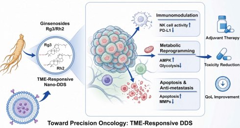
Cancer remains a major global health challenge, highlighting the urgent need to develop safer and more effective therapeutic drugs. Ginseng (Panax ginseng C.A. Mey.), as a medicinal plant widely used in traditional Chinese medicine, has attracted much attention for its extensive anticancer potential and a variety of biologically active ingredients (especially ginseng saponins and polysaccharides). This article reviews the latest research progress on the pharmacological activity and molecular mechanism of ginseng extract in cancer treatment, focusing on its inhibition of tumor growth, inducing cell apoptosis, inhibiting epithelial-mesenchymal transformation and metastasis, reshaping the tumor microenvironment, enhancing antitumor immunity, regulating metabolic reprogramming, and regulating epigenetics. In addition, this article also explores their potential to overcome multi-drug resistance and enhance the efficacy of traditional chemotherapy drugs. In recent years, a variety of drug delivery systems, including polymer nanoparticles, lipids, and micelles, have been developed to improve the bioavailability, stability, and tumor targeting of these compounds. This article also focuses on the current progress and challenges in clinical transformation, especially in the development and application of ginseng saponin preparations. In a word, this article provides a comprehensive review of ginseng-derived drugs from the perspective of pharmacology and translational medicine, and looks forward to the future direction of their reasonable development in cancer treatment.
Keywords: ginseng, ginsenosides, cancer therapy, anticancer mechanisms, drug delivery, translational potential
1. Introduction
Cancer is one of the leading causes of morbidity and mortality worldwide, and is still a huge public health and socio-economic burden. It is caused by the accumulation of genetic and epigenetic changes in somatic cells, leading to uncontrolled proliferation, local invasion, and metastatic diffusion[1]. In 2022, an estimated 20 million new cancer cases and 9.7 million cancer-related deaths were reported globally, and the annual number of new cases is projected to reach 35 million by 2050[2]. Common types of cancer include breast cancer, lung cancer, colorectal cancer (CRC), and prostate cancer (PCa), which still account for a large proportion of cancer incidence and mortality. In view of the increasing global burden of cancer, the development of safe and effective therapeutic drugs remains a top priority in the field of oncology[3].
Traditional treatment strategies, including chemotherapy and radiotherapy, have improved the clinical efficacy of many cancers, but their application is often limited by systemic toxicity, treatment-related complications, and drug resistance[4-7]. In recent years, immunotherapy and targeted therapy have expanded the field of treatment; however, these methods still face many challenges, such as high cost, limited patient response, adverse reactions, and acquired drug resistance[8]. These limitations have prompted people to continue to pay attention to new anticancer drugs and complementary treatment strategies that are safer, more applicable, and more flexible.
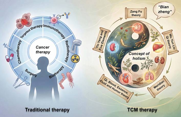
Natural products have long served as an important source of anticancer agents, and traditional Chinese medicine (TCM) has received increasing attention in this context[9]. Owing to its multitarget characteristics and systemic mode of action, TCM has been explored not only as a source of bioactive compounds, but also as an adjunctive approach for alleviating treatment-related toxicity, improving quality of life, and supporting comprehensive cancer management[10, 11] (Fig. 1). More and more evidence shows that natural products derived from TCM can interfere with many aspects of tumor progression, including proliferation, apoptosis, angiogenesis, metastasis and tumor microenvironment regulation[12-14]. In particular, Scutellaria baicalensis Georgi (Huangqin), Sophora flavescens Ait. (Kushen), Salvia miltiorrhiza Bunge (Danshen), Ganoderma lucidum (Curtis) P. Karst. (Lingzhi), and Curcuma longa L. (Jianghuang) have exhibited notable anticancer potential in various in vitro and in vivo experimental models[15-19]. These medicinal plants provide important resources and a theoretical basis for the development of novel antitumor agents.
Comparison between traditional and TCM treatments for cancer.
Among these candidates, ginseng (Panax ginseng C.A. Mey.) has attracted much attention because of its broad-spectrum anticancer properties. Traditional pharmacology shows that ginseng has the effect of toning qi and blood, stabilizing the pulse, nourishing the spleen and nourishing the lungs, and nourishing the blood[20, 21]. As a highly valued herb in TCM, ginseng and its active components, such as ginsenosides, polysaccharides, and volatile oils, have been reported to regulate multiple biological processes involved in cancer development, including cell proliferation, metabolism, migration, invasion, and angiogenesis[22, 23]. In particular, ginsenosides and ginseng polysaccharides (GPS) have shown great potential in regulating tumor immunity, enhancing immune surveillance, and reducing toxic side effects associated with traditional anticancer therapies (such as bone marrow suppression and organ damage)[24, 25]. This dual potential of both antitumor activity and auxiliary therapeutic effect makes ginseng a promising candidate drug in comprehensive cancer treatment.
Recently, several reviews have discussed specific applications of ginseng-derived compounds in oncology, including their combination with chemotherapy[26], potential role in cancer cachexia[27], activity against cancer stem cells[28], and application in ginseng-based nanotherapeutic systems[29]. These studies have substantially expanded our understanding of individual therapeutic contexts. Even so, a comprehensive synthesis that integrates chemical diversity with mechanistic evidence, resistance modulation, formulation strategies, and clinical translation has yet to emerge. Here, we provide such an integrated overview. The available evidence is organized around key biological processes that govern tumor initiation, progression, and therapeutic response, allowing a more mechanism-oriented interpretation of the field. We further compare the level of experimental support across different classes of ginseng constituents and examine how pharmacokinetic characteristics influence delivery design and clinical applicability. This review summarizes current knowledge of the pharmacological activities and molecular mechanisms underlying the anticancer effects of ginseng and its major bioactive constituents. It also discusses recent advances in clinical investigation together with innovative drug delivery approaches aimed at improving efficacy and bioavailability. Finally, we consider the major barriers to clinical translation and outline future directions that may facilitate the development of ginseng-derived compounds as effective anticancer therapeutics.
2. Chemical components of ginseng
Ginseng is a well-known medicinal herb in TCM with a long history of clinical use. It contains a wide range of chemical constituents, including ginsenosides, GPS, volatile oils, proteins, amino acids, and peptides[30]. In addition to the traditionally used roots and rhizomes, ginseng stems and leaves are also rich in ginsenosides and other bioactive or nutritional constituents, highlighting their potential as underutilized resources for the development of ginseng-derived products[31]. Among these, ginsenosides and GPS are generally regarded as the major bioactive components associated with the pharmacological activities of ginseng, including its anticancer effects[32].
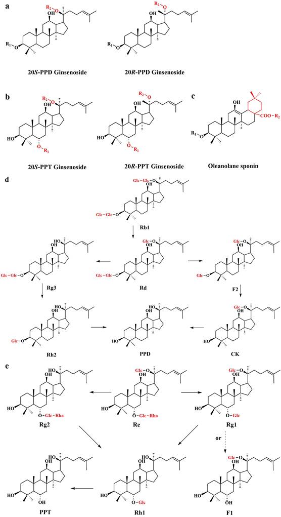
Ginsenosides are the principal active constituents of ginseng and exhibit diverse pharmacological properties, including anti-inflammatory, antioxidant, antitumor, and antifatigue activities[33]. To date, more than 140 ginsenoside monomers have been isolated and identified from ginseng. Based on differences in their aglycone structures, ginsenosides can be classified into protopanaxadiol (PPD), protopanaxatriol (PPT), and oleanolic acid (OA) types. From a pharmacological perspective, these compounds are further distinguished as prototype or rare ginsenosides according to their abundance and metabolic origin[34]. Rare ginsenosides have attracted particular interest in oncology. Among them, Rg3 and Rh2 are the most extensively studied, owing to their potent anticancer activities and increasingly well-defined mechanisms of action. Rg3 has entered adjunctive clinical use in China as a component of several anticancer preparations[35]. Fig. 2 and Table 1 provide an overview of the classification of ginsenosides, the major scaffold structures, and the interconversion between the major saponins.
Representative classification of major ginsenosides, metabolites, and aglycones
| Classification | Prototype ginsenosides | Rare ginsenosides and metabolites |
|---|---|---|
| PPD | Ginsenoside Rb1, Rb2, Rc, Rd | Ginsenoside Rg3, Rg5, Rh2, Rk1, F2, CK, aPPD |
| PPT | Ginsenoside Rg1, Rf, Re | Ginsenoside Rh1, Rh4, Rg2, Rk3, F1, aPPT |
| OA | Ginsenoside Ro | Not listed |
Note: Ginsenosides are classified according to their aglycone skeletons into protopanaxadiol (PPD)-, protopanaxatriol (PPT)-, and oleanolic acid (OA)-type compounds. Prototype ginsenosides are naturally abundant parent saponins, whereas rare ginsenosides and metabolites are generally present at low levels or are generated through processing, deglycosylation, or intestinal microbial biotransformation. CK, compound K; aPPD, protopanaxadiol aglycone; aPPT, protopanaxatriol aglycone. “Not listed” indicates that no representative rare OA-type compound was included in this summary table.
The scaffold structure of ginsenosides and the interconversion between saponins. a. 20S-Protopanaxadiol (PPD)-type saponins and 20R-Protopanaxadiol (PPD)-type saponins; b. 20S-Protopanaxatriol (PPT)-type saponins and 20R-Protopanaxatriol (PPT)-type saponins; c. Oleanolic acid (OA); d. Major metabolic processes of PPD-type ginsenosides; e. Major metabolic processes of PPT-type ginsenosides.
GPS are another important class of bioactive constituents. According to monosaccharide composition, GPS can be broadly divided into neutral polysaccharides and acidic pectic polysaccharides. Neutral fractions mainly include glucans and arabinogalactans, whereas acidic fractions are typically rich in galacturonic acid[36]. In addition to their antioxidant properties, GPS have been reported to exert immunomodulatory and antitumor effects. These activities are thought to be mediated, at least in part, through the activation of immune cells and the enhancement of immune surveillance, thereby contributing to the anticancer potential of ginseng[37].
3. Biological mechanisms of ginseng-derived constituents in cancer therapy
Cancer progression is increasingly recognized as a dynamic systems-level process rather than the consequence of isolated molecular abnormalities. Continuous proliferation, resistance to programmed cell death, metastatic plasticity, immune escape, metabolic adaptation, epigenetic remodeling, and interaction with the host microenvironment jointly determine the evolution and therapeutic response of tumors[38]. These interrelated biological processes provide a useful framework for understanding the pharmacological effects of ginseng extract, whose reported activity goes far beyond direct cytotoxicity.
Among the many components that have been identified in ginseng plants, research on the mechanism mainly focuses on ginseng saponins. Rare saponins, especially Rg3, Rh2, Rg5, Rk1 and Rk3, as well as intestinal microbial metabolite compounds K (CK), constitute the field with the richest experimental evidence. In addition, people are also paying more and more attention to GPS, glycosides, chemically modified derivatives, and standardized plant preparations, which occupy a unique position in the field of pharmacology. Importantly, the functions of these compounds should not be considered interchangeably. Structural diversity, glycosylation mode, and microbial biotransformation significantly affect cell uptake, target accessibility, and biological activity, resulting in significant differences between prototype ginsenosides, rare ginsenosides, microbial metabolites, and complex preparations.
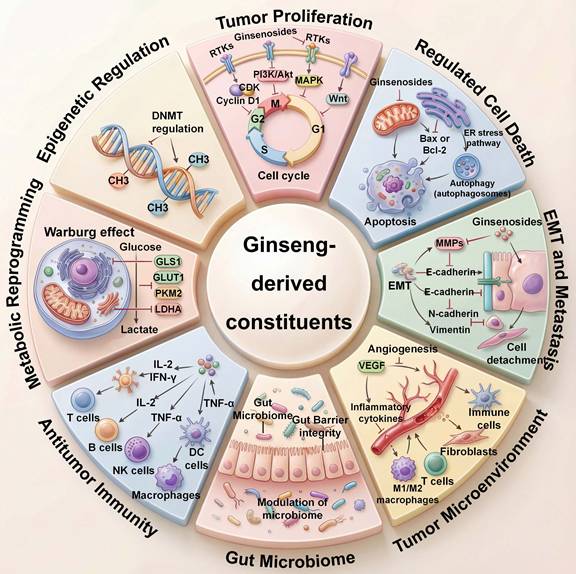
Although the structure of ginseng extract is diverse, most of the ingredients are repeatedly concentrated in a relatively limited number of regulatory networks. Phosphatidylinositol 3-kinase (PI3K)/Akt, mitogen-activated protein kinase (MAPK), nuclear factor-κB (NF-κB), signal transducer and activator of transcription 3 (STAT3), mechanistic target of rapamycin (mTOR), oxidative stress signaling, epithelial-mesenchymal transition (EMT), and programmed cell death protein 1/programmed death-ligand 1 (PD-1/PD-L1) signaling pathways have been found in a variety of tumor types and experimental systems[22, 39]. Rather than representing independent mechanisms, these pathways function as regulatory hubs linking diverse hallmarks of cancer. Therefore, the biological activity of ginseng extract is best interpreted as the synergistic regulation of interrelated signal networks rather than the isolation of a single pathway. These comprehensive effects cover tumor cell survival, metastatic plasticity, tumor-immune interaction, metabolic adaptation, epigenetic regulation, and microbiome-related processes, as shown in Fig. 3.
Multi-target anticancer mechanisms of ginseng-derived constituents in tumor regulation. Ginseng-derived constituents exert pleiotropic anticancer effects by regulating tumor cells, immune components, metabolism, and the tumor microenvironment. These compounds inhibit tumor proliferation through modulation of PI3K/Akt/mTOR, MAPK, Wnt, and cell-cycle signaling pathways; induce apoptosis via reactive oxygen species (ROS)-mediated mitochondrial dysfunction and caspase-dependent pathways; and suppress epithelial-mesenchymal transition (EMT), angiogenesis, and metastasis through regulation of matrix metalloproteinases (MMPs), vascular endothelial growth factor (VEGF), and adhesion molecules. Furthermore, ginseng compounds remodel antitumor immunity by activating immune effector cells and regulating inflammatory cytokines, while affecting tumor metabolism through inhibition of the Warburg effect and glycolytic reprogramming. Their regulation of intestinal flora further promotes systemic anticancer activity and highlights the multidimensional therapeutic potential of ginseng extract.
3.1 Regulation of tumor-cell survival
The ability of malignant cells to maintain survival under continuous proliferative, metabolic, and therapeutic stress represents one of the defining characteristics of cancer[40]. Cell-cycle progression, mitochondrial integrity, oxidative homeostasis, and multiple forms of regulated cell death are tightly coordinated components of this adaptive program rather than independent biological events[41]. Current evidence indicates that ginseng-derived constituents interfere with several of these processes simultaneously, thereby reducing the capacity of tumor cells to maintain long-term viability.
Disruption of cell-cycle progression is one of the most consistently observed biological responses to structurally diverse ginseng-derived compounds. Although the underlying molecular mechanisms vary across tumor types, these compounds commonly restrict proliferative capacity by modulating cyclins, cyclin-dependent kinases, checkpoint proteins, and key regulators of mitotic progression. Representative examples include Rg1-mediated inhibition of Haspin-dependent histone H3 phosphorylation[42] and SIRT1/TSC2 signaling[43], Rb1-induced cell-cycle arrest accompanied by mitochondrial apoptosis[44, 45], Rg3-mediated regulation of the circ_03074/miR-516b-5p/KPNA4 axis and of KIF20A expression and CDC25A proteasomal degradation[46, 47]. Comparable antiproliferative activity has also been described for Rh2[48-53], Re/Rf[54, 55], 20S-PPD[56-58], and panaxadiol[59], despite substantial structural differences among these compounds. Such consistency across multiple chemical scaffolds suggests that restriction of proliferative capacity represents a shared pharmacological characteristic rather than an isolated property of individual ginsenosides.
Limiting proliferation alone is insufficient to eliminate malignant cells, making induction of regulated cell death another major component of the anticancer response. Mitochondrial apoptosis remains the best-characterized mechanism, with repeated evidence demonstrating activation of caspases, disruption of mitochondrial membrane integrity, accumulation of ROS, and modulation of B-cell lymphoma 2 (Bcl-2) family proteins[60, 61]. Rare ginsenosides, particularly Rg3[62-66] and Rh2[67-74], consistently regulate these interconnected processes across diverse tumor models, whereas structurally related compounds including CK[75-77] and several aglycones[78-80] exhibit comparable pro-apoptotic activities through partially overlapping signaling pathways. However, Rg3 has also been reported to activate mTORC1 and promote cell growth under specific experimental conditions, indicating a context-dependent effect[81]. Although individual studies frequently emphasize distinct upstream regulators, most ultimately converge on restoration of mitochondrial susceptibility to programmed cell death.
Accumulating evidence further indicates that ginseng-derived constituents influence forms of regulated cell death extending beyond apoptosis. Autophagy has received the greatest attention, reflecting its dual role in supporting cellular adaptation under stress while also contributing to irreversible cell destruction under sustained perturbation. Depending on the biological context, Rh2[82-84], Rg3[66], Rk1[85], and Rk3[86, 87] regulate autophagic flux primarily through AMP-activated protein kinase (AMPK)/mTOR and PI3K/Akt-associated pathways. Ferroptosis has emerged as another promising mechanism, particularly in studies showing that CK induces FOXO-associated ferroptosis[88]. Direct evidence for pyroptosis remains insufficient. Current studies mainly indicate that CK[89] and GPS[90, 91] can regulate NLRP3-related inflammasome signaling, but whether this response culminates in canonical pyroptotic cell death requires further investigation.
In summary, these observations show that the main effect of ginseng extract is not to induce any single form of cell death. On the contrary, they destroy the interrelated regulatory network for tumor cells to survive under continuous biological stress. Therefore, cell cycle block, apoptosis, autophagy, iron death, and related reactions represent different biological manifestations of the same upstream disturbance, rather than independent pharmacological effects. This network perspective also explains why compounds with different structures repeatedly produce overlapping phenotypes even though they act on some different molecular pathways.
3.2 Regulation of tumor plasticity and metastatic progression
Metastatic dissemination represents one of the greatest obstacles to the success of cancer treatment and the main cause of cancer-related death. However, metastasis is no longer regarded as a simple sequence of tumor-cell detachment, migration, invasion, and colonization. On the contrary, it is now understood as a dynamic process driven by tumor plasticity. In the process, malignant cells constantly adjust their phenotypes through interaction with extracellular matrix components, matrix cell groups, vascular networks, and immune signals[92]. Therefore, therapeutic inhibition of metastasis not only needs to inhibit pathways related to migration, but also needs to destroy the adaptive procedures for tumor cells to acquire invasive, stem cell-like, and drug-resistant states.
Among these adaptive programs, EMT has been widely investigated in studies of ginseng-derived constituents[93]. Although EMT was initially described as a binary transition between epithelial and mesenchymal states, accumulating evidence indicates that tumor cells frequently occupy intermediate or hybrid phenotypes characterized by simultaneous epithelial stability and mesenchymal plasticity. These transitional states promote invasion, metastatic competence, cancer stemness[94, 95], and can contribute to resistance to anticancer therapies[96]. Ginseng-derived compounds appear to restrict this phenotypic flexibility by restoring epithelial characteristics and reducing mesenchymal activation. Consistent changes in E-cadherin, N-cadherin, vimentin, Snail, and related regulators have been reported across multiple cancer models[97], although these markers should be interpreted as indicators of phenotypic transition rather than definitive evidence of EMT reversal.
The anti-metastatic activity of ginseng-derived constituents involves coordinated regulation of several interconnected biological modules. Suppression of extracellular matrix remodeling represents one major mechanism, with repeated evidence showing reduced activity of matrix metalloproteinases (MMPs), particularly MMP-2 and MMP-9[98-104], following treatment with multiple ginseng-derived compounds. These effects are frequently accompanied by inhibition of upstream signaling pathways involving PI3K/Akt, MAPK, Wnt/β-catenin, epidermal growth factor receptor (EGFR), transforming growth factor-β (TGF-β)/Smad, and STAT3[105-109], all of which contribute to maintenance of invasive phenotypes. Rg3 provides one of the most extensively studied examples, demonstrating inhibition of EMT-associated programs across ovarian cancer[110-113], osteosarcoma[108], nasopharyngeal carcinoma[99, 114], and colorectal cancer models[115, 116]. Importantly, the downstream mechanisms vary among tumor contexts, involving lncRNA-mediated regulation, β-catenin signaling, or EGFR-associated transcriptional control. These findings suggest that the biological outcome of EMT suppression is more conserved than the specific molecular route used to achieve it.
Beyond tumor-cell intrinsic regulation, ginseng-derived constituents also influence the surrounding tumor microenvironment. Rh2 and related compounds have been shown to modify cytokine networks[48, 117-119], reduce pro-invasive signaling[120], and regulate macrophage polarization[121, 122], thereby limiting stromal support for tumor dissemination (Table 2). The ability to simultaneously affect malignant cells and their microenvironment may explain why certain rare ginsenosides produce stronger anti-metastatic phenotypes than prototype compounds. Rather than simply suppressing migration in isolated cell assays, these compounds appear to interfere with the reciprocal communication between tumor cells and stromal components that sustains metastatic progression.
Integrated anticancer mechanisms and evidence profiles of representative ginseng-derived constituents
| Representative constituent | Anticancer process | Key molecular pathways or mechanisms | Representative experimental evidence and evidence status | Major references |
|---|---|---|---|---|
| Ginsenoside aglycones 20S-PPD, 20S-PPT, panaxadiol, and panaxatriol | Tumor growth and regulated cell death | Cell-cycle arrest; PI3K/Akt/mTOR and HIF-1α/STAT3 inhibition; mitochondrial/caspase-dependent apoptosis; ROS/ER stress and autophagy | In vitro: several tumor-cell models. In vivo: selected xenograft models. Status: predominantly preclinical; evidence is concentrated in a limited number of aglycones | [58, 59, 79, 80, 266] |
| Metastasis, immune escape, and epigenetic regulation | 20S-PPD: SIRT1- and EGFR/MAPK-related EMT and MMP-2/9 inhibition; panaxadiol: HIF-1α/STAT3/PD-L1 suppression; panaxatriol: METTL3/m6A-STUB1 regulation | In vitro: EMT, invasion, checkpoint, and epigenetic assays. In vivo: selected metastasis models. Status: immune and epigenetic evidence remains limited | [59, 105, 164, 267] | |
| Rg1 | Cell-cycle regulation, metastasis, immunity, and microbiome modulation | Haspin/histone H3 and SIRT1/TSC2 regulation; TGF-β1/EMT and MMP-2/9 inhibition; enhanced T-cell function and macrophage/dendritic-cell regulation; gut microbiota remodeling | In vitro: cancer-cell and immune-cell studies. In vivo: adoptive immunotherapy and microbiota-related animal models. Status: multidomain but context-dependent; direct cancer-specific microbiome evidence is limited | [42, 43, 98, 118, 143, 174, 268] |
| Rb1 | Tumor growth, apoptosis, TME inflammation, and angiogenesis | p53/Bax/cytochrome c/caspase-mediated mitochondrial apoptosis; hypoxia-induced EMT inhibition; reduced TNF-α and IL-6; miR-33a/PEDF/PPAR-γ-mediated antiangiogenesis | In vitro: tumor-cell and endothelial models. In vivo: cancer-cachexia and angiogenesis-related models. Status: evidence is distributed across a relatively small number of models | [44, 45, 119, 126, 269] |
| Rd | Tumor growth, apoptosis, EMT, and stemness | G0/G1 arrest; p53/Bax- and caspase-mediated apoptosis; H19/miR-675-5p/CDH1, miR-18a/Smad2, and EGFR/Akt-related EMT regulation | In vitro: lung, gastric, tongue, breast, and colorectal cancer models. In vivo: selected xenografts; some human-tissue association. Status: moderate preclinical support without clinical intervention evidence | [106, 270-274] |
| Rb2 | EMT, metastasis, and cancer stemness | Suppression of EGFR/Akt/SOX2 and TGF-β/Smad signaling; reduced EMT, MMP activity, and stemness-associated genes | In vitro: colorectal cancer migration, EMT, and stemness assays. In vivo: xenograft models and human CRC samples. Status: focused mainly on colorectal cancer | [107, 275] |
| Other prototype ginsenosidesRe and Rf | Tumor growth and cell-cycle regulation | Re: MITF-related melanoma growth inhibition; Rf: G2/M arrest and mitochondrial apoptosis | In vitro: melanoma and osteosarcoma models. In vivo: zebrafish and xenograft evidence for Re; Rf evidence is mainly cellular. Status: model-specific and mechanistically narrow | [54, 55] |
| Rg3 | Tumor growth and regulated cell death | Cell-cycle arrest; PI3K/Akt and NF-κB inhibition; ROS-associated mitochondrial apoptosis; ER stress and autophagy; context-dependent mTORC1 regulation | In vitro: multiple solid and hematological tumor models. In vivo: several xenograft and precancerous-lesion models. Status: extensive preclinical evidence, but effects vary with stereochemistry, dose, and tumor context | [47, 62, 66, 81, 110, 276, 277] |
| EMT, metastasis, TME remodeling, and angiogenesis | H19- and EMT-related regulation; MMP suppression; inhibition of stemness, VEGF signaling, endothelial activation, and vasculogenic mimicry | In vitro: migration, invasion, stemness, and endothelial assays. In vivo: xenograft and zebrafish angiogenesis models. Status: broad but predominantly preclinical evidence | [108, 113, 115, 116, 128, 131, 278] | |
| Antitumor immunity | Immunogenic cell death; EGFR/GSK-3β-mediated PD-L1 degradation; enhanced dendritic-cell, T-cell, and NK-cell activity | In vitro: tumor-immune-cell and checkpoint assays. In vivo: tumor-bearing animal models. Status: among the more developed immune-related ginsenoside mechanisms; clinical immune validation remains limited | [134, 135, 135, 140, 203] | |
| Metabolic and epigenetic regulation | H19/miR-324-5p/PKM2, STAT3/HK2, and DNMT3A/miR-532-3p/HK2 axes; regulation of DNA methylation and non-coding RNAs | In vitro: glycolytic and epigenetic studies. In vivo: selected xenograft validation. Status: mechanistic causality and metabolic-flux confirmation vary among studies | [156-158, 163] | |
| Rh2 | Tumor growth and regulated cell death | Akt, PBK/TOPK, Axl, Wnt, NF-κB/MAPK, and PI3K/Akt/mTOR regulation; mitochondrial ROS, ER stress, autophagy, and intrinsic/extrinsic apoptosis | In vitro: extensive evidence across solid and hematological malignancies. In vivo: multiple xenograft models. Status: broad preclinical support; 20S-Rh2 generally shows greater activity than 20R-Rh2 | [48, 50, 51, 67, 74, 83, 279, 280] |
| EMT, metastasis, and TME remodeling | EMT and MMP inhibition; IL-6/STAT3 suppression; reduced invasive signaling; M2-to-M1 macrophage repolarization | In vitro: migration, invasion, and macrophage-related assays. In vivo: selected tumor-bearing models. Status: preclinical evidence is supportive but heterogeneous across tumor types | [120, 121, 281] | |
| Antitumor immunity | PD-L1 suppression; enhanced NK-cell surveillance through the ERp5/NKG2D/MICA axis | In vitro: checkpoint and NK-cell recognition studies. In vivo: selected breast and lung tumor models. Status: mechanistically defined but supported by relatively few independent studies | [139, 141] | |
| Metabolic and epigenetic regulation | HIF-1α/PDK4-mediated shift from glycolysis to oxidative phosphorylation; STAT3/c-Myc inhibition; lncRNA, EZH2, and m⁶A-related regulation | In vitro: metabolic, transcriptomic, and epigenetic studies. In vivo: limited xenograft validation. Status: exposure-effect and PK/PD relationships remain insufficiently defined | [68, 71, 155, 167, 168, 282] | |
| CK | Tumor growth and regulated cell death | Cell-cycle arrest; PI3K/Akt/NF-κB and Akt/mTOR/c-Myc inhibition; apoptosis, programmed necrosis, FOXO1-related ferroptosis, and STAT3/ER-stress-related death | In vitro: gastric, prostate, liver, and hematological tumor models. In vivo: selected xenograft and leukemia models. Status: strongest support is for regulated cell death | [76, 77, 88, 283-286] |
| Invasion and immune escape | SDF-1/MMP-2/9/PKCα/ERK-related invasion inhibition; STAT3/PD-L1-associated tumor-cell regulation | In vitro: invasion and checkpoint-related assays. In vivo: limited confirmation. Status: substantially less developed than the cell-death evidence | [75, 287] | |
| Metabolic and microbiome-associated regulation | Inhibition of glycolysis and glutamine utilization; reduced ATP production; remodeling of gut microbiota | In vitro: glycolytic and glutamine-metabolism studies. In vivo: AOM/DSS-induced colitis-associated CRC model. Status: promising but causal microbiome-efficacy relationships remain unresolved | [160, 172, 286] | |
| Rg5 | Tumor growth and regulated cell death | Akt/Bcl-2 and PI3K/Akt/mTOR inhibition; DNA damage; caspase-dependent apoptosis and autophagy | In vitro: esophageal, retinal, cervical, and breast cancer models. In vivo: selected xenograft models. Status: consistent preclinical activity but limited mechanistic replication | [288-292] |
| EMT and metastasis | NF-κB/EphA2-related migration inhibition; suppression of TGF-β1-induced EMT, MMP-2/9, anoikis resistance, and stem-like properties | In vitro: migration, invasion, and EMT assays. In vivo: limited animal confirmation. Status: anti-metastatic evidence remains less extensive than cell-death evidence | [100, 109] | |
| Rk1 | Tumor growth and regulated cell death | G0/G1 arrest; ER stress; caspase-dependent apoptosis; AMPK/mTOR-mediated cytotoxic autophagy | In vitro: cervical, breast, lung, and liver cancer models. In vivo: xenograft and chemically induced HCC models. Status: multidomain preclinical evidence with limited independent replication | [85, 138, 293, 294] |
| Metastasis, immune escape, and metabolism | Inhibition of EMT and MMP activity; NF-κB/PD-L1 suppression; ERK/c-Myc-mediated inhibition of glutamine metabolism | In vitro: EMT, checkpoint, and metabolic assays. In vivo: selected tumor models. Status: each domain is supported by relatively few studies | [109, 138, 159] | |
| Rk3 | Tumor growth and regulated cell death | PI3K/Akt/mTOR-related cell-cycle arrest, mitochondrial apoptosis, and autophagy | In vitro: lung, liver, and esophageal cancer models. In vivo: xenograft and chemically induced HCC models. Status: evidence is focused mainly on apoptosis and autophagy | [86, 87, 295] |
| Other rare ginsenosidesRh1, Rg2, and Rg18 | Tumor growth, regulated cell death, and metastasis | Rh1: mitochondrial ROS/ER stress and RhoA/ROCK1-related apoptosis; Rg2/Rg18: ROS/AMPK- and cell-cycle-related growth inhibition; Rh1: CK2α-HHEX/CCL20/CCR6-associated metastasis inhibition | In vitro: several tumor-cell models. In vivo: selected Rh1 tumor models. Status: compound-specific evidence with limited mechanistic breadth | [296-302] |
| Other rare ginsenosidesRh4, F1, and F2 | Immune, metabolic, and microbiome-associated regulation | Rh4: HDAC2/JAK/STAT- or Akt-related PD-L1 suppression and microbiota-associated bile acid regulation; F1: IGF-1-dependent NK-cell activation; F2: miR-193a-5p/β-catenin/c-Myc/HK2-mediated glycolysis inhibition | In vitro: checkpoint, NK-cell, and metabolic assays. In vivo: selected immune and microbiome models. Status: evidence is mechanism-specific and remains limited for each compound | [136, 137, 142, 162, 171] |
| GPS and related fractions | Direct tumor growth inhibition and regulated cell death | Growth inhibition; mitochondrial apoptosis; NLRP3 activation; autophagy-related radiosensitization | In vitro: breast, bone, and colorectal tumor models. In vivo: selected tumor-bearing models. Status: direct cytotoxic evidence is less extensive than host-mediated evidence | [90, 91, 303] |
| Metastasis and antitumor immunity | Macrophage and NK-cell activation; complement responses; dendritic-cell and cytotoxic T-lymphocyte activation; enhanced anti-PD-1/PD-L1 responses | In vitro: immune-cell activation studies. In vivo: multiple mouse tumor models. Status: broad host-mediated activity, but structural heterogeneity complicates comparison | [146-148, 150, 304] | |
| Metabolic and microbiome-associated regulation | Regulation of the kynurenine/tryptophan ratio; remodeling of gut microbial composition | In vivo: tumor-bearing and microbiome-related mouse models. Status: causal links among polysaccharide structure, microbiota change, and tumor control remain incompletely defined | [150, 173] | |
| Ginsenoside derivatives and related compounds1C, C3DM, 2-deoxy-Rh2, Rh2E2, Rh2-O, 4-XL-PPD, and related derivatives | Tumor growth and regulated cell death | Cell-cycle inhibition; ROS/ER stress; mitochondrial apoptosis; p53/MDM2 regulation; glycolysis-associated cytotoxicity | In vitro: multiple tumor-cell models. In vivo: selected xenograft and intracranial tumor models. Status: emerging structure-activity evidence; direct potency comparisons remain difficult | [161, 305-313] |
| Metastasis and metabolic regulation | EMT and MMP inhibition; c-Jun/COX2/PGE2-associated invasion suppression; interference with glycolysis and cellular energy metabolism | In vitro: invasion and metabolic assays. In vivo: limited confirmation for selected derivatives. Status: early-stage preclinical evidence | [103, 104, 161, 308, 311] |
Note: This table summarizes representative rather than exhaustive evidence for the anticancer activities of major ginseng-derived constituents. Mechanisms were grouped according to the principal biological processes discussed in Sections 3.1-3.5, while individual pathways and targets were retained only when supported by representative experimental studies. Evidence status was qualitatively assessed according to the number of independent studies, diversity of tumor models, availability of in vivo confirmation, genetic or rescue validation, and clinical evidence; it does not represent a formal systematic evidence-grading or risk-of-bias assessment. Statements such as “limited,” “moderate,” or “predominantly preclinical” indicate the current breadth and maturity of evidence rather than the absence of biological activity. References are representative and are not intended to include every published study. CK, Compound K; GPS, ginseng polysaccharides; PPD, protopanaxadiol; PPT, protopanaxatriol; TME, tumor microenvironment; EMT, epithelial-mesenchymal transition; MMP, matrix metalloproteinase; ROS, reactive oxygen species; ER, endoplasmic reticulum; NK, natural killer; CRC, colorectal cancer; HCC, hepatocellular carcinoma; PK/PD, pharmacokinetics/pharmacodynamics; AOM/DSS, azoxymethane/dextran sulfate sodium; m6A, N6-methyladenosine; lncRNA, long non-coding RNA.
Angiogenic remodeling represents another critical component of metastatic expansion. Tumor vascularization not only supplies nutrients and oxygen but also provides routes for systemic dissemination[123, 124]. Several ginseng-derived constituents inhibit angiogenic processes by reducing vascular endothelial growth factor (VEGF) signaling, endothelial-cell activation, microvessel formation, and vasculogenic mimicry[125, 126]. Among these, Rg3 has generated the strongest evidence, with anti-angiogenic activity demonstrated across endothelial models[127, 128], zebrafish assays[129], and xenograft systems[130, 131]. The reproducibility of these findings across experimental platforms suggests that interference with vascular adaptation may represent one of the more translationally relevant mechanisms associated with ginseng-derived compounds.
Despite extensive mechanistic research, there are still some important limitations. At present, most studies rely on wound healing experiments, Transwell invasive systems, or subcutaneous xenotransplant models, which can only reflect some of the characteristics of metastatic biology. There are still relatively few methods that are more physiologically relevant, such as spontaneous transfer models, in situ transplants, organoids of patient origin, and immunoactive systems. In addition, changes in EMT markers, MMP expression, or angiogenic factors may not prove the persistent inhibition of metastasis in vivo.
Taken together, the anti-metastatic activity of ginseng-derived constituents is best understood as the regulation of tumor plasticity, rather than inhibition of a single metastatic pathway. EMT, extracellular matrix remodeling, angiogenesis, and immune-tumor interactions are interrelated components in the adaptive procedures for the survival and proliferation of malignant cells. By limiting this plasticity, ginseng extract may reduce the ability of tumors to transition between proliferation, invasion, and drug resistance. Therefore, future research should go beyond static marker analysis and focus on building longitudinal, spatial, and clinically related models that can capture the transition evolution process.
3.3 Remodeling of tumor-immune interactions
The outcome of cancer progression depends not only on the intrinsic characteristics of malignant cells, but also on the balance between immune surveillance and immunosuppression in the tumor microenvironment. Effective antitumor immunity requires coordinated antigen identification, activation of cytotoxic immune cell groups, and maintenance of an immune microenvironment that can promote tumor clearance[132]. Cancer cells disrupt this balance through multiple strategies, including impaired antigen presentation, induction of immune checkpoints, secretion of immunosuppressive cytokines, and recruitment of regulatory immune populations[133]. Increasing evidence indicates that ginseng-derived constituents influence several components of this tumor-immune interaction network.
Immune checkpoint regulation is one of the most widely studied mechanisms. PD-L1 is the main mediator of tumor immune escape, and its expression is regulated by a variety of upstream pathways, including EGFR, STAT3, NF-κB, hypoxia-inducible factor 1-alpha (HIF-1α), and PI3K/Akt signaling. Some rare ginseng saponins can interfere with these regulatory loops, thus reducing the expression of PD-L1 or enhancing immunorecognition. Rg3 has attracted much attention because it can promote immunogenic tumor cell death[134] and enhance T cell activation by regulating the stability and glycosylation of PD-L1[135]. Other compounds, including Rh4[136, 137], Rk1[138], Rh2[139], and panaxadiol[59], affect the expression of PD-L1 through different molecular mechanisms, indicating that checkpoint regulation is a convergence effect rather than compound specificity.
In addition to regulating immune checkpoints, ginseng extract can also directly affect immune effector function. Studies have reported that ginseng saponins Rg3, Rh2, and F1 can enhance the activity of natural killer (NK) cells by regulating activated receptors, stress response routes, and immunometric molecules[140-142]. Similarly, Rg1 can improve cytotoxic T cell response and enhance the efficacy of sequential immunotherapy[143]. These effects are particularly important because the direct tumor inhibition effect of ginseng saponins may complement the recovery of immune-mediated tumor removal function. Therefore, the anticancer activity of these compounds cannot be fully explained by the autonomic mechanism of tumor cells alone.
GPS represent a unique immunological category[144, 145]. Unlike many small-molecule ginsenosides, GPS usually has limited direct cytotoxicity but has a wider impact on immune cell communication. Its activity includes macrophage activation, dendritic cell maturation, NK cell stimulation, T cell response regulation, and inflammatory cytokine network regulation. This host-oriented mechanism of action may explain why GPS shows therapeutic potential when immune reconstruction is more important than direct tumor killing[146-149]. In addition, microbiota-dependent immune regulation further expands their biological relevance by linking intestinal microbial composition with systemic antitumor immunity[150].
Existing evidence supports such a model: ginseng extract reshapes the immune microenvironment of tumors through a variety of complementary mechanisms. Small-molecule ginseng saponins mainly affect the signal transduction of immune checkpoints and the activation of immune effector cells, while GPS provide a wider range of immune support functions, involving innate and adaptive immunity. These unique and complementary activities provide a theoretical basis for the combined application of ginseng extract with modern anticancer therapies, including immunocheckpoint inhibitors, chemotherapy, and cell therapy.
However, the current evidence is still limited, mainly due to the lack of immune activity models and clinical verification. Many studies still rely on single-layer culture of tumor cells, which cannot reproduce complex immune interactions in patients. Future research should prioritize the construction of models containing the complete immune system, spatial immunospectroscopy analysis, and combined treatment strategies to determine whether ginseng extract can significantly improve the efficacy of existing immunotherapy.
3.4 Systems-level regulation: metabolic adaptation, epigenetic plasticity, and microbiome interactions
Cancer cells will constantly adapt to the fluctuations of nutritional supply, oxidative stress, immune stress, and therapeutic intervention. This adaptability is due to extensive metabolic remodeling[151], epigenetic reprogramming[152], and interactions with the host microbiome[153, 154]. Unlike mechanisms that directly regulate the proliferation or apoptosis of tumor cells, these processes affect the broader biological environment of tumor evolution. More and more evidence shows that ginseng extract can affect these systemic regulatory levels, thus providing a broader explanation for its diverse anticancer activity.
Metabolic reprogramming is one of the most consistently observed effects. Tumor cells usually rely on enhanced glycolysis, increased lactic acid production, and altered nutrient utilization to maintain rapid growth and survival under adverse conditions. Some rare ginseng saponins interfere with these metabolic adaptations by targeting key regulatory factors of glucose metabolism. For example, Rh2 inhibits glycolysis dependence in non-small cell lung cancer (NSCLC) by regulating the HIF-1α/PDK4 axis[71] and STAT3/c-Myc signaling[155], with associated regulation of glucose transport and glycolytic enzymes, including glucose transporter type 1 (GLUT1), pyruvate kinase isozyme type M2 (PKM2), and lactate dehydrogenase A (LDHA). Similar metabolic effects have been reported for Rg3 through regulation of glycolysis-associated signaling involving H19/miRNA networks[156], STAT3/hexokinase 2 (HK2)[157], and related metabolic regulators[158]. Instead of targeting a single metabolic enzyme, these compounds weaken the metabolic flexibility required for tumor cells to maintain growth under stress conditions.
In addition to glucose metabolism, ginseng-derived constituents also influence alternative nutrient pathways that support malignant progression. Rk1 suppresses glutamine utilization in hepatocellular carcinoma through inhibition of ERK/c-Myc signaling[159], whereas CK reduces glutamine consumption and glutaminase activity in triple-negative breast cancer models[160]. Other derivatives, including 2-Deoxy-Rh2[161], further demonstrate that structural modification can enhance metabolic interference by affecting both glycolytic activity and mitochondrial respiration. Related evidence from F2 also links ginseng-derived compounds to glycolytic regulation[162]. In short, these findings show that the regulation of metabolism by ginseng extract involves the synergistic disruption of interrelated energy-producing pathways, rather than simply suppressing the Warburg effect.
Epigenetic regulation provides another explanation for the extensive biological effects of ginseng extract. Tumor progression depends not only on genetic changes, but also on reversible changes in transcription processes that determine cell properties, plasticity, and therapeutic responses. Many studies have shown that ginseng extract can affect DNA methylation, histone-related regulation, RNA modification, and non-coding RNA networks. For example, 20S-Rg3 is associated with whole-genome methylation changes in breast cancer, including the regulation of KDM5A-related pathways[163]. Panaxatriol enhances METTL3-mediated N6-methyladenosine (m6A) modification of STUB1 and suppresses autophagy-related tumor cell survival[164]. It is reported that ginseng extract can also inhibit DNA methyltransferase activity and restore the expression of genes silenced by abnormal methylation in colorectal cancer models[165]. These findings show that ginseng extract may affect cancer biology by changing the regulatory mechanism that controls gene expression.
RNA-based epigenetic regulation has become another important mechanism, especially for Rh2 and its related compounds. Studies show that Rh2 is related to the regulation of long-chain non-coding RNA, microRNA network, and m6A modification pathway. Through these mechanisms, Rh2 affects transcription procedures related to immunomodulation[166], tumor-cell proliferation[167, 168], autophagy[169], and stress adaptation. Although its precise molecular target is not fully clear, these findings expand the biological function of ginseng extract, making it beyond the traditional signaling pathway and suggesting that the regulation of cell identity may help it play an anticancer role.
Intestinal microbiota is another regulatory interface that connects the chemical composition of ginseng with the physiology of the host. Many ginseng saponins have limited oral bioavailability and undergo extensive microbial transformation in the intestine to produce metabolites with enhanced biological activity, including CK. On the contrary, ginseng-derived compounds can reshape the microbial community and change the microbial metabolites involved in inflammation, immunity, bile acid metabolism, and tumor development[170]. This two-way relationship distinguishes ginseng from many traditional anticancer drugs, because its biological activity is not only affected by direct interaction with tumor cells, but also by microbial processing and host-microbiota communication.
Emerging research provides more and more support for microbiome-related anticancer mechanisms. It is reported that Rh4 can inhibit the progression of colorectal cancer by regulating bile acid metabolism dependent on microbiota[171]. CK suppresses inflammation-associated colorectal tumorigenesis while simultaneously altering intestinal microbial composition[172]. GPS partially affects tumor development through microbiome remodeling and immune activation[173]. Rg1 has been associated with immune-cell recovery through modulation of the intestinal microbiota[174], while Ro has shown microbiota-associated anti-liver-cancer activity[175]. Although most of these findings are still in the preclinical stage, they raise an important possibility that the composition of the microbiome may affect the efficacy and variability of ginseng-derived interventions.
In summary, metabolic adaptation, epigenetic plasticity, and microbiome regulation should not be regarded as independent mechanisms. On the contrary, they represent interrelated cancer regulatory levels and jointly determine how tumor cells respond to environmental pressure and treatment pressure. Ginseng-derived constituents seem to be able to influence this system-level network by regulating cell metabolism, transcription status, and communication between hosts and microorganisms. Future research on integrating metabolomics, epigenome, microbiome analysis, and pharmacokinetic analysis is crucial to clarify how these regulatory levels interact and how they can be applied to treatment.
3.5 Mechanistic convergence and evidence hierarchy
The preceding sections illustrate the remarkable diversity of biological effects attributed to ginseng-derived constituents. However, this diversity should not be interpreted as evidence of the infinite complexity of the mechanism. On the contrary, in different compound categories and tumor models, there is a consistent pattern: components with different structures repeatedly affect a relatively small number of regulatory networks, which coordinate a variety of cancer characteristics. Understanding this convergence is crucial to distinguish between true biological principles and observations that depend on specific situations.
At present, rare saponins provide the strongest evidence of mechanism in individual compounds. Compared with prototype saponins, the enhancement of their biological activity is usually due to differences in glycosylation, polarity, membrane interaction, and metabolic transformation[176]. Rg3, Rh2, and CK are the most in-depth examples of research, and their evidence covers tumor cell survival, metastatic plasticity, immunomodulation, metabolic adaptation, and microbiome-related effects. Therefore, these compounds are important models for understanding how structural modification affects pharmacological activity.
Prototype ginsenosides remain biologically relevant but generally display more moderate and context-dependent effects. Their importance may not be limited to direct anticancer activity, because they can act as precursors of microbial transformation and participate in the regulation of inflammation, immune response, and tumor-related signaling pathways. Chemical modification of derivatives and synthetic analogues further shows that structural optimization can change their effectiveness, selectivity, and action routes. However, activity enhancement does not necessarily mean an increase in therapeutic potential, because efficacy enhancement may be accompanied by reduced selectivity or adverse changes in pharmacokinetic properties.
GPS and complex preparations occupy a distinct pharmacological space. Comparative analyses of Panax GPS indicate that their structural heterogeneity is associated with diverse antitumor and immunoregulatory activities[177]. Their anticancer effects are less frequently associated with direct tumor-cell killing and more closely linked to immune regulation, macrophage activation, NK-cell function, and microbiome-mediated host responses. This difference highlights an important principle: different types of ginseng extracts may not compete with each other, but regulate complementary components in tumor biology. Small-molecule ginseng saponins may primarily affect the inherent vulnerability of tumor cells, while GPS may enhance the host-mediated antitumor defense mechanism.
Despite encouraging findings, the strength of mechanistic evidence varies considerably. Several pathways repeatedly appear in the literature, including PI3K/Akt, MAPK, NF-κB, STAT3, mTOR, EMT-associated regulators, PD-L1, VEGF, and MMPs. At the network level, several recurrent upstream-downstream relationships can be identified across different ginseng-derived constituents. Growth-factor and receptor-associated signals frequently converge on the EGFR/PI3K/Akt/mTOR axis, thereby supporting tumor-cell proliferation and survival and modulating autophagy in a context-dependent manner[178, 179]. Excessive oxidative stress and persistent endoplasmic reticulum stress can converge on mitochondrial dysfunction, loss of mitochondrial membrane potential, cytochrome c release, and subsequent caspase activation[180]. Hypoxia-associated HIF-1α/STAT3 cooperation regulates glycolytic and angiogenic transcriptional programs and can contribute to immune-effector dysfunction and resistance to PD-1 blockade[181, 182]. Meanwhile, TGF-β/Smad and Wnt/β-catenin-related transcriptional programs promote epithelial-mesenchymal plasticity, altered cell adhesion, invasion, and metastatic progression[183, 184]. At the host level, intestinal microbial transformation and bioconversion alter the formation and systemic exposure of less-glycosylated metabolites such as CK, thereby linking chemical structure and metabolism with pharmacokinetic behavior and potentially therapeutic efficacy[185, 186]. Together, these regulatory axes provide a mechanistic framework for interpreting the diverse phenotypes described in the preceding sections. However, alterations in these molecules should generally be interpreted as biological responses rather than definitive proof of primary target engagement. Many studies rely on pharmacological concentrations exceeding achievable systemic exposure, particularly for poorly absorbed ginsenosides[187]. In addition, rescue experiments, genetic validation, target occupancy studies, and pharmacokinetic correlation remain relatively uncommon.
A more rigorous evidence hierarchy is therefore required. Mechanisms supported by multiple experimental systems, genetic validation, and in vivo confirmation should be distinguished from observations based primarily on marker changes in cell culture. For example, the attenuation of Rh2-induced Bax translocation and apoptosis following voltage-dependent anion channel 1 (VDAC1) knockdown provides comparatively stronger evidence for a causal target-effect relationship than changes in protein expression alone[73]. Similarly, effects demonstrated in immunocompetent orthotopic models should carry greater translational weight than findings obtained from simplified xenograft systems. Establishing such a hierarchy will be critical for identifying which mechanisms represent genuine therapeutic opportunities and which reflect secondary cellular responses.
Overall, the current evidence supports a revised conceptual framework for understanding the anticancer activity of ginseng-derived constituents. These compounds should not be considered as collections of independent pathway inhibitors but rather as chemically diverse network modulators that influence interconnected regulatory systems governing tumor survival, plasticity, immunity, metabolism, and host interaction. This perspective reconciles the apparent mechanistic diversity of the literature and provides a foundation for future studies aimed at target validation, rational combination strategies, and clinical translation. The integrated mechanisms and evidence characteristics of representative ginseng-derived constituents are summarized in Table 2.
4. Ginseng-derived bioactive constituents: Multitarget regulators of tumor multidrug resistance and chemosensitization
Multidrug resistance (MDR) remains a major barrier to effective chemotherapy and is closely associated with treatment failure and tumor relapse[188, 189]. The molecular underpinnings of MDR are multifaceted, encompassing the overexpression of ATP-binding cassette (ABC) transporters such as P-glycoprotein (P-gp) and breast cancer resistance protein (BCRP), dysregulation of autophagy and apoptosis pathways, the influence of tumor-associated macrophages, and the pervasive immunosuppression within the TME[190-192]. Natural products have attracted attention in this setting because they can regulate multiple resistance-related pathways[193, 194]. Among them, ginseng-derived constituents have been investigated as chemosensitizing adjuvants that may interfere with resistance pathways while, in some settings, also mitigating treatment-related toxicity[195].
A major mechanism involves regulation of drug efflux and compensatory survival pathways. Rh2 inhibits P-gp expression and restores paclitaxel sensitivity in resistant breast cancer models when incorporated into a nano-in-thermogel system[196]. Rg3 also affects transporter-related resistance. In breast cancer models, 20S-Rg3 combined with near-infrared photothermal therapy suppresses PI3K/Akt/mTOR signaling and reduces BCRP, P-gp, and multidrug resistance-associated protein 1 (MRP1) expression[197]. In lung cancer xenografts, 20S-Rg3 enhances cisplatin activity by downregulating P-gp, MRP1, and LRP1[198]. These findings suggest that ginsenosides may improve chemotherapy response partly by limiting drug efflux and weakening survival signaling.
Ginseng-derived compounds also regulate intracellular programs that support treatment resistance. In glioblastoma (GBM), 20S-Rg3 increases temozolomide sensitivity by suppressing MGMT, a DNA repair protein closely related to temozolomide resistance[199]. Rg1 reverses bortezomib resistance in multiple myeloma (MM) through AMPK/mTOR-mediated autophagy regulation[200]. 20S-PPT synergizes with EGFR tyrosine kinase inhibitors in resistant NSCLC by inhibiting EGFR phosphorylation and downstream signaling[201]. Panaxynol targets Hsp90 and suppresses acquired chemoresistance in NSCLC[202].
Resistance can also be shaped by immune escape, metabolic adaptation, and extracellular matrix remodeling. 20R-Rg3 counteracts chemotherapy-induced PD-L1 upregulation by inhibiting NF-κB p65 and Akt signaling, thereby restoring T-cell cytotoxicity in NSCLC models[203]. In tamoxifen-resistant breast cancer, 20S-Rg3 targets 6-phosphofructo-2-kinase/fructose-2,6-biphosphatase 3 (PFKFB3) and interferes with glycolytic adaptation[204]. In pancreatic ductal adenocarcinoma, Rh2 reverses gemcitabine resistance by downregulating laminin subunit gamma 2 (LAMC2), linking extracellular matrix remodeling with chemoresistance[205]. These findings extend the anti-MDR profile of ginsenosides beyond classical transporter inhibition. The expected benefits may vary depending on the treatment method. In combined chemotherapy treatment, ginseng extract may restore intracellular drug exposure and apoptosis ability; while in immunotherapy-oriented combined treatment, its potential value is mainly reflected in reducing PD-L1-mediated immune escape or improving effector cell function. However, at present, most of the evidence of combined treatment is still in the preclinical stage, and the optimal dose, treatment sequence, pharmacokinetic interaction, and additional toxicity of these combined treatments have not been fully clarified.
Current evidence suggests that ginseng-derived constituents may improve anticancer efficacy through coordinated effects on drug efflux, apoptosis, autophagy, DNA repair, metabolic adaptation, and immune-or matrix-associated resistance. Among them, Rg3 and Rh2 are the most frequently studied compounds in MDR reversal and chemosensitization. Some sensitizing effects, however, are observed in specific resistant cell models, combination systems, or nanoformulations, and should therefore be interpreted in relation to drug exposure, formulation design, and tumor context.
5. Clinical translation and advanced delivery strategies for ginseng in oncology
The integration of ginseng-derived preparations into modern oncology has been explored mainly through adjunctive clinical use and pharmaceutical formulation development. Clinical translation has progressed particularly in China, where ginsenoside-containing preparations such as Shenyi Capsule have been used as adjunctive agents in cancer treatment[206]. Furthermore, ginseng extracts are integral components of widely used composite formulations. Aidi injection, a composite formulation containing ginseng, has shown mitochondria-associated pro-apoptotic activity in experimental hepatocellular carcinoma (HCC) models[207]. Similarly, GPS injections have been evaluated as adjunctive preparations for alleviating treatment-associated immune suppression and reducing radiotherapy-or chemotherapy-related adverse effects[147]. However, most available evidence still comes from domestic studies, systematic reviews, meta-analyses, or health technology assessments. Small sample sizes, heterogeneous regimens, variable endpoints, and insufficient high-quality multicenter randomized trials limit the overall strength of evidence. Therefore, these preparations should currently be regarded as adjunctive or supportive agents rather than alternatives to standard anticancer therapy.
In terms of guideline positioning, ginseng-related preparations are more commonly used in Chinese integrative oncology practice, whereas their recognition in international oncology guidelines remains limited. Notably, recent ASCO-SIO guidance mentions American ginseng mainly for cancer-related fatigue, rather than positioning ginseng-derived preparations as standard antitumor agents[208]. This gap highlights the need for stronger clinical validation, standardized preparations, and clearer indications.
At the same time, it should be considered in combination with the pharmacological activity summarized in Section 3 and the pharmacokinetic behavior of each ginseng saponin. Rg3 is a typical example: although it can regulate cell apoptosis, angiogenesis, immune response, and drug resistance, its poor water solubility, low membrane permeability, and low bioavailability limit its drug development and systemic exposure[209]. Rh2 is affected by poor solubility, intestinal metabolism, and P-gp-related efflux[210], and CK is constrained by poor aqueous solubility, insufficient absorption, and limited and variable tissue exposure[211]. Therefore, drug delivery systems (DDS) should match the main obstacles of each active ingredient, and not just regard it as a general-purpose nanotechnology.
Compared with traditional methods such as oral mixed suspension, simple injection, or non-targeted preparations, advanced DDS provide more targeted solutions for improving bioavailability, stability, controlled release, and tumor accumulation. Liposomes and precursor liposomes are especially suitable for improving the dispersion and oral bioavailability of ginseng saponin Rg3[212]. Polymer- or albumin-based nanoparticles are more suitable when the main goal is to protect unstable compounds, achieve surface ligand-mediated targeting, prolong residence time, or combine delivery with chemotherapy drugs[213]. Ginsenoside-based liposomes represent a further step, because amphiphilic ginsenosides such as Rg3 and Rh2 may function not only as therapeutic cargos but also as bioactive membrane components, thereby linking delivery improvement with intrinsic pharmacological activity[214]. Polymer micelle and emulsion systems are mainly suitable for hydrophobic or orally restricted compounds, such as CK, Rh1, and Rh2. The solubility, intestinal absorption, CYP450 metabolism, and P-gp excretion of these compounds are the main concerns. The main delivery platforms are illustrated in Fig. 4, while the compound-specific relationships between pharmacokinetic barriers and delivery strategies are summarized in Table 3.
Representative delivery systems, pharmacokinetic limitations, and translational relevance of ginseng-derived agents
| Agent | Main barrier | Delivery strategy | Key advantage | Translational relevance | Ref. |
|---|---|---|---|---|---|
| Rg3 | Poor solubility, low permeability, and limited bioavailability | Rg3 proliposomes, Rg3-based liposomes | Improves dispersion, formulation state, oral bioavailability, and tumor/TME delivery | Supports Rg3-related apoptosis, anti-angiogenesis, immune regulation, and chemosensitization | [240, 242] |
| Limited tumor accumulation and insufficient co-delivery efficiency | PEG-GO-FA/ICG-Rg3, Rg3-PLGA@TMV, Rg3-PTX-LPs, G/R-MLP | Enhances tumor targeting, controlled release, photodynamic effect, and co-delivery | Strengthens cancer stemness inhibition, TME remodeling, anti-metastatic activity, and combination chemotherapy | [215, 216, 241, 243] | |
| Rh2 | Poor solubility, intestinal metabolism, P-gp-related efflux, and limited tumor exposure | Rh2-functionalized liposomes, PTX-Rh2-lipo, LTL@Rh2@Lipo-GE11, Rh2-containing cocktail liposomes | Provides nanoscale delivery and combines delivery enhancement with the intrinsic antitumor activity of Rh2 | Enhances apoptosis induction, antimetastatic activity, immune modulation, and chemotherapy combination | [214, 244, 245, 247] |
| Poor aqueous solubility and limited cellular uptake | Rh2 nanoniosomal formulation, BSA-Rh2 NPs, Rh2-M | Improves solubility, stability, cellular uptake, and tumor targeting | Enhances antitumor efficacy in prostate, lung, colon, and other cancer models | [219, 220, 251] | |
| CK | Poor aqueous solubility, insufficient absorption, and limited tissue retention | CK mixed micelles, APD-CK micelles | Improves solubilization, drug loading, retention, cellular uptake, and liver targeting | Supports CK-mediated apoptosis and liver cancer-targeted delivery | [211, 250] |
| Poor solubility and limited delivery efficiency | CK-OCMC NPs, DCY51T-AuCK NPs, GCKT-Lipo | Enhances water solubility, cellular uptake, formulation stability, and photothermal synergy | Strengthens caspase-mediated apoptosis and combination antitumor efficacy | [225, 226, 248] | |
| Rh1 / Rh2 | Low oral bioavailability related to intestinal metabolism and efflux transport | Rh1-SMEs, ECG-MEs | Improves oral absorption, systemic exposure, and multicomponent delivery | Supports systemic antitumor exposure and enhances Rh2-related apoptosis in xenograft models | [253, 254] |
| Rg3 / GLP / oridonin | Need for multicomponent oral delivery and TME-related modulation | RGO-SMEDDS | Improves self-emulsifying delivery and multicomponent exposure | Supports immune restoration, anti-angiogenesis, and EGFR/Akt/GSK-3-related HCC inhibition | [255] |
| Rg5 | Poor solubility and weak tumor selectivity | FA-Rg5-BSA NPs | Improves water dispersion, folate receptor-mediated uptake, and tumor targeting | Supports Rg5-related antiproliferative and pro-apoptotic activity | [221] |
| Rh1 / Rh2/PPD | Poor aqueous solubility and limited circulation stability | PEG-Rh1/Rh2 conjugates, PEG-PPD conjugates | Improves aqueous dispersion, circulation time, pH-dependent release, and tumor-site delivery | Enhances the anticancer potential of aglycone- or rare ginsenoside-based agents | [217, 218] |
| Rg1 / Rb1 / PPD combination | Limited tumor selectivity, insufficient co-delivery, and chemotherapy-related toxicity | Dox@Rg1 NPs, Rb1/PTX/PPD co-loaded GPP NPs | Improves tumor targeting, co-delivery efficiency, and reduces off-target toxicity | Enhances chemotherapy efficacy and supports TME modulation | [222, 223] |
| GsE / ginseng extract | Limited tumor retention and insufficient TME-directed delivery | ICG/CMSN@GsE, GDNPs | Enhances tumor retention, macrophage specificity, and immune/TME regulation | Links delivery improvement with macrophage reprogramming, T-cell activation, and cold-to-hot TME conversion | [93, 224, 236-238] |
| Ginseng-derived exosome-like nanoparticles | Limited tissue penetration and glioma targeting | GENs | Crosses the BBB and improves glioma-targeted delivery | Inhibits glioma progression and regulates tumor-associated macrophages | [239] |
| Ginsenoside-based CDs, Re-based CDs, Ginseng-extract-derived CDs | Limited cellular uptake and need for low-toxicity nanoscale delivery | GS-CDs, Re-CDs, GCDs | Improves cellular uptake, provides low toxicity, and supports bioimaging | Promotes apoptosis and ferroptosis; Re-CDs improve Re uptake | [231-233] |
| Rb3 / Rb1 / Rg1 | Limited intracellular delivery and insufficient immunotherapy-related activity | Rb3-CNT, Rb1/Rg1-CNT | Enhances drug loading, cellular interaction, and antiproliferative activity | Supports TNBC immunotherapy-related activity and improves anticancer effects | [234, 235] |
| Ginseng extracts / CK / plant-mediated preparations | Limited stability, weak delivery efficiency, and need for multifunctional platforms | GBAu NPs, GBAg NPs, AgNPs, P.g AgNPs, HGRCm-ZnO NPs, DCY51T-AuCK NPs | Provides nanoscale delivery and adds photothermal, antioxidant, or apoptotic effect | Supports extract-based antitumor activity, apoptosis induction, and potential combination therapy | [226-230] |
Note: This table presents representative rather than exhaustive delivery systems and links the principal physicochemical or pharmacokinetic limitations of each ginseng-derived agent with the corresponding formulation strategy, reported pharmaceutical advantages, and potential translational relevance. Most evidence is derived from preclinical studies; therefore, the translational relevance described here indicates experimentally supported potential rather than established clinical benefit. Formulation abbreviations are retained from the original studies. CK, Compound K; TME, tumor microenvironment; P-gp, P-glycoprotein; NP, nanoparticle; PEG, polyethylene glycol; GO, graphene oxide; FA, folic acid; ICG, indocyanine green; PLGA, poly(lactic-co-glycolic acid); TMV, tumor-derived microvesicle; PTX, paclitaxel; BSA, bovine serum albumin; OCMC, O-carboxymethyl chitosan; TPGS, D-α-tocopheryl polyethylene glycol 1000 succinate; SME, self-microemulsion; ME, microemulsion; SMEDDS, self-microemulsifying drug delivery system; GLP, Ganoderma lucidum polysaccharide; GDNP, ginseng-derived nanoparticle; GEN, ginseng-derived exosome-like nanoparticle; BBB, blood-brain barrier; HCC, hepatocellular carcinoma.
Nanotechnology-based delivery strategies for enhancing the bioactivity of ginseng-derived compounds. Ginseng-derived bioactive compounds, especially ginseng saponins Rg3 and Rh2, have a variety of biological activities, but their applications are limited by poor water solubility, low bioavailability, and insufficient delivery efficiency. Nanotechnology-based drug delivery systems (nano-DDS), including liposomes, polymer micelles, nanoemulsions, nanoparticles, and carbon-based nanocarriers, have been developed to improve the pharmacological properties of these natural compounds. Through packaging, self-assembly, and chemical modification strategies, these nano-platforms can improve the stability, solubility, controlled release, and targeted delivery ability of compounds, thus enhancing the therapeutic potential of bioactive compounds derived from ginseng. These advances provide a promising way to transform ginseng ingredients into effective biomedical applications.
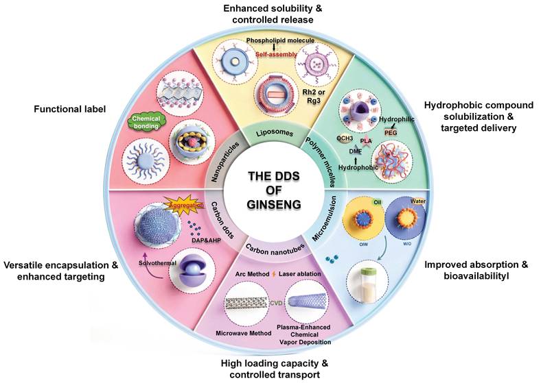
5.1 Nanoparticle-based platforms for enhanced targeting and efficacy
Nanoplatforms provide a flexible strategy for matching the physicochemical and pharmacokinetic limitations of ginseng-derived compounds with specific delivery designs. For poorly soluble ginsenosides, nanocarriers can improve dispersion and apparent solubility. For unstable or rapidly cleared compounds, they may protect the payload and prolong systemic exposure. For compounds requiring tumor-selective delivery, surface modification, biomimetic coating, or ligand-mediated targeting can enhance tumor accumulation and cellular uptake[213].
Polymeric, albumin-based, and biomimetic nanoparticles (NPs) mainly address poor solubility, insufficient stability, limited tumor accumulation, and the need for co-delivery. Rg3-loaded functionalized graphene oxide (PEG-GO-FA/ICG-Rg3)[215] and tumor-cell-membrane-coated PLGA NPs (Rg3-PLGA@TMV) have been used to improve tumor targeting, controlled release, immune activation, or co-delivery with doxorubicin[216]. PEGylated ginsenoside conjugates, including PEG-Rh1/Rh2 and PEG-PPD, improve water solubility, circulation behavior, and antitumor performance[217, 218]. Other representative systems, including an Rh2-loaded nanoniosomal formulation[219], BSA-Rh2 NPs[220], and folic acid-targeted Rg5 albumin NPs[221], further support the value of protein-or vesicle-like carriers for increasing cellular uptake and tumor selectivity.
Several self-assembled or composite nanoparticle systems also connect delivery improvement with biological activity. Dox@Rg1 NPs enhance the antitumor activity of doxorubicin while reducing cardiotoxicity[222], and Rb1-stabilized PTX/PPD co-loaded NPs (GPP NPs) improve tumor targeting and TME modulation[223]. Mesoporous silica NPs incorporating ginseng-derived components (ICG/CMSN@GsE) increase drug retention and macrophage-related targeting[224], while CK-loaded carboxymethyl chitosan calcium NPs (CK-OCMC NPs) improve CK uptake and caspase activation in PCa cells[225]. These examples suggest that nanoparticle design can be used not only to improve drug exposure, but also to integrate ginseng-derived compounds with chemotherapy, phototherapy, or TME-directed strategies.
Metallic nanoparticles and carbon-based systems provide additional delivery or imaging-related possibilities, although their translational profile requires careful evaluation. CK-loaded gold NPs prepared using Lactobacillus kimchicus DCY51T enhance CK-related apoptosis and may support photothermal therapy[226], while the gold and silver GBAu NPs and GBAg NPs, mediated by ginseng berry, have shown antioxidant, antimicrobial, and cytotoxic activities in experimental models[227]. Silver nanoparticles derived from ginseng root[228], or fresh leaf extracts (P.g AgNPs)[229], as well as ginseng-root-derived zinc oxide NPs[230], have been associated with cytotoxicity, EGFR-related inhibition, or mitochondria-mediated apoptosis in cancer models. Carbon dots (CDs) and carbon nanotubes (CNTs) also broaden this field. Ginsenoside-based CDs[231, 232] and ginseng-extract-derived CDs[233] show antitumor or immune-related activity, whereas CNT-based formulations carrying Rb1/Rg1[234] or Rb3[235] have enhanced anti-proliferation effects in the experimental model. Compared with polymer or lipid matrix systems, these platforms may provide additional optical, photothermal, or structural advantages, but their long-term safety, biodegradation, and manufacturability need stricter assessment.
Unlike inert carriers, ginseng-derived nanoparticles (GDNPs) and ginseng-derived exosome-like nanoparticles (GENs) contain natural lipids, proteins, and other bioactive components that may contribute directly to their biological effects. GDNPs have been reported to inhibit EMT through pentose phosphate pathway regulation[93], reprogram macrophages to alleviate T-cell exhaustion[236], and convert immunosuppressive tumors toward a more T-cell-inflamed phenotype[237]. Other studies show that GDNPs promote M2-to-M1 macrophage polarization and induce ROS-related apoptosis[238]. In preclinical models, GENs have been reported to cross the blood-brain barrier, accumulate in glioma, and modulate tumor-associated macrophages[239]. These vesicle-like systems are particularly related to TME remodeling and immunomodulation, but their component definition, batch consistency, and quality control are still the core issues for further development.
5.2 Liposomes: biomimetic and self-assembling systems
Liposomes are phospholipid bilayer vesicles that can encapsulate poorly soluble compounds, improve dispersion, protect unstable payloads, and reduce nonspecific toxicity. Surface modification can further enhance the targeting of tumors. In the ginseng-related delivery system, liposomes are particularly attractive because amphiphilic ginseng saponins (such as Rh2) can partially replace cholesterol-like stabilizing components and directly participate in membrane assembly. This forms a "carrier-drug" design, in which ginseng saponins not only contribute to drug delivery, but also have therapeutic activity[214].
Rg3-based liposomes provide representative examples of this strategy. Folate-modified Rg3 liposomes improve solubility and targeting in TNBC models[240]. Rg3-PTX liposomes (Rg3-PTX-LPS) deliver paclitaxel and remodel the TME by promoting M2-to-M1 macrophage repolarization through IL-6/STAT3-related regulation[241]. Other Rg3-based liposomal systems have been applied to glioma and TNBC, including Rg3-based glioma liposomes[242] and gambogic-acid-loaded biomimetic liposomes (G/R-MLP)[243], which enhance tumor targeting, migration inhibition, and therapeutic efficacy. These systems are particularly useful for combinations where cytotoxic delivery and TME modulation are both desired.
Liposomes based on ginseng saponin Rh2 further clarify the structural value of ginseng saponins in lipid carriers. PTX-Rh2 liposomes (PTX-Rh2-lipo)[244], LTL@Rh2@Lipo-GE11[245], and ACG-loaded Rh2 liposomes (ACGs+Rh2)-Lipo[246] containing ginseng saponin ACG have improved Drug delivery efficiency and tumor targeting in lung cancer, breast cancer, and glioma models. Compared with traditional cholesterol- or PEG-based liposomes, liposomes containing ginseng saponin Rh2 may have higher membrane stability and inherent antitumor activity. Mixed liposomes containing Rh2 and other natural products also showed enhanced anti-lung cancer effects[247]. In addition, TPGS-modified CK liposomes (GCKT-Lipo) improve the water solubility and antitumor efficacy of CK, and support the use of liposomes to treat poorly soluble microbial metabolites[248].
Therefore, the liposomal system is most suitable for ginseng derivatives that require increased solubility, membrane stabilization, tumor targeting, or combined administration. Compared with simple encapsulation, ginseng-based liposomes have the additional advantage of combining carrier function with pharmacological activity, especially suitable for Rg3, Rh2, and CK.
5.3 Polymeric micelles for improved bioavailability
Polymeric micelles are self-assembled nanoscale carriers formed from amphiphilic polymers in aqueous solution. Their hydrophobic core can encapsulate poorly soluble compounds, while the hydrophilic shell improves dispersion, reduces premature clearance, and prolongs circulation time[249]. These features make micelles particularly suitable for hydrophobic ginsenosides or metabolites whose application is limited by poor aqueous solubility and low oral bioavailability.
CK and Rh2 are representative examples. CK has broad anticancer activity, but its poor solubility and limited bioavailability restrict its in vivo performance. A54 peptide-modified deoxycholic acid-O-carboxymethyl chitosan micelles loaded with CK (APD-CK) improve liver targeting and antitumor activity[250]. Rh2 mixed micelles (Rh2-M) prepared by thin-film dispersion enhance cellular uptake, tumor targeting, and anticancer activity[251]. These studies show that micellar systems can improve the pharmaceutical performance of hydrophobic ginsenosides without requiring covalent structural modification.
Polymeric micelles are most suitable as exposure enhancement systems for compounds such as CK and Rh2. Their main value is to improve solubility, whole-body exposure, and tissue targeting. Compared with more complex multifunctional nanoparticles, micelles may provide a simpler preparation strategy, but stability, drug delivery efficiency, release behavior, and repeatability of amplified production still need to be considered.
5.4 Nanoemulsion-and microemulsion-based systems
Nanoemulsions and microemulsions are colloidal delivery systems composed of an oil phase, aqueous phase, surfactant, or cosurfactant. Microemulsions are thermodynamically stable, whereas nanoemulsions are kinetically stable and generally require external energy for their preparation. They are useful for improving solubilization, oral absorption, and combination delivery of lipophilic compounds[252]. Unlike polymeric nanoparticles or liposomes, emulsion-based systems are particularly suitable for oral or multicomponent formulations in which solubilization and intestinal absorption are the main barriers.
This strategy is relevant to Rh1 and Rh2, whose oral bioavailability is limited by intestinal CYP450-mediated metabolism and P-gp efflux[253]. A multicomponent microemulsion containing etoposide, coix seed oil, and Rh2 (ECG-MEs) improved the antitumor efficacy of Rh2 in A549 xenograft models, with increased apoptosis and tumor growth inhibition[254]. Another example is a self-microemulsifying delivery system co-loading Rg3, ganoderma lucidum polysaccharide (GLP), and oridonin (RGO-SMEDDS). This system was reported to reduce immunosuppressive cytokine production and M2-like macrophage polarization, inhibit VEGF-related angiogenesis, and suppress EGFR/Akt/GSK-3β signaling in HCC models[255].
The emulsion system is popular because it can improve solubility, oral absorption rate, and combined administration ability at the same time. As for ginseng extracts, the emulsion system is especially suitable for multi-component preparations aimed at enhancing drug exposure and integrating immunomodulatory or anti-angiogenic effects. Its transformation application potential will depend on the further optimization of the robustness, stability, and repeatability of the preparation.
6. Discussion and future perspectives
More and more experimental evidence shows that ginseng extract has a wide range of anticancer potential, which can be achieved by coordinating and regulating tumor cell survival, metastatic behavior, immune response, metabolic adaptation, epigenetic status, and therapeutic drug resistance. However, the future development of ginseng-based oncology should move beyond the accumulation of mechanistic observations and toward a more integrated framework that connects chemical identity, molecular mechanism, pharmacological exposure, and clinical outcome. Therefore, the core challenge is not only to discover new anticancer active ingredients, but also to determine which ingredients can produce repeatable effects, through which causal mechanisms, and under what treatment conditions these effects can be transformed into clinical benefits. For immunotherapy-oriented combined therapy, this transformation should also take into account the existing challenges in biomarker selection, model correlation, joint program design, and clinical implementation[256].
The main premise of this transformation is to improve the definition and standardization of chemical components. The biological activity of ginseng products is significantly affected by the species source, cultivation environment, processing technology, extraction method, and preparation strategy. This variability is an important reason for the inconsistency of experimental results and complicates the comparison between different studies. For ginseng saponins and GPS, this problem is particularly prominent because their chemical composition will change due to steaming, fermentation, intestinal microbial transformation, or structural modification[170, 176]. Therefore, future research should combine comprehensive chemical characterization with pharmacological evaluation, including standardized preparation methods, quantitative analysis of biologically active ingredients, and multi-component quality control methods[257]. The establishment of a reliable chemical framework will lay the foundation for repeatable mechanism research and reasonable clinical development.
Beyond chemical standardization, a deeper understanding of molecular mechanisms is required. The multitarget nature of ginseng-derived compounds may represent an intrinsic advantage in cancer biology because malignant progression involves interconnected processes rather than isolated pathways. Nevertheless, at present, many studies are still mainly at the descriptive level, only reporting changes in signaling molecules such as PI3K/Akt, MAPK, NF-κB, STAT3, mTOR, and autophagy-related pathways, without fully clarifying their causal contribution to treatment results[258]. Future research should therefore shift from pathway cataloging toward mechanistic validation by incorporating genetic manipulation, rescue experiments, target identification approaches, multi-omics integration, and clinically relevant models such as patient-derived organoids and xenografts. Such strategies will help distinguish between the main molecular targets and downstream reactions, and improve the rational design of ginseng-derived therapies.
Successful clinical transformation also depends on overcoming pharmacological limitations. Many bioactive ginseng saponins show strong anticancer activity in experimental systems, but due to poor solubility, limited absorption, rapid metabolism, or insufficient tumor accumulation, whole-body exposure is insufficient. Advanced delivery strategies provide promising solutions by improving pharmacokinetic behavior and achieving more accurate interaction with tumor tissue. However, the future development of DDS should not focus solely on increasing drug concentration. On the contrary, the design of the preparation should be combined with the biological mechanism to determine how, where, and when the ginseng-derived molecules work. Issues such as production scale, preparation stability, long-term safety, metabolic pathways, and the potential impact of therapeutic drug resistance are still key issues that need to be systematically evaluated[259-261].
Emerging technologies may provide new opportunities to redefine the therapeutic potential of ginseng-derived compounds. Tumor microenvironment-responsive delivery systems, biomimetic nanoparticles, and exosome-based platforms may enable controlled release and improved combination strategies with chemotherapy or immunotherapy[259]. At the mechanistic level, unexplored areas such as mitochondrial epigenetic regulation, mitochondrial DNA-related signal transmission, and metabolic-epigenetic interaction may reveal other dimensions of ginseng-mediated cancer regulation[262, 263]. At the same time, the progress of high-throughput sequencing, spatial multi-omics, artificial intelligence-based drug discovery, and computational screening technology may accelerate the identification of key targets and optimize the development of saponin derivatives[264, 265].
Ultimately, the next generation of ginseng oncology research requires a closer integration of natural product chemistry, molecular oncology, pharmacology, and clinical medicine. Mechanism research should give priority to the identification of causal targets, not pathway association. Drug research and development should regard delivery optimization as an important part of treatment plan design, rather than post-remedial measures. Clinical research should focus on clear indications, reasonable combined drug strategies, pharmacological administration schemes, and clinically significant efficacy indicators. Through this integrated framework, ginseng-derived drugs are expected to gradually develop from empirical auxiliary products to mechanism-oriented and evidence-based tumor treatments.
7. Conclusion
Ginseng-derived compounds represent a unique class of natural product-based candidates with diverse but interconnected anticancer activities. More and more evidence shows that ginseng saponins, GPS, and their related derivatives regulate cancer progression through synergy in tumor cell survival, metastatic plasticity, immunoregulation, metabolic adaptation, epigenetic regulation, and therapeutic drug resistance. These compounds do not act as traditional cytotoxic drugs, but seem to affect multiple biological levels, together shaping tumor behavior and therapeutic response. Among the most extensively investigated constituents, Rg3, Rh2, and CK provide the strongest mechanistic evidence, but their relative therapeutic value still depends on drug exposure, tumor microenvironment, and clinical verification.
Although remarkable progress has been made in the experiment, ginseng-derived agents remain in an intermediate stage between mechanistic discovery and clinical application. Their most realistic applications in the near future may be in adjuvant treatment, enhancing therapeutic effects, regulating immunity or metabolism, and combined treatment strategies, rather than replacing existing anticancer therapies. Advances in drug delivery technology, chemical standardization, and pharmacokinetic optimization may help overcome the current limitations in bioavailability, target exposure, and preparation consistency.
Future progress will require an integrated development framework that links chemical characterization, causal mechanism verification, transformational pharmacology, and clinical evaluation. Identifying active ingredients, identifying clinically relevant targets, establishing exposure-reaction relationships, and carrying out rigorously designed clinical trials are crucial to determine where ginseng extracts play a significant therapeutic role. Through such mechanism-guided approaches, ginseng may gradually evolve from a traditional medicinal resource into a scientifically defined and clinically relevant component of modern oncology.
Abbreviations
ABC: ATP-binding cassette;
AMPK: AMP-activated protein kinase;
Bcl-2: B-cell lymphoma 2;
BCRP: breast cancer resistance protein;
CDs: carbon dots;
CK: compound K;
CNTs: carbon nanotubes;
CRC: colorectal cancer;
DDS: drug delivery systems;
EGFR: epidermal growth factor receptor;
EMT: epithelial-mesenchymal transition;
GBM: glioblastoma;
GDNPs: ginseng-derived nanoparticles;
GENs: ginseng-derived exosome-like nanoparticles;
GLP: Ganoderma lucidum polysaccharide;
GLUT1: glucose transporter type 1;
GPS: ginseng polysaccharides;
HCC: hepatocellular carcinoma;
HIF-1α: hypoxia-inducible factor 1-alpha;
HK2: hexokinase 2;
LAMC2: laminin subunit gamma 2;
LDHA: lactate dehydrogenase A;
m6A: N6-methyladenosine;
MAPK: mitogen-activated protein kinase;
MDR: multidrug resistance;
MM: myeloma;
MMPs: matrix metalloproteinases;
MRP1: multidrug resistance-associated protein 1;
mTOR: mechanistic target of rapamycin;
NF-κB: nuclear factor-κB;
NK: natural killer cells;
NPs: nanoparticles;
NSCLC: non-small-cell lung cancer;
OA: oleanolic acid;
P-gp: P-glycoprotein;
PCa: prostate cancer;
PD-1: programmed cell death protein 1;
PD-L1: programmed death-ligand 1;
PFKFB3: 6-phosphofructo-2-kinase/fructose-2,6-bisphosphatase 3;
PI3K: phosphatidylinositol 3-kinase;
PKM2: pyruvate kinase isozyme type M2;
PPD: protopanaxadiol;
PPT: protopanaxatriol;
ROS: reactive oxygen species;
STAT3: signal transducer and activator of transcription 3;
TCM: traditional Chinese medicine;
TGF-β: transforming growth factor-β;
TME: tumor microenvironment;
VDAC1: voltage-dependent anion channel 1;
VEGF: vascular endothelial growth factor.
Acknowledgements
This work was supported by the grants of the National Natural Science Foundation of China (No. 82474070) and Jilin Provincial Science and Technology Development Plan (No. YDZJ202401311ZYTS).
Author contributions
Jia-hui Li: Conceptualization, Methodology, Data Curation, Writing-Original Draft; Yan-fang Xian: Investigation, Formal analysis, Writing-Review & Editing; De-wen Liu: Conceptualization, Writing-Review & Editing; Hong-yuan Li: Supervision, Writing-Review & Editing; Wei Li: Conceptualization, Writing-Review & Editing, Supervision, Funding acquisition.
Competing Interests
The authors have declared that no competing interest exists.
References
1. Mullard A. Addressing cancer's grand challenges. Nat Rev Drug Discov. 2020;19:825-6
2. Bray F, Laversanne M, Sung H, Ferlay J, Siegel RL, Soerjomataram I. et al. Global cancer statistics 2022: GLOBOCAN estimates of incidence and mortality worldwide for 36 cancers in 185 countries. CA Cancer J Clin. 2024;74:229-63
3. Shih Y-CT, Owsley KM, Nicholas LH, Yabroff KR, Bradley CJ. Cancer's Lasting Financial Burden: Evidence From a Longitudinal Assessment. J Natl Cancer Inst. 2022;114:1020-8
4. Newman NB, Sidhu MK, Baby R, Moss RA, Nissenblatt MJ, Chen T. et al. Long-Term Bone Marrow Suppression During Postoperative Chemotherapy in Rectal Cancer Patients After Preoperative Chemoradiation Therapy. Int J Radiat Oncol Bio Phys. 2016;94:1052-60
5. Chen H, Yang G, Ma J. Ocular toxicity associated with anti-HER2 agents in breast cancer: A pharmacovigilance analysis using the FAERS database. Int J Cancer. 2024;154:1616-25
6. Wang H, Jia H, Gao Y, Zhang H, Fan J, Zhang L. et al. Serum metabolic traits reveal therapeutic toxicities and responses of neoadjuvant chemoradiotherapy in patients with rectal cancer. Nat Commun. 2022;13:7802
7. Méndez-Blanco C, Fondevila F, García-Palomo A, González-Gallego J, Mauriz JL. Sorafenib resistance in hepatocarcinoma: role of hypoxia-inducible factors. Exp Mol Med. 2018;50:1-9
8. Soleimani N, Javadi MM. Future prospects of bacteria-mediated cancer therapies: Affliction or opportunity? Microb Pathog. 2022;172:105795
9. China Association of Chinese Medicine, Beijing, China. Top 10 academic advances in traditional Chinese medicine in 2024. Sci Tradit Chin Med. 2025;3:292-5
10. Zhang X, Qiu H, Li C, Cai P, Qi F. The positive role of traditional Chinese medicine as an adjunctive therapy for cancer. Biosci Trends. 2021;15:283-98
11. Liu S-H, Chen P-S, Huang C-C, Hung Y-T, Lee M-Y, Lin W-H. et al. Unlocking the Mystery of the Therapeutic Effects of Chinese Medicine on Cancer. Front Pharmacol. 2021;11:601785
12. Ghosh S, Das SK, Sinha K, Ghosh B, Sen K, Ghosh N. et al. The Emerging Role of Natural Products in Cancer Treatment. Arch Toxicol. 2024;98:2353-91
13. Kong F, Wang C, Zhao L, Liao D, Wang X, Sun B. et al. Traditional Chinese medicines for non-small cell lung cancer: Therapies and mechanisms. Chin Herb Med. 2023;15:509-15
14. Zhao W, Zheng X, Tang PY, Li H, Liu X, Zhong J. et al. Advances of antitumor drug discovery in traditional Chinese medicine and natural active products by using multi-active components combination. Med Res Rev. 2023;43:1778-808
15. Wang L, Feng T, Su Z, Pi C, Wei Y, Zhao L. Latest research progress on anticancer effect of baicalin and its aglycone baicalein. Arch Pharm Res. 2022;45:535-57
16. Chen F, Pan Y, Xu J, Liu B, Song H. Research progress of matrine's anticancer activity and its molecular mechanism. J Ethnopharmacol. 2022;286:114914
17. Jia Y, Yao D, Bi H, Duan J, Liang W, Jing Z. et al. Salvia miltiorrhiza Bunge (Danshen) based nano-delivery systems for anticancer therapeutics. Phytomedicine. 2024;128:155521
18. Wang M, Yu F. Research Progress on the Anticancer Activities and Mechanisms of Polysaccharides From Ganoderma. Front Pharmacol. 2022;13:891171
19. Luo Y, Zhu L, Ren Z, Xiao J, Hao E, Lu J. et al. Curcumae Rhizoma: An anti-cancer traditional Chinese medicine. Chin Herb Med. 2025;17:428-47
20. Sajana M, Gopenath TS, Kanthesh BM. A comprehensive review of phytochemical approaches in treatment of acute myeloid leukemia: Associated pathways and molecular mechanisms. Chin Herb Med. 2025;17:41-55
21. Fang W, Huang K, Hu J. Potential of Panax ginseng for bone health and osteoporosis management. Chin Herb Med. 2025;17:673-84
22. Yang Y, Nan Y, Du Y, Liu W, Ning N, Chen G. et al. Ginsenosides in cancer: Proliferation, metastasis, and drug resistance. Biomed Pharmacother. 2024;177:117049
23. Xu Y, Bian S, Shang L, Wang X, Bai X, Zhang W. Phytochemistry, pharmacological effects and mechanism of action of volatile oil from Panax ginseng C.A.Mey: a review. Front Pharmacol. 2024;15:1436624
24. Zhang H, Zhang L, Yang C, Zhang Y, Li J, Zhang X. et al. Prevention Effect of Protopanaxadiol-Type Saponins Saponins and Protopanaxatriol-Type Saponins on Myelosuppression Mice Induced by Cyclophosphamide. Front Pharmacol. 2022;13:845034
25. Han J, Wang Y, Cai E, Zhang L, Zhao Y, Sun N. et al. Study of the Effects and Mechanisms of Ginsenoside Compound K on Myelosuppression. J Agric Food Chem. 2019;67:1402-8
26. Zhang Y, Shao C, Li J, Bai Y, Li G, Ren T. Research progress of ginseng active ingredients combined with chemotherapy in cancer therapy. J Ginseng Res. 2026;50:100971
27. Ho PT, Rini IA, Hoang PT, Lee TK, Lee S. Exploring the potential of ginseng-derived compounds in treating cancer cachexia. J Ginseng Res. 2025;49:631-9
28. Lee J-S, Lee H-Y. Ginseng-derived compounds as potential anticancer agents targeting cancer stem cells. J Ginseng Res. 2024;48:266-75
29. Tiwari P, Park K-I. Ginseng-Based Nanotherapeutics in Cancer Treatment: State-of-the-Art Progress, Tackling Gaps, and Translational Achievements. Curr Issues Mol Biol. 2025;47:250
30. Ru W, Wang D, Xu Y, He X, Sun Y-E, Qian L. et al. Chemical constituents and bioactivities of Panax ginseng (C. A. Mey.). Drug Discov Ther. 2015;9:23-32
31. Liu Y, Li W. From bioactivity to application: Utilization of non-medicinal ginseng stems and leaves. Food Biosci. 2026;77:108308
32. Li S-L, Zhang S, Fu L, Chen C, Zhang W-H, Li W. Chemical structure and beneficial roles of active biomacromolecules from Ginseng: a narrative review. Acupunct Herb Med. 2025;5:366-78
33. Hou M, Wang R, Zhao S, Wang Z. Ginsenosides in Panax genus and their biosynthesis. Acta Pharm Sin B. 2021;11:1813-34
34. Yao W, Guan Y. Ginsenosides in cancer: A focus on the regulation of cell metabolism. Biomed Pharmacother. 2022;156:113756
35. Xu W, Lyu W, Duan C, Ma F, Li X, Li D. Preparation and bioactivity of the rare ginsenosides Rg3 and Rh2: An updated review. Fitoterapia. 2023;167:105514
36. Ji L, Jie Z, Ying X, Yue Q, Zhou Y, Sun L. Structural characterization of alkali-soluble polysaccharides from Panax ginseng C. A. Meyer. R Soc Open Sci. 2018;5:171644
37. Ji X, Hou C, Shi M, Yan Y, Liu Y. An Insight into the Research Concerning Panax ginseng C. A. Meyer Polysaccharides: A Review. Food Rev Int. 2022;38:1149-65
38. Hanahan D. Hallmarks of Cancer: New Dimensions. Cancer Discov. 2022;12:31-46
39. Mou C, Wang Y, Kim M-Y, Cho JY. Ginsenosides from Panax ginseng: Their mechanisms, microbiome determinants, and translational strategies in therapeutic activity against cancer. J Ginseng Res. 2026;50:101029
40. Matthews HK, Bertoli C, De Bruin RAM. Cell cycle control in cancer. Nat Rev Mol Cell Biol. 2022;23:74-88
41. Carneiro BA, El-Deiry WS. Targeting apoptosis in cancer therapy. Nat Rev Clin Oncol. 2020;17:395-417
42. Hong J, Gwon D, Jang C-Y. Ginsenoside Rg1 suppresses cancer cell proliferation through perturbing mitotic progression. J Ginseng Res. 2022;46:481-8
43. Tang Y-L, Zhang C-G, Liu H, Zhou Y, Wang Y-P, Li Y. et al. Ginsenoside Rg1 Inhibits Cell Proliferation and Induces Markers of Cell Senescence in CD34+CD38- Leukemia Stem Cells Derived from KG1α Acute Myeloid Leukemia Cells by Activating the Sirtuin 1 (SIRT1)/Tuberous Sclerosis Complex 2 (TSC2) Signaling Pathway. Med Sci Monit. 2020;26:e918207
44. An L, Yu Y, He L, Xiao X, Li P. Ginsenoside Rb1 Deters Cell Proliferation, Induces Apoptosis, Alleviates Oxidative Stress, and Antimetastasis in Oral Squamous Carcinoma Cells. Appl Biochem Biotechnol. 2024;196:7642-56
45. Feng L, Liu X, Sun K, Sun Y, Wu W, Chen C. et al. Ginsenoside Rb1 Inhibits the Proliferation of Lung Cancer Cells by Inducing the Mitochondrial-mediated Apoptosis Pathway. Anticancer Agents Med Chem. 2024;24:928-41
46. Wang T, Zhang C, Wang S. Ginsenoside Rg3 inhibits osteosarcoma progression by reducing circ_0003074 expression in a miR-516b-5p/KPNA4-dependent manner. J Orthop Surg. 2021;16:724
47. Zhang R, Li L, Li H, Bai H, Suo Y, Cui J. et al. Ginsenoside 20(S)-Rg3 reduces KIF20A expression and promotes CDC25A proteasomal degradation in epithelial ovarian cancer. J Ginseng Res. 2024;48:40-51
48. Hu Q-R, Pan Y, Wu H-C, Dai Z-Z, Huang Q-X, Luo T. et al. The ways for ginsenoside Rh2 to fight against cancer: the molecular evidences in vitro and in vivo. J Ginseng Res. 2023;47:173-82
49. Li K-F, Kang C-M, Yin X-F, Li H-X, Chen Z-Y, Li Y. et al. Ginsenoside Rh2 inhibits human A172 glioma cell proliferation and induces cell cycle arrest status via modulating Akt signaling pathway. Mol Med Rep. 2018;17:3062-8
50. Zhang H, Yi J-K, Huang H, Park S, Kwon W, Kim E. et al. 20 (S)-ginsenoside Rh2 inhibits colorectal cancer cell growth by suppressing the Axl signaling pathway in vitro and in vivo. J Ginseng Res. 2022;46:396-407
51. Li S, Han W, He Q, Wang Y, Jin G, Zhang Y. Ginsenoside Rh2 suppresses colon cancer growth by targeting the miR-150-3p/SRCIN1/Wnt axis. Acta Biochim Biophys Sin. 2023;55:633-48
52. Li C, Gao H, Feng X, Bi C, Zhang J, Yin J. Ginsenoside Rh2 impedes proliferation and migration and induces apoptosis by regulating NF-κB, MAPK, and PI3K/Akt/mTOR signaling pathways in osteosarcoma cells. J Biochem Mol Toxicol. 2020;34:e22597
53. Shi X, Yang J, Wei G. Ginsenoside 20(S)-Rh2 exerts anti-cancer activity through the Akt/GSK3β signaling pathway in human cervical cancer cells. Mol Med Rep. 2018;17:4811-6
54. Hwang SJ, Bang HJ, Lee H-J. Ginsenoside Re inhibits melanogenesis and melanoma growth by downregulating microphthalmia-associated transcription factor. Biomed Pharmacother. 2023;165:115037
55. Shangguan W-J, Li H, Zhang Y-H. Induction of G2/M phase cell cycle arrest and apoptosis by ginsenoside Rf in human osteosarcoma MG-63 cells through the mitochondrial pathway. Oncol Rep. 2014;31:305-13
56. Zhang H, Xu H-L, Wang Y-C, Lu Z-Y, Yu X-F, Sui D-Y. 20(S)-Protopanaxadiol-Induced Apoptosis in MCF-7 Breast Cancer Cell Line through the Inhibition of PI3K/AKT/mTOR Signaling Pathway. Int J Mol Sci. 2018;19:1053
57. Jo H, Jang D, Park SK, Lee M-G, Cha B, Park C. et al. Ginsenoside 20(S)-protopanaxadiol induces cell death in human endometrial cancer cells via apoptosis. J Ginseng Res. 2021;45:126-33
58. Song C, Shen T, Kim HG, Hu W, Cho JY. 20(S)-Protopanaxadiol from Panax ginseng Induces Apoptosis and Autophagy in Gastric Cancer Cells by Inhibiting Src. Am J Chin Med. 2023;51:205-21
59. Wang Z, Li MY, Zhang ZH, Zuo HX, Wang JY, Xing Y. et al. Panaxadiol inhibits programmed cell death-ligand 1 expression and tumour proliferation via hypoxia-inducible factor (HIF)-1α and STAT3 in human colon cancer cells. Pharmacol Res. 2020;155:104727
60. Ashrafizadeh M, Tavakol S, Mohammadinejad R, Ahmadi Z, Yaribeygi H, Jamialahmadi T. et al. Paving the Road Toward Exploiting the Therapeutic Effects of Ginsenosides: An Emphasis on Autophagy and Endoplasmic Reticulum Stress. Adv Exp Med Bio. 2021:137-60
61. Li X, Cao D, Sun S, Wang Y. Anticancer therapeutic effect of ginsenosides through mediating reactive oxygen species. Front Pharmacol. 2023;14:1215020
62. Lv S, Chen X, Chen Y, Gong D, Mao G, Shen C. et al. Ginsenoside Rg3 induces apoptosis and inhibits proliferation by down-regulating TIGAR in rats with gastric precancerous lesions. BMC Complement Med Ther. 2022;22:188
63. Sun HY, Lee JH, Han Y-S, Yoon YM, Yun CW, Kim JH. et al. Pivotal Roles of Ginsenoside Rg3 in Tumor Apoptosis Through Regulation of Reactive Oxygen Species. Anticancer Res. 2016;36:4647-54
64. Xie Q, Wen H, Zhang Q, Zhou W, Lin X, Xie D. et al. Inhibiting PI3K-AKt signaling pathway is involved in antitumor effects of ginsenoside Rg3 in lung cancer cell. Biomed Pharmacother. 2017;85:16-21
65. Yuan Z, Jiang H, Zhu X, Liu X, Li J. Ginsenoside Rg3 promotes cytotoxicity of Paclitaxel through inhibiting NF-κB signaling and regulating Bax/Bcl-2 expression on triple-negative breast cancer. Biomed Pharmacother. 2017;89:227-32
66. Bian S, Zhao Y, Li F, Lu S, Wang S, Bai X. et al. 20(S)-Ginsenoside Rg3 Promotes HeLa Cell Apoptosis by Regulating Autophagy. Mol Basel Switz. 2019;24:3655
67. Ge G, Yan Y, Cai H. Ginsenoside Rh2 Inhibited Proliferation by Inducing ROS Mediated ER Stress Dependent Apoptosis in Lung Cancer Cells. Biol Pharm Bull. 2017;40:2117-24
68. Jeong D, Ham J, Park S, Kim HW, Kim H, Ji HW. et al. Ginsenoside Rh2 Suppresses Breast Cancer Cell Proliferation by Epigenetically Regulating the Long Noncoding RNA C3orf67-AS1. Am J Chin Med. 2019;47:1643-58
69. Peng K, Luo T, Li J, Huang J, Dong Z, Liu J. et al. Ginsenoside Rh2 inhibits breast cancer cell growth via ERβ-TNFα pathway. Acta Biochim Biophys Sin. 2022;54:647-56
70. Zhang J, Li W, Yuan Q, Zhou J, Zhang J, Cao Y. et al. Transcriptome Analyses of the Anti-Proliferative Effects of 20(S)-Ginsenoside Rh2 on HepG2 Cells. Front Pharmacol. 2019;10:1331
71. Liu X, Li J, Huang Q, Jin M, Huang G. Ginsenoside Rh2 shifts tumor metabolism from aerobic glycolysis to oxidative phosphorylation through regulating the HIF1-α/PDK4 axis in non-small cell lung cancer. Mol Med Camb Mass. 2024;30:56
72. Wang J, Bian S, Wang S, Yang S, Zhang W, Zhao D. et al. Ginsenoside Rh2 represses autophagy to promote cervical cancer cell apoptosis during starvation. Chin Med. 2020;15:118
73. Liu Y, Wang J, Qiao J, Liu S, Wang S, Zhao D. et al. Ginsenoside Rh2 inhibits HeLa cell energy metabolism and induces apoptosis by upregulating voltage-dependent anion channel 1. Int J Mol Med. 2020;46:1695-706
74. Li H, Han C, Chen C, Han G, Li Y. (20S) Ginsenoside Rh2-Activated, Distinct Apoptosis Pathways in Highly and Poorly Differentiated Human Esophageal Cancer Cells. Mol Basel Switz. 2022;27:5602
75. Lee J-H, Lee D-Y, Lee H-J, Im E, Sim D-Y, Park J-E. et al. Inhibition of STAT3/PD-L1 and Activation of miR193a-5p Are Critically Involved in Apoptotic Effect of Compound K in Prostate Cancer Cells. Cells. 2021;10:2151
76. Zhang X, Zhang S, Sun Q, Jiao W, Yan Y, Zhang X. Compound K Induces Endoplasmic Reticulum Stress and Apoptosis in Human Liver Cancer Cells by Regulating STAT3. Molecules. 2018;23:1482
77. Hou Y, Meng X, Sun K, Zhao M, Liu X, Yang T. et al. Anti-cancer effects of ginsenoside CK on acute myeloid leukemia in vitro and in vivo. Heliyon. 2022;8:e12106
78. Yu R, Zhang Y, Xu Z, Wang J, Chen B, Jin H. Potential antitumor effects of panaxatriol against DU-15 human prostate cancer cells is mediated via mitochondrial mediated apoptosis, inhibition of cell migration and sub-G1 cell cycle arrest. J BUON. 2018;23:200-4
79. Jin HR, Du CH, Wang C-Z, Yuan C-S, Du W. Ginseng metabolite protopanaxadiol interferes with lipid metabolism and induces endoplasmic reticulum stress and p53 activation to promote cancer cell death. Phytother Res PTR. 2019;33:610-7
80. Li Y, Wang P, Zou Z, Pan Q, Li X, Liang Z. et al. Ginsenoside (20S)-protopanaxatriol induces non-protective autophagy and apoptosis by inhibiting Akt/mTOR signaling pathway in triple-negative breast cancer cells. Biochem Biophys Res Commun. 2021;583:184-91
81. Liu W, Zhang S-X, Ai B, Pan H-F, Zhang D, Jiang Y. et al. Ginsenoside Rg3 Promotes Cell Growth Through Activation of mTORC1. Front Cell Dev Biol. 2021;9:730309
82. Liu Y, Yu S, Xing X, Qiao J, Yin Y, Wang J. et al. Ginsenoside Rh2 stimulates the production of mitochondrial reactive oxygen species and induces apoptosis of cervical cancer cells by inhibiting mitochondrial electron transfer chain complex. Mol Med Rep. 2021;24:873
83. Liu Y, Wang X, Qiao J, Wang J, Jiang L, Wang C. et al. Ginsenoside Rh2 Induces HeLa Apoptosis through Upregulating Endoplasmic Reticulum Stress-Related and Downstream Apoptotic Gene Expression. Molecules. 2022;27:7865
84. Bian S, Liu M, Yang S, Lu S, Wang S, Bai X. et al. 20(S)-Ginsenoside Rh2-induced apoptosis and protective autophagy in cervical cancer cells by inhibiting AMPK/mTOR pathway. Biosci Biotechnol Biochem. 2021;86:92-103
85. Wu H, Qu L, Bai X, Zhu C, Liu Y, Duan Z. et al. Ginsenoside Rk1 induces autophagy-dependent apoptosis in hepatocellular carcinoma by AMPK/mTOR signaling pathway. Food Chem Toxicol. 2024;186:114587
86. Qu L, Liu Y, Deng J, Ma X, Fan D. Ginsenoside Rk3 is a novel PI3K/AKT-targeting therapeutics agent that regulates autophagy and apoptosis in hepatocellular carcinoma. J Pharm Anal. 2023;13:463-82
87. Liu H, Zhao J, Fu R, Zhu C, Fan D. The ginsenoside Rk3 exerts anti-esophageal cancer activity in vitro and in vivo by mediating apoptosis and autophagy through regulation of the PI3K/Akt/mTOR pathway. PloS One. 2019;14:e0216759
88. Chen J, Wang Z, Fu J, Cai Y, Cheng H, Cui X. et al. Ginsenoside compound K induces ferroptosis via the FOXO pathway in liver cancer cells. BMC Complement Med Ther. 2024;24:174
89. Hwang S, Park M-S, Koo AJ, Yoo E, Song S-H, Kim H-K. et al. Compound K Promotes Megakaryocytic Differentiation by NLRP3 Inflammasome Activation. Biomolecules. 2024;14:1257
90. Zhou H, Yan Y, Zhang X, Zhao T, Xu J, Han R. Ginseng polysaccharide inhibits MDA-MB-231 cell proliferation by activating the inflammatory response. Exp Ther Med. 2020;20:229
91. Tian X, Zhang C, Wang D, Li X, Wang Q. Ginseng polysaccharide promotes the apoptosis of colon cancer cells via activating the NLRP3 inflammasome. Immunopharmacol Immunotoxicol. 2024;46:715-26
92. Yuan S, Almagro J, Fuchs E. Beyond genetics: driving cancer with the tumour microenvironment behind the wheel. Nat Rev Cancer. 2024;24:274-86
93. Yang L, Jin W, Tang X, Zhang S, Ma R, Zhao D. et al. Ginseng-derived nanoparticles inhibit lung cancer cell epithelial mesenchymal transition by repressing pentose phosphate pathway activity. Front Oncol. 2022;12:942020
94. Malagoli Tagliazucchi G, Wiecek AJ, Withnell E, Secrier M. Genomic and microenvironmental heterogeneity shaping epithelial-to-mesenchymal trajectories in cancer. Nat Commun. 2023;14:789
95. Cui J, Zhang C, Lee J-E, Bartholdy BA, Yang D, Liu Y. et al. MLL3 loss drives metastasis by promoting a hybrid epithelial-mesenchymal transition state. Nat Cell Biol. 2023;25:145-58
96. Debaugnies M, Rodríguez-Acebes S, Blondeau J, Parent M-A, Zocco M, Song Y. et al. RHOJ controls EMT-associated resistance to chemotherapy. Nature. 2023;616:168-75
97. Dong A, Blanpain C. Identification, functional insights and therapeutic targeting of EMT tumour states. Nat Rev Cancer. 2026;26:8-26
98. Lee SY. Ginsenoside Rg1 Drives Stimulations of Timosaponin AIII-Induced Anticancer Effects in Human Osteosarcoma Cells. Evid-Based Complement Altern Med. 2020;2020:8980124
99. Wang D, Wu C, Liu D, Zhang L, Long G, Hu G. et al. Ginsenoside Rg3 Inhibits Migration and Invasion of Nasopharyngeal Carcinoma Cells and Suppresses Epithelial Mesenchymal Transition. BioMed Res Int. 2019;2019:8407683
100. Song L, Yang F, Wang Z, Yang L, Zhou Y. Ginsenoside Rg5 inhibits cancer cell migration by inhibiting the nuclear factor-κB and erythropoietin-producing hepatocellular receptor A2 signaling pathways. Oncol Lett. 2021;21:452
101. Chen Y, Zhang Y, Song W, Zhang Y, Dong X, Tan M. Ginsenoside Rh2 Inhibits Migration of Lung Cancer Cells under Hypoxia via mir-491. Anticancer Agents Med Chem. 2019;19:1633-41
102. Kim JH, Kim M, Yun S-M, Lee S, No JH, Suh DH. et al. Ginsenoside Rh2 induces apoptosis and inhibits epithelial-mesenchymal transition in HEC1A and Ishikawa endometrial cancer cells. Biomed Pharmacother. 2017;96:871-6
103. Huang Q, Zhang H, Bai LP, Law BYK, Xiong H, Zhou X. et al. Novel ginsenoside derivative 20(S)-Rh2E2 suppresses tumor growth and metastasis in vivo and in vitro via intervention of cancer cell energy metabolism. Cell Death Dis. 2020;11:621
104. Hu Q-R, Huang Q-X, Hong H, Pan Y, Luo T, Li J. et al. Ginsenoside Rh2 and its octyl ester derivative inhibited invasion and metastasis of hepatocellular carcinoma via the c-Jun/COX2/PGE2 pathway. Phytomedicine. 2023;121:155131
105. Peng B, He R, Xu Q, Yang Y, Hu Q, Hou H. et al. Ginsenoside 20(S)-protopanaxadiol inhibits triple-negative breast cancer metastasis in vivo by targeting EGFR-mediated MAPK pathway. Pharmacol Res. 2019;142:1-13
106. Phi LTH, Sari IN, Wijaya YT, Kim KS, Park K, Cho AE. et al. Ginsenoside Rd Inhibits the Metastasis of Colorectal Cancer via Epidermal Growth Factor Receptor Signaling Axis. IUBMB Life. 2019;71:601-10
107. Dai G, Sun B, Gong T, Pan Z, Meng Q, Ju W. Ginsenoside Rb2 inhibits epithelial-mesenchymal transition of colorectal cancer cells by suppressing TGF-β/Smad signaling. Phytomedicine. 2019;56:126-35
108. Mao X, Jin Y, Feng T, Wang H, Liu D, Zhou Z. et al. Ginsenoside Rg3 Inhibits the Growth of Osteosarcoma and Attenuates Metastasis through the Wnt/β-Catenin and EMT Signaling Pathway. Evid-Based Complement Altern Med. 2020;2020:6065124
109. Kim H, Choi P, Kim T, Kim Y, Song BG, Park Y-T. et al. Ginsenosides Rk1 and Rg5 inhibit transforming growth factor-β1-induced epithelial-mesenchymal transition and suppress migration, invasion, anoikis resistance, and development of stem-like features in lung cancer. J Ginseng Res. 2021;45:134-48
110. Zhao L, Sun W, Zheng A, Zhang Y, Fang C, Zhang P. Ginsenoside Rg3 suppresses ovarian cancer cell proliferation and invasion by inhibiting the expression of lncRNA H19. Acta Biochim Pol. 2021;68:575-82
111. Liu T, Zhao L, Hou H, Ding L, Chen W, Li X. Ginsenoside 20(S)-Rg3 suppresses ovarian cancer migration via hypoxia-inducible factor 1 alpha and nuclear factor-kappa B signals. Tumour Biol. 2017;39:1010428317692225
112. Zheng X, Chen W, Hou H, Li J, Li H, Sun X. et al. Ginsenoside 20(S)-Rg3 induced autophagy to inhibit migration and invasion of ovarian cancer. Biomed Pharmacother. 2017;85:620-6
113. Li J, Lu J, Ye Z, Han X, Zheng X, Hou H. et al. 20(S)-Rg3 blocked epithelial-mesenchymal transition through DNMT3A/miR-145/FSCN1 in ovarian cancer. Oncotarget. 2017;8:53375-86
114. Hu G, Luo N, Guo Q, Wang D, Peng P, Liu D. et al. Ginsenoside Rg3 Sensitizes Nasopharyngeal Carcinoma Cells to Radiation by Suppressing Epithelial Mesenchymal Transition. Radiat Res. 2023;199:460-7
115. Li X, Liu W, Geng C, Li T, Li Y, Guo Y. et al. Ginsenoside Rg3 Suppresses Epithelial-Mesenchymal Transition via Downregulating Notch-Hes1 Signaling in Colon Cancer Cells. Am J Chin Med. 2021;49:217-35
116. Phi LTH, Wijaya YT, Sari IN, Kim KS, Yang Y-G, Lee M-W. et al. 20(R)-Ginsenoside Rg3 Influences Cancer Stem Cell Properties and the Epithelial-Mesenchymal Transition in Colorectal Cancer via the SNAIL Signaling Axis. OncoTargets Ther. 2019;12:10885-95
117. Li J, Li F, Jin D. Ginsenosides are Promising Medicine for Tumor and Inflammation: A Review. Am J Chin Med. 2023;51:883-908
118. Wang L, Zhang Y, Song Z, Liu Q, Fan D, Song X. Ginsenosides: a potential natural medicine to protect the lungs from lung cancer and inflammatory lung disease. Food Funct. 2023;14:9137-66
119. Lu S, Zhang Y, Li H, Zhang J, Ci Y, Han M. Ginsenoside Rb1 can ameliorate the key inflammatory cytokines TNF-α and IL-6 in a cancer cachexia mouse model. BMC Complement Med Ther. 2020;20:11
120. Yuan Y, Wang J, Xu M, Zhang Y, Wang Z, Liang L. et al. 20(S)-ginsenoside Rh2 as agent for the treatment of LMN-CRC via regulating epithelial-mesenchymal transition. Biosci Rep. 2020;40:BSR20191507
121. Li H, Huang N, Zhu W, Wu J, Yang X, Teng W. et al. Modulation the crosstalk between tumor-associated macrophages and non-small cell lung cancer to inhibit tumor migration and invasion by ginsenoside Rh2. BMC Cancer. 2018;18:579
122. Baatar D, Siddiqi MZ, Im WT, Ul Khaliq N, Hwang SG. Anti-Inflammatory Effect of Ginsenoside Rh2 -Mix on Lipopolysaccharide-Stimulated RAW 264.7 Murine Macrophage Cells. J Med Food. 2018;21:951-60
123. Lv X, Li J, Zhang C, Hu T, Li S, He S. et al. The role of hypoxia-inducible factors in tumor angiogenesis and cell metabolism. Genes Dis. 2017;4:19-24
124. Nakhjavani M, Smith E, Yeo K, Tomita Y, Price TJ, Yool A. et al. Differential antiangiogenic and anticancer activities of the active metabolites of ginsenoside Rg3. J Ginseng Res. 2024;48:171-80
125. Zheng S-W, Xiao S-Y, Wang J, Hou W, Wang Y-P. Inhibitory Effects of Ginsenoside Ro on the Growth of B16F10 Melanoma via Its Metabolites. Molecules. 2019;24:2985
126. Lu H, Zhou X, Kwok H-H, Dong M, Liu Z, Poon P-Y. et al. Ginsenoside-Rb1-Mediated Anti-angiogenesis via Regulating PEDF and miR-33a through the Activation of PPAR-γ Pathway. Front Pharmacol. 2017;8:783
127. Meng L, Ji R, Dong X, Xu X, Xin Y, Jiang X. Antitumor activity of ginsenoside Rg3 in melanoma through downregulation of the ERK and Akt pathways. Int J Oncol. 2019;54:2069-79
128. Tang Y-C, Zhang Y, Zhou J, Zhi Q, Wu M-Y, Gong F-R. et al. Ginsenoside Rg3 targets cancer stem cells and tumor angiogenesis to inhibit colorectal cancer progression in vivo. Int J Oncol. 2018;52:127-38
129. Keung M-H, Chan L-S, Kwok H-H, Wong RN-S, Yue PY-K. Role of microRNA-520h in 20(R)-ginsenoside-Rg3-mediated angiosuppression. J Ginseng Res. 2016;40:151-9
130. Zeng Z, Nian Q, Chen N, Zhao M, Zheng Q, Zhang G. et al. Ginsenoside Rg3 inhibits angiogenesis in gastric precancerous lesions through downregulation of Glut1 and Glut4. Biomed Pharmacother. 2022;145:112086
131. Cai X, Wang Z, Lin S, Chen H, Bu H. Ginsenoside Rg3 suppresses vasculogenic mimicry by impairing DVL3-maintained stemness via PAAD cell-derived exosomal miR-204 in pancreatic adenocarcinoma. Phytomedicine. 2024;126:155402
132. Mellman I, Chen DS, Powles T, Turley SJ. The cancer-immunity cycle: Indication, genotype, and immunotype. Immunity. 2023;56:2188-205
133. Roerden M, Spranger S. Cancer immune evasion, immunoediting and intratumour heterogeneity. Nat Rev Immunol. 2025;25:353-69
134. Son K-J, Choi KR, Lee SJ, Lee H. Immunogenic Cell Death Induced by Ginsenoside Rg3: Significance in Dendritic Cell-based Anti-tumor Immunotherapy. Immune Netw. 2016;16:75-84
135. Wang W, Kong M, Shen F, Li P, Chen C, Li Y. et al. Ginsenoside Rg3 targets glycosylation of PD-L1 to enhance anti-tumor immunity in non-small cell lung cancer. Front Immunol. 2024;15:1434078
136. Dong F, Qu L, Duan Z, He Y, Ma X, Fan D. Ginsenoside Rh4 inhibits breast cancer growth through targeting histone deacetylase 2 to regulate immune microenvironment and apoptosis. Bioorganic Chem. 2023;135:106537
137. Deng X, Zhao J, Qu L, Duan Z, Fu R, Zhu C. et al. Ginsenoside Rh4 suppresses aerobic glycolysis and the expression of PD-L1 via targeting AKT in esophageal cancer. Biochem Pharmacol. 2020;178:114038
138. Hu M, Yang J, Qu L, Deng X, Duan Z, Fu R. et al. Ginsenoside Rk1 induces apoptosis and downregulates the expression of PD-L1 by targeting the NF-κB pathway in lung adenocarcinoma. Food Funct. 2020;11:456-71
139. Chen Y, Zhang Y, Song W, Zhang Y, Dong X, Tan M. Ginsenoside Rh2 Improves the Cisplatin Anti-tumor Effect in Lung Adenocarcinoma A549 Cells via Superoxide and PD-L1. Anticancer Agents Med Chem. 2020;20:495-503
140. Lee Y, Park A, Park Y-J, Jung H, Kim T-D, Noh J-Y. et al. Ginsenoside 20(R)-Rg3 enhances natural killer cell activity by increasing activating receptor expression through the MAPK/ERK signaling pathway. Int Immunopharmacol. 2022;107:108618
141. Yang C, Qian C, Zheng W, Dong G, Zhang S, Wang F. et al. Ginsenoside Rh2 enhances immune surveillance of natural killer (NK) cells via inhibition of ERp5 in breast cancer. Phytomedicine. 2024;123:155180
142. Kwon H-J, Lee H, Choi G-E, Kwon SJ, Song AY, Kim SJ. et al. Ginsenoside F1 Promotes Cytotoxic Activity of NK Cells via Insulin-Like Growth Factor-1-Dependent Mechanism. Front Immunol. 2018;9:2785
143. Liu Y, An L, Yang C, Wang X, Huang R, Zhang X. Ginsenoside Rg1 improves anti-tumor efficacy of adoptive cell therapy by enhancing T cell effector functions. Blood Sci. 2023;5:170-9
144. Hu Y, He Y, Niu Z, Shen T, Zhang J, Wang X. et al. A review of the immunomodulatory activities of polysaccharides isolated from Panax species. J Ginseng Res. 2022;46:23-32
145. Jean Baptiste S, Le THY, Le TKV, Vu DN, Nguyen DD. Anti-cancer Immune-modulatory Activities of Panax Genus Extracts and Bioactive Compounds. Food Rev Int. 2022;38:1461-84
146. Son S-U, Hong KR, Shin K-S. Immunostimulating and Anticancer Activities of the Pectic Polysaccharide from Panax ginseng Leaves Treated with High Pressure/Enzyme Process. Curr Issues Mol Biol. 2025;47:257
147. Zhao Q, Bai L, Zhu D, Li T, Xu J, Xu Y. et al. Clinical efficacy and potential mechanism of ginseng polysaccharides in the treatment of non-small cell lung cancer based on meta-analysis associated with network pharmacology. Heliyon. 2024;10:e27152
148. Lee D-Y, Park CW, Lee SJ, Park H-R, Kim SH, Son S-U. et al. Anti-Cancer Effects of Panax ginseng Berry Polysaccharides via Activation of Immune-Related Cells. Front Pharmacol. 2019;10:1411
149. Bai S, Zhang G, Han Y, Ma J, Bai B, Gao J. et al. Ginsenosides and Polysaccharides from Ginseng Co-Fermented with Multi-Enzyme-Coupling Probiotics Improve In Vivo Immunomodulatory Effects. Nutrients. 2023;15:2434
150. Huang J, Liu D, Wang Y, Liu L, Li J, Yuan J. et al. Ginseng polysaccharides alter the gut microbiota and kynurenine/tryptophan ratio, potentiating the antitumour effect of antiprogrammed cell death 1/programmed cell death ligand 1 (anti-PD-1/PD-L1) immunotherapy. Gut. 2022;71:734-45
151. Li Y-J, Zhang C, Martincuks A, Herrmann A, Yu H. STAT proteins in cancer: orchestration of metabolism. Nat Rev Cancer. 2023;23:115-34
152. Laisné M, Lupien M, Vallot C. Epigenomic heterogeneity as a source of tumour evolution. Nat Rev Cancer. 2025;25:7-26
153. Lythgoe MP, Mullish BH, Frampton AE, Krell J. Polymorphic microbes: a new emerging hallmark of cancer. Trends Microbiol. 2022;30:1131-4
154. Hajjar R, Mars RAT, Kashyap PC. Harnessing the microbiome for cancer therapy. Nat Rev Microbiol. 2026;24:392-407
155. Sun X, Zhao P, Li H, Liu Y, Wang T, Cheng Y. Ginsenoside Rh2 Inhibits Glycolysis through the STAT3/c-MYC Axis in Non-Small-Cell Lung Cancer. J Oncol. 2021;2021:9715154
156. Zheng X, Zhou Y, Chen W, Chen L, Lu J, He F. et al. Ginsenoside 20(S)-Rg3 Prevents PKM2-Targeting miR-324-5p from H19 Sponging to Antagonize the Warburg Effect in Ovarian Cancer Cells. Cell Physiol Biochem. 2018;51:1340-53
157. Li J, Liu T, Zhao L, Chen W, Hou H, Ye Z. et al. Ginsenoside 20(S)-Rg3 inhibits the Warburg effect through STAT3 pathways in ovarian cancer cells. Int J Oncol. 2015;46:775-81
158. Zhou Y, Zheng X, Lu J, Chen W, Li X, Zhao L. Ginsenoside 20(S)-Rg3 Inhibits the Warburg Effect Via Modulating DNMT3A/ MiR-532-3p/HK2 Pathway in Ovarian Cancer Cells. Cell Physiol Biochem. 2018;45:2548-59
159. Lu H, Yin H, Qu L, Ma X, Fu R, Fan D. Ginsenoside Rk1 regulates glutamine metabolism in hepatocellular carcinoma through inhibition of the ERK/c-Myc pathway. Food Funct. 2022;13:3793-811
160. Zhang B, Fu R, Duan Z, Shen S, Zhu C, Fan D. Ginsenoside CK induces apoptosis in triple-negative breast cancer cells by targeting glutamine metabolism. Biochem Pharmacol. 2022;202:115101
161. Gao H, Liang D, Li C, Xu G, Jiang M, Li H. et al. 2-Deoxy-Rh2: A novel ginsenoside derivative, as dual-targeting anti-cancer agent via regulating apoptosis and glycolysis. Biomed Pharmacother. 2020;124:109891
162. Shin N, Lee H-J, Sim DY, Ahn C-H, Park S-Y, Koh W. et al. Anti-Warburg Mechanism of Ginsenoside F2 in Human Cervical Cancer Cells via Activation of miR193a-5p and Inhibition of β-Catenin/c-Myc/Hexokinase 2 Signaling Axis. Int J Mol Sci. 2024;25:9418
163. Ham J, Lee S, Lee H, Jeong D, Park S, Kim SJ. Genome-Wide Methylation Analysis Identifies NOX4 and KDM5A as Key Regulators in Inhibiting Breast Cancer Cell Proliferation by Ginsenoside Rg3. Am J Chin Med. 2018;46:1333-55
164. Li Y, Luo B, Lin X, Bai D, Li L, Gao D. et al. 20(R)-Panaxatriol enhances METTL3-mediated m6A modification of STUB1 to inhibit autophagy and exert antitumor effects in Triple-Negative Breast Cancer cells. Phytomedicine. 2024;130:155537
165. Okuno K, Pratama MY, Li J, Tokunaga M, Wang X, Kinugasa Y. et al. Ginseng mediates its anticancer activity by inhibiting the expression of DNMTs and reactivating methylation-silenced genes in colorectal cancer. Carcinogenesis. 2023;44:394-403
166. Lee H, Lee S, Jeong D, Kim SJ. Ginsenoside Rh2 epigenetically regulates cell-mediated immune pathway to inhibit proliferation of MCF-7 breast cancer cells. J Ginseng Res. 2018;42:455-62
167. Park JE, Kim HW, Yun SH, Kim SJ. Ginsenoside Rh2 upregulates long noncoding RNA STXBP5-AS1 to sponge microRNA-4425 in suppressing breast cancer cell proliferation. J Ginseng Res. 2021;45:754-62
168. Hu C, Yang L, Wang Y, Zhou S, Luo J, Gu Y. Ginsenoside Rh2 reduces m6A RNA methylation in cancer via the KIF26B-SRF positive feedback loop. J Ginseng Res. 2021;45:734-43
169. Yang D, Li X, Zhang X. Ginsenoside Rh2 induces DNA damage and autophagy in vestibular schwannoma is dependent of LAMP2 transcriptional suppression. Biochem Biophys Res Commun. 2020;522:300-7
170. Chen Z, Zhang Z, Liu J, Qi H, Li J, Chen J. et al. Gut Microbiota: Therapeutic Targets of Ginseng Against Multiple Disorders and Ginsenoside Transformation. Front Cell Infect Microbiol. 2022;12:853981
171. Bai X, Duan Z, Deng J, Zhang Z, Fu R, Zhu C. et al. Ginsenoside Rh4 inhibits colorectal cancer via the modulation of gut microbiota-mediated bile acid metabolism. J Adv Res. 2025;72:37-52
172. Shao L, Guo Y-P, Wang L, Chen M-Y, Zhang W, Deng S. et al. Effects of ginsenoside compound K on colitis-associated colorectal cancer and gut microbiota profiles in mice. Ann Transl Med. 2022;10:408
173. Xie N-N, Wu C-Y, Ge Q, Zhou J, Long F, Mao Q. et al. Structure-specific antitumor effects and potential gut microbiota-involved mechanisms of ginseng polysaccharides on B16F10 melanoma-bearing mice. Food Funct. 2023;14:796-809
174. Yousuf S, Liu H, Yingshu Z, Zahid D, Ghayas H, Li M. et al. Ginsenoside Rg1 modulates intestinal microbiota and supports re-generation of immune cells in dexamethasone-treated mice. Acta Microbiol Immunol Hung. 2022;69:259-69
175. Ai Z, Liu S, Zhang J, Hu Y, Tang P, Cui L. et al. Ginseng Glucosyl Oleanolate from Ginsenoside Ro, Exhibited Anti-Liver Cancer Activities via MAPKs and Gut Microbiota In Vitro/Vivo. J Agric Food Chem. 2024;72:7845-60
176. Hu Y, Li Y, Cao Y, Shen Y, Zou X, Liu J. et al. Advancements in enzymatic biotransformation and bioactivities of rare ginsenosides: A review. J Biotechnol. 2024;392:78-89
177. Qi H, Zhang Z, Liu J, Chen Z, Huang Q, Li J. et al. Comparisons of Isolation Methods, Structural Features, and Bioactivities of the Polysaccharides from Three Common Panax Species: A Review of Recent Progress. Molecules. 2021;26:4997
178. Whitehead CE, Ziemke EK, Frankowski-McGregor CL, Mumby RA, Chung J, Li J. et al. A first-in-class selective inhibitor of EGFR and PI3K offers a single-molecule approach to targeting adaptive resistance. Nat Cancer. 2024;5:1250-66
179. Grüninger PK, Uhl F, Herzog H, Gentile G, Andrade-Martinez M, Schmidt T. et al. Functional characterization of the PI3K/AKT/MTOR signaling pathway for targeted therapy in B-precursor acute lymphoblastic leukemia. Cancer Gene Ther. 2022;29:1751-60
180. Dulloo I, Atakpa-Adaji P, Yeh Y-C, Levet C, Muliyil S, Lu F. et al. iRhom pseudoproteases regulate ER stress-induced cell death through IP3 receptors and BCL-2. Nat Commun. 2022;13:1257
181. Dinarello A, Betto RM, Diamante L, Tesoriere A, Ghirardo R, Cioccarelli C. et al. STAT3 and HIF1α cooperatively mediate the transcriptional and physiological responses to hypoxia. Cell Death Discov. 2023;9:226
182. Ma S, Zhao Y, Lee WC, Ong L-T, Lee PL, Jiang Z. et al. Hypoxia induces HIF1α-dependent epigenetic vulnerability in triple negative breast cancer to confer immune effector dysfunction and resistance to anti-PD-1 immunotherapy. Nat Commun. 2022;13:4118
183. Lee JH, Sánchez-Rivera FJ, He L, Basnet H, Chen FX, Spina E. et al. TGF-β and RAS jointly unmask primed enhancers to drive metastasis. Cell. 2024;187:6182-6199.e29
184. Li Y, Liu C, Zhang X, Huang X, Liang S, Xing F. et al. CCT5 induces epithelial-mesenchymal transition to promote gastric cancer lymph node metastasis by activating the Wnt/β-catenin signalling pathway. Br J Cancer. 2022;126:1684-94
185. Kim HJ, Oh TK, Kim YH, Lee J, Moon JM, Park YS. et al. Pharmacokinetics of Ginsenoside Rb1, Rg3, Rk1, Rg5, F2, and Compound K from Red Ginseng Extract in Healthy Korean Volunteers. Evid Based Complement Alternat Med. 2022;2022:1-10
186. Deng M-S, Huang S, Xu Y-N, Shao L, Wang Z-G, Chen L-J. et al. In vivo pharmacokinetics of ginsenoside compound K mediated by gut microbiota. PLOS ONE. 2024;19:e0307286
187. Sun M, Li Y, Zhu M, Luo H, Teng Y. Ginsenosides in Modern Pharmaceutics: Mechanisms, Applications, Challenges, and Perspectives. Biomolecules. 2026;16:350
188. Attwaters M. The ins and outs of tumour resistance. Nat Rev Gastroenterol Hepatol. 2025;22:7
189. Khalili-Tanha G, Moghbeli M. Long non-coding RNAs as the critical regulators of doxorubicin resistance in tumor cells. Cell Mol Biol Lett. 2021;26:39
190. Li S, Sheng J, Zhang D, Qin H. Targeting tumor-associated macrophages to reverse antitumor drug resistance. Aging. 2024;16:10165-96
191. Hu X, Wen L, Li X, Zhu C. Relationship Between Autophagy and Drug Resistance in Tumors. Mini-Rev Med Chem. 2023;23:1072-8
192. Pogrebniak KL, Curtis C. Harnessing Tumor Evolution to Circumvent Resistance. Trends Genet. 2018;34:639-51
193. Zhang W, Li S, Li C, Li T, Huang Y. Remodeling tumor microenvironment with natural products to overcome drug resistance. Front Immunol. 2022;13:1051998
194. Lepeltier E, Rijo P, Rizzolio F, Popovtzer R, Petrikaite V, Assaraf YG. et al. Nanomedicine to target multidrug resistant tumors. Drug Resist Updat. 2020;52:100704
195. Xu J-F, Wan Y, Tang F, Chen L, Yang Y, Xia J. et al. Emerging Significance of Ginsenosides as Potentially Reversal Agents of Chemoresistance in Cancer Therapy. Front Pharmacol. 2021;12:720474
196. Long J, Hu W, Ren T, Wang X, Lu C, Pan X. et al. Combating multidrug resistance of breast cancer with ginsenoside Rh2-irrigated nano-in-thermogel. Int J Pharm. 2024;650:123718
197. Chang Y, Fu Q, Lu Z, Jin Q, Jin T, Zhang M. Ginsenoside Rg3 combined with near-infrared photothermal reversal of multidrug resistance in breast cancer MCF-7/ADR cells. Food Sci Nutr. 2024;12:5750-61
198. Liu C, Gong Q, Chen T, Lv J, Feng Z, Liu P. et al. Treatment with 20(S)-ginsenoside Rg3 reverses multidrug resistance in A549/DDP xenograft tumors. Oncol Lett. 2018;15:4376-82
199. Chen Z, Wei X, Shen L, Zhu H, Zheng X. 20(S)-ginsenoside-Rg3 reverses temozolomide resistance and restrains epithelial-mesenchymal transition progression in glioblastoma. Cancer Sci. 2019;110:389-400
200. Lin L, Chen D, Li S, Wang T. Ginsenoside Rg1 inhibits multiple myeloma and overcomes bortezomib resistance through AMPK-mTOR pathway. Heliyon. 2024;10:e33935
201. Huang Q, Wang Q, Li D, Wei X, Jia Y, Zhang Z. et al. Co-administration of 20(S)-protopanaxatriol (g-PPT) and EGFR-TKI overcomes EGFR-TKI resistance by decreasing SCD1 induced lipid accumulation in non-small cell lung cancer. J Exp Clin Cancer Res. 2019;38:129
202. Le HT, Nguyen HT, Min H-Y, Hyun SY, Kwon S, Lee Y. et al. Panaxynol, a natural Hsp90 inhibitor, effectively targets both lung cancer stem and non-stem cells. Cancer Lett. 2018;412:297-307
203. Jiang Z, Yang Y, Yang Y, Zhang Y, Yue Z, Pan Z. et al. Ginsenoside Rg3 attenuates cisplatin resistance in lung cancer by downregulating PD-L1 and resuming immune. Biomed Pharmacother. 2017;96:378-83
204. Zhao W, Ma J, Zhang Q, Zhang H, Ma W, Li S. et al. Ginsenoside Rg3 overcomes tamoxifen resistance through inhibiting glycolysis in breast cancer cells. Cell Biol Int. 2024;48:496-509
205. Wu Y, Zhang J, Tian Y, Chi Shing Cho W, Xu H-X, Lin Z-X. et al. 20(S)-Ginsenoside Rh2 overcomes gemcitabine resistance in pancreatic cancer by inhibiting LAMC2-Modulated ABC transporters. J Adv Res. 2024 S2090123224003904
206. Wu C, Li P, Yang H, Zou Y, Hu P, Yang T. et al. Efficacy and Safety of Shenyi Capsule (Ginsenoside Rg3) as Adjuvant Therapy for Cancer: An Overview of Systematic Reviews and Meta-Analyses. Integr Cancer Ther. 2025;24:15347354251396519
207. Lan H-Y, An P, Liu Q-P, Chen Y-Y, Yu Y-Y, Luan X. et al. Aidi injection induces apoptosis of hepatocellular carcinoma cells through the mitochondrial pathway. J Ethnopharmacol. 2021;274:114073
208. Bower JE, Lacchetti C, Alici Y, Barton DL, Bruner D, Canin BE. et al. Management of Fatigue in Adult Survivors of Cancer: ASCO-Society for Integrative Oncology Guideline Update. J Clin Oncol. 2024;42:2456-87
209. Yao Z-W, Zhu H. Pharmacological mechanisms and drug delivery systems of ginsenoside Rg3: A comprehensive review. Pharmacol Res. 2025;216:107799
210. Hu Q, Hong H, Zhang Z, Feng H, Luo T, Li J. et al. Methods on improvements of the poor oral bioavailability of ginsenosides: Pre-processing, structural modification, drug combination, and micro- or nano- delivery system. J Ginseng Res. 2023;47:694-705
211. Jin X, Yang Q, Cai N. Preparation of ginsenoside compound-K mixed micelles with improved retention and antitumor efficacy. Int J Nanomedicine. 2018;13:3827-38
212. Nguyen D-T, Kim M-H, Baek M-J, Kang N-W, Kim D-D. Preparation and evaluation of proliposomes formulation for enhancing the oral bioavailability of ginsenosides. J Ginseng Res. 2024;48:417-24
213. Yu X, Lu Y, Chen J, Deng Y, Liu H. Unlocking ginsenosides' therapeutic power with polymer-based delivery systems: current applications and future perspectives. Front Pharmacol. 2025;16:1629803
214. Hong C, Liang J, Xia J, Zhu Y, Guo Y, Wang A. et al. One Stone Four Birds: A Novel Liposomal Delivery System Multi-functionalized with Ginsenoside Rh2 for Tumor Targeting Therapy. Nanomicro Lett. 2020;12:129
215. Lu S-L, Wang Y-H, Liu G-F, Wang L, Li Y, Guo Z-Y. et al. Graphene Oxide Nanoparticle-Loaded Ginsenoside Rg3 Improves Photodynamic Therapy in Inhibiting Malignant Progression and Stemness of Osteosarcoma. Front Mol Biosci. 2021;8:663089
216. Zhang S, Zheng B, Wei Y, Liu Y, Yang L, Qiu Y. et al. Bioinspired ginsenoside Rg3 PLGA nanoparticles coated with tumor-derived microvesicles to improve chemotherapy efficacy and alleviate toxicity. Biomater Sci. 2024;12:2672-88
217. Mathiyalagan R, Wang C, Kim YJ, Castro-Aceituno V, Ahn S, Subramaniyam S. et al. Preparation of Polyethylene Glycol-Ginsenoside Rh1 and Rh2 Conjugates and Their Efficacy against Lung Cancer and Inflammation. Molecules. 2019;24:4367
218. Mathiyalagan R, Kim YJ, Wang C, Jin Y, Subramaniyam S, Singh P. et al. Protopanaxadiol aglycone ginsenoside-polyethylene glycol conjugates: synthesis, physicochemical characterizations, and in vitro studies. Artif Cells Nanomedicine Biotechnol. 2016;44:1803-9
219. Zare-Zardini H, Alemi A, Taheri-Kafrani A, Hosseini SA, Soltaninejad H, Hamidieh AA. et al. Assessment of a New Ginsenoside Rh2 Nanoniosomal Formulation for Enhanced Antitumor Efficacy on Prostate Cancer: An in vitro Study. Drug Des Devel Ther. 2020;14:3315-24
220. Singh P, Kim YJ, Singh H, Ahn S, Castro-Aceituno V, Yang DC. In situ preparation of water-soluble ginsenoside Rh2-entrapped bovine serum albumin nanoparticles: in vitro cytocompatibility studies. Int J Nanomedicine. 2017;12:4073-84
221. Dong Y, Fu R, Yang J, Ma P, Liang L, Mi Y. et al. Folic acid-modified ginsenoside Rg5-loaded bovine serum albumin nanoparticles for targeted cancer therapy in vitro and in vivo. Int J Nanomedicine. 2019;14:6971-88
222. Li C, Gou X, Gao H. Doxorubicin nanomedicine based on ginsenoside Rg1 with alleviated cardiotoxicity and enhanced antitumor activity. Nanomed. 2021;16:2587-604
223. Lu L, Ao H, Fu J, Li M, Guo Y, Guo Y. et al. Ginsenoside Rb1 stabilized and paclitaxel / protopanaxadiol co-loaded nanoparticles for synergistic treatment of breast tumor. Biomed Pharmacother. 2023;163:114870
224. Gu W, Guo W, Ren Z, Zhang Y, Han M, Zhao Q. et al. A bioactive nanocomposite integrated specific TAMs target and synergistic TAMs repolarization for effective cancer immunotherapy. Bioact Mater. 2024;38:472-85
225. Zhang J, Zhou J, Yuan Q, Zhan C, Shang Z, Gu Q. et al. Characterization of ginsenoside compound K loaded ionically cross-linked carboxymethyl chitosan-calcium nanoparticles and its cytotoxic potential against prostate cancer cells. J Ginseng Res. 2021;45:228-35
226. Kim Y-J, Perumalsamy H, Markus J, Balusamy SR, Wang C, Ho Kang S. et al. Development of Lactobacillus kimchicus DCY51T-mediated gold nanoparticles for delivery of ginsenoside compound K: in vitro photothermal effects and apoptosis detection in cancer cells. Artif Cells Nanomedicine Biotechnol. 2019;47:30-44
227. Jiménez Pérez ZE, Mathiyalagan R, Markus J, Kim Y-J, Kang HM, Abbai R. et al. Ginseng-berry-mediated gold and silver nanoparticle synthesis and evaluation of their in vitro antioxidant, antimicrobial, and cytotoxicity effects on human dermal fibroblast and murine melanoma skin cell lines. Int J Nanomedicine. 2017;12:709-23
228. Sreekanth TVM, Nagajyothi PC, Muthuraman P, Enkhtaivan G, Vattikuti SVP, Tettey CO. et al. Ultra-sonication-assisted silver nanoparticles using Panax ginseng root extract and their anti-cancer and antiviral activities. J Photochem Photobiol B. 2018;188:6-11
229. Castro-Aceituno V, Ahn S, Simu SY, Singh P, Mathiyalagan R, Lee HA. et al. Anticancer activity of silver nanoparticles from Panax ginseng fresh leaves in human cancer cells. Biomed Pharmacother. 2016;84:158-65
230. Jin Y, Rupa EJ, Nahar J, Ling L, Puja AM, Akter R. et al. Hydroponic Ginseng ROOT Mediated with CMC Polymer-Coated Zinc Oxide Nanoparticles for Cellular Apoptosis via Downregulation of BCL-2 Gene Expression in A549 Lung Cancer Cell Line. Molecules. 2023;28:906
231. Jiang Y, Xiao L, Wang J, Tian T, Liu G, Zhao Y. et al. Carbon nanodots constructed by ginsenosides and their high inhibitory effect on neuroblastoma. J Nanobiotechnology. 2023;21:244
232. Yao H, Li J, Song Y, Zhao H, Wei Z, Li X. et al. Synthesis of ginsenoside Re-based carbon dots applied for bioimaging and effective inhibition of cancer cells. Int J Nanomedicine. 2018;13:6249-64
233. Wang Z, Han J, Guo Z, Wu H, Liu Y, Wang W. et al. Ginseng-based carbon dots inhibit the growth of squamous cancer cells by increasing ferroptosis. Front Oncol. 2023;13:1097692
234. Lahiani MH, Eassa S, Parnell C, Nima Z, Ghosh A, Biris AS. et al. Carbon nanotubes as carriers of Panax ginseng metabolites and enhancers of ginsenosides Rb1 and Rg1 anti-cancer activity. Nanotechnology. 2017;28:015101
235. Luo X, Wang H, Ji D. Carbon nanotubes (CNT)-loaded ginsenosides Rb3 suppresses the PD-1/PD-L1 pathway in triple-negative breast cancer. Aging. 2021;13:17177-89
236. Lv Y, Li M, Weng L, Huang H, Mao Y, Yang DA. et al. Ginseng-derived nanoparticles reprogram macrophages to regulate arginase-1 release for ameliorating T cell exhaustion in tumor microenvironment. J Exp Clin Cancer Res. 2023;42:322
237. Han X, Wei Q, Lv Y, Weng L, Huang H, Wei Q. et al. Ginseng-derived nanoparticles potentiate immune checkpoint antibody efficacy by reprogramming the cold tumor microenvironment. Mol Ther J Am Soc Gene Ther. 2022;30:327-40
238. Cao M, Yan H, Han X, Weng L, Wei Q, Sun X. et al. Ginseng-derived nanoparticles alter macrophage polarization to inhibit melanoma growth. J Immunother Cancer. 2019;7:326
239. Kim J, Zhu Y, Chen S, Wang D, Zhang S, Xia J. et al. Anti-glioma effect of ginseng-derived exosomes-like nanoparticles by active blood-brain-barrier penetration and tumor microenvironment modulation. J Nanobiotechnology. 2023;21:253
240. Miao L, Ma H, Dong T, Zhao C, Gao T, Wu T. et al. Ginsenoside Rg3 liposomes regulate tumor microenvironment for the treatment of triple negative breast cancer. Drug Dev Ind Pharm. 2023;49:139-48
241. Zhu Y, Wang A, Zhang S, Kim J, Xia J, Zhang F. et al. Paclitaxel-loaded ginsenoside Rg3 liposomes for drug-resistant cancer therapy by dual targeting of the tumor microenvironment and cancer cells. J Adv Res. 2023;49:159-73
242. Zhu Y, Liang J, Gao C, Wang A, Xia J, Hong C. et al. Multifunctional ginsenoside Rg3-based liposomes for glioma targeting therapy. J Control Release. 2021;330:641-57
243. Lan J, Chen L, Li Z, Liu L, Zeng R, He Y. et al. Multifunctional Biomimetic Liposomes with Improved Tumor-Targeting for TNBC Treatment by Combination of Chemotherapy, Antiangiogenesis and Immunotherapy. Adv Healthc Mater. 2024;13:e2400046
244. Hong C, Wang A, Xia J, Liang J, Zhu Y, Wang D. et al. Ginsenoside Rh2-Based Multifunctional Liposomes for Advanced Breast Cancer Therapy. Int J Nanomedicine. 2024;19:2879-88
245. Gu H, Shi R, Xu C, Lv W, Hu X, Xu C. et al. EGFR-Targeted Liposomes Combined with Ginsenoside Rh2 Inhibit Triple-Negative Breast Cancer Growth and Metastasis. Bioconjug Chem. 2023;34:1157-65
246. Ao H, Song H, Li J, Wang X. Enhanced anti-glioma activity of annonaceous acetogenins based on a novel liposomal co-delivery system with ginsenoside Rh2. Drug Deliv. 2024;31:2324716
247. Jin X, Yang Q, Cai N, Zhang Z. A cocktail of betulinic acid, parthenolide, honokiol and ginsenoside Rh2 in liposome systems for lung cancer treatment. Nanomed. 2020;15:41-54
248. Yang L, Xin J, Zhang Z, Yan H, Wang J, Sun E. et al. TPGS-modified liposomes for the delivery of ginsenoside compound K against non-small cell lung cancer: formulation design and its evaluation in vitro and in vivo. J Pharm Pharmacol. 2016;68:1109-18
249. Sun X, Wang G, Zhang H, Hu S, Liu X, Tang J. et al. The Blood Clearance Kinetics and Pathway of Polymeric Micelles in Cancer Drug Delivery. ACS Nano. 2018;12:6179-92
250. Zhang J, Jiang Y, Li Y, Li W, Zhou J, Chen J. et al. Micelles modified with a chitosan-derived homing peptide for targeted intracellular delivery of ginsenoside compound K to liver cancer cells. Carbohydr Polym. 2020;230:115576
251. Xia X, Tao J, Ji Z, Long C, Hu Y, Zhao Z. Increased antitumor efficacy of ginsenoside Rh2 via mixed micelles: in vivo and in vitro evaluation. Drug Deliv. 2020;27:1369-77
252. Vladisavljević GT. Preparation of microemulsions and nanoemulsions by membrane emulsification. Colloids Surf Physicochem Eng Asp. 2019;579:123709
253. Yang F, Zhou J, Hu X, Yu SK, Liu C, Pan R. et al. Preparation and evaluation of self-microemulsions for improved bioavailability of ginsenoside-Rh1 and Rh2. Drug Deliv Transl Res. 2017;7:731-7
254. Qu D, Guo M, Qin Y, Wang L, Zong B, Chen Y. et al. A multicomponent microemulsion using rational combination strategy improves lung cancer treatment through synergistic effects and deep tumor penetration. Drug Deliv. 2017;24:1179-90
255. He S, Tian S, He X, Le X, Ning Y, Chen J. et al. Multiple targeted self-emulsifying compound RGO reveals obvious anti-tumor potential in hepatocellular carcinoma. Mol Ther Oncolytics. 2021;22:604-16
256. Emens LA, Romero PJ, Anderson AC, Bruno TC, Capitini CM, Collyar D. et al. Challenges and opportunities in cancer immunotherapy: a Society for Immunotherapy of Cancer (SITC) strategic vision. J Immunother Cancer. 2024;12:e009063
257. Wang Y, Yang Z, Wu J, Ma X, Zhou L, Li X. et al. Development and challenges of mass spectrometry database for traditional Chinese medicine: A review. Sci Tradit Chin Med. 2025;3:210-21
258. Zhuang Z, Li F, Lv D, Duan H, Chen L, Chen P. et al. Regulation of Autophagy Signaling Pathways by Ginseng Saponins: A Review. Chem Biodivers. 2024;21:e202400934
259. Mahmud MdM, Pandey N, Winkles JA, Woodworth GF, Kim AJ. Toward the scale-up production of polymeric nanotherapeutics for cancer clinical trials. Nano Today. 2024;56:102314
260. Kashyap BK, Singh VV, Solanki MK, Kumar A, Ruokolainen J, Kesari KK. Smart Nanomaterials in Cancer Theranostics: Challenges and Opportunities. ACS Omega. 2023;8:14290-320
261. Zhu Y-S, Wu J, Zhi F. Advances in conjugate drug delivery System: Opportunities and challenges. Int J Pharm. 2024;667:124867
262. Donato L, Mordà D, Scimone C, Alibrandi S, D'Angelo R, Sidoti A. From powerhouse to regulator: The role of mitoepigenetics in mitochondrion-related cellular functions and human diseases. Free Radic Biol Med. 2024;218:105-19
263. Chen K, Lu P, Beeraka NM, Sukocheva OA, Madhunapantula SV, Liu J. et al. Mitochondrial mutations and mitoepigenetics: Focus on regulation of oxidative stress-induced responses in breast cancers. Semin Cancer Biol. 2022;83:556-69
264. Zhou G, Rusnac D-V, Park H, Canzani D, Nguyen HM, Stewart L. et al. An artificial intelligence accelerated virtual screening platform for drug discovery. Nat Commun. 2024;15:7761
265. Liu Q, Zhang D, Wang B, Zhao W, Zhang T, Sutcharitchan C. et al. Network pharmacology: Advancing the application of large language models in traditional Chinese medicine research. Sci Tradit Chin Med. 2025;3:113-23
266. Wanderi C, Kim E, Chang S, Choi C, Choi K. Ginsenoside 20(S)-Protopanaxadiol Suppresses Viability of Human Glioblastoma Cells via Down-regulation of Cell Adhesion Proteins and Cell-cycle Arrest. Anticancer Res. 2016;36:925-32
267. Wang Y, Xu H, Fu W, Lu Z, Guo M, Wu X. et al. 20(S)-Protopanaxadiol Inhibits Angiotensin II-Induced Epithelial- Mesenchymal Transition by Downregulating SIRT1. Front Pharmacol. 2019;10:475
268. Yu M, Yu X, Guo D, Yu B, Li L, Liao Q. et al. Ginsenoside Rg1 attenuates invasion and migration by inhibiting transforming growth factor-β1-induced epithelial to mesenchymal transition in HepG2 cells. Mol Med Rep. 2015;11:3167-73
269. Liu D, Liu T, Teng Y, Chen W, Zhao L, Li X. Ginsenoside Rb1 inhibits hypoxia-induced epithelial-mesenchymal transition in ovarian cancer cells by regulating microRNA-25. Exp Ther Med. 2017;14:2895-902
270. Wan X, Jin X, Wu X, Dong D, Yang H, Tan R. et al. Ginsenoside Rd reduces cell proliferation of non-small cell lung cancer cells by p53-mitochondrial apoptotic pathway. Heliyon. 2024;10:e32483
271. Chian S, Zhao Y, Xu M, Yu X, Ke X, Gao R. et al. Ginsenoside Rd reverses cisplatin resistance in non-small-cell lung cancer A549 cells by downregulating the nuclear factor erythroid 2-related factor 2 pathway. Anticancer Drugs. 2019;30:838-45
272. Tian Y-Z, Liu Y-P, Tian S-C, Ge S-Y, Wu Y-J, Zhang B-L. Antitumor activity of ginsenoside Rd in gastric cancer via up-regulation of Caspase-3 and Caspase-9. Pharmazie. 2020;75:147-50
273. Chang L, Wang D, Kan S, Hao M, Liu H, Yang Z. et al. Ginsenoside Rd inhibits migration and invasion of tongue cancer cells through H19/miR-675-5p/CDH1 axis. J Appl Oral Sci. 2022;30:e20220144
274. Wang P, Du X, Xiong M, Cui J, Yang Q, Wang W. et al. Ginsenoside Rd attenuates breast cancer metastasis implicating derepressing microRNA-18a-regulated Smad2 expression. Sci Rep. 2016;6:33709
275. Phi LTH, Wijaya YT, Sari IN, Yang Y-G, Lee YK, Kwon HY. The anti-metastatic effect of ginsenoside Rb2 in colorectal cancer in an EGFR/SOX2-dependent manner. Cancer Med. 2018;7:5621-31
276. Hong S, Cai W, Huang Z, Wang Y, Mi X, Huang Y. et al. Ginsenoside Rg3 enhances the anticancer effect of 5-FU in colon cancer cells via the PI3K/AKT pathway. Oncol Rep. 2020;44:1333-42
277. Wu K, Li N, Sun H, Xu T, Jin F, Nie J. Endoplasmic reticulum stress activation mediates Ginseng Rg3-induced anti-gallbladder cancer cell activity. Biochem Biophys Res Commun. 2015;466:369-75
278. Zhang L, Shan X, Chen Q, Xu D, Fan X, Yu M. et al. Downregulation of HDAC3 by ginsenoside Rg3 inhibits epithelial-mesenchymal transition of cutaneous squamous cell carcinoma through c-Jun acetylation. J Cell Physiol. 2019;234:22207-19
279. Xia T, Zhang J, Zhou C, Li Y, Duan W, Zhang B. et al. 20(S)-Ginsenoside Rh2 displays efficacy against T-cell acute lymphoblastic leukemia through the PI3K/Akt/mTOR signal pathway. J Ginseng Res. 2020;44:725-37
280. Yang J, Yuan D, Xing T, Su H, Zhang S, Wen J. et al. Ginsenoside Rh2 inhibiting HCT116 colon cancer cell proliferation through blocking PDZ-binding kinase/T-LAK cell-originated protein kinase. J Ginseng Res. 2016;40:400-8
281. Han S, Jeong AJ, Yang H, Bin Kang K, Lee H, Yi EH. et al. Ginsenoside 20(S)-Rh2 exerts anti-cancer activity through targeting IL-6-induced JAK2/STAT3 pathway in human colorectal cancer cells. J Ethnopharmacol. 2016;194:83-90
282. Li Q, Li B, Dong C, Wang Y, Li Q. 20(S)-Ginsenoside Rh2 suppresses proliferation and migration of hepatocellular carcinoma cells by targeting EZH2 to regulate CDKN2A-2B gene cluster transcription. Eur J Pharmacol. 2017;815:173-80
283. Wan Y, Liu D, Xia J, Xu J-F, Zhang L, Yang Y. et al. Ginsenoside CK, rather than Rb1, possesses potential chemopreventive activities in human gastric cancer via regulating PI3K/AKT/NF-κB signal pathway. Front Pharmacol. 2022;13:977539
284. Kwak CW, Son YM, Gu MJ, Kim G, Lee IK, Kye YC. et al. A Bacterial Metabolite, Compound K, Induces Programmed Necrosis in MCF-7 Cells via GSK3β. J Microbiol Biotechnol. 2015;25:1170-6
285. Choi E, Kim E, Kim JH, Yoon K, Kim S, Lee J. et al. AKT1-targeted proapoptotic activity of compound K in human breast cancer cells. J Ginseng Res. 2019;43:692-8
286. Shin N, Lee H-J, Sim DY, Im E, Park JE, Park WY. et al. Apoptotic effect of compound K in hepatocellular carcinoma cells via inhibition of glycolysis and Akt/mTOR/c-Myc signaling. Phytother Res. 2021;35:3812-20
287. Kim H, Roh HS, Kim JE, Park SD, Park WH, Moon J-Y. Compound K attenuates stromal cell-derived growth factor 1 (SDF-1)-induced migration of C6 glioma cells. Nutr Res Pract. 2016;10:259-64
288. Cui Y, Su Y, Deng L, Wang W. Ginsenoside-Rg5 Inhibits Retinoblastoma Proliferation and Induces Apoptosis through Suppressing BCL2 Expression. Chemotherapy. 2018;63:293-300
289. Zhang D, Wang A, Feng J, Zhang Q, Liu L, Ren H. Ginsenoside Rg5 induces apoptosis in human esophageal cancer cells through the phosphoinositide-3 kinase/protein kinase B signaling pathway. Mol Med Rep. 2019;19:4019-26
290. Gao X-F, Zhang J-J, Gong X-J, Li K-K, Zhang L-X, Li W. Ginsenoside Rg5: A Review of Anticancer and Neuroprotection with Network Pharmacology Approach. Am J Chin Med. 2022;50:2033-56
291. Liu Y, Fan D. Ginsenoside Rg5 induces apoptosis and autophagy via the inhibition of the PI3K/Akt pathway against breast cancer in a mouse model. Food Funct. 2018;9:5513-27
292. Liang L-D, He T, Du T-W, Fan Y-G, Chen D-S, Wang Y. Ginsenoside-Rg5 induces apoptosis and DNA damage in human cervical cancer cells. Mol Med Rep. 2015;11:940-6
293. Li Q, Sun H, Liu S, Tang J, Liu S, Yin P. et al. Ginsenoside Rk1 inhibits HeLa cell proliferation through an endoplasmic reticulum signaling pathway. J Ginseng Res. 2023;47:645-53
294. Hong Y, Fan D. Ginsenoside Rk1 induces cell cycle arrest and apoptosis in MDA-MB-231 triple negative breast cancer cells. Toxicology. 2019;418:22-31
295. Duan Z, Deng J, Dong Y, Zhu C, Li W, Fan D. Anticancer effects of ginsenoside Rk3 on non-small cell lung cancer cells: in vitro and in vivo. Food Funct. 2017;8:3723-36
296. Jin Y, Huynh DTN, Heo K-S. Ginsenoside Rh1 inhibits tumor growth in MDA-MB-231 breast cancer cells via mitochondrial ROS and ER stress-mediated signaling pathway. Arch Pharm Res. 2022;45:174-84
297. Nahar J, Boopathi V, Murugesan M, Rupa EJ, Yang DC, Kang SC. et al. Investigating the Anticancer Activity of G-Rh1 Using In Silico and In Vitro Studies (A549 Lung Cancer Cells). Molecules. 2022;27:8311
298. Duan Z, Wei B, Deng J, Mi Y, Dong Y, Zhu C. et al. The anti-tumor effect of ginsenoside Rh4 in MCF-7 breast cancer cells in vitro and in vivo. Biochem Biophys Res Commun. 2018;499:482-7
299. Wu Q, Deng J, Fan D, Duan Z, Zhu C, Fu R. et al. Ginsenoside Rh4 induces apoptosis and autophagic cell death through activation of the ROS/JNK/p53 pathway in colorectal cancer cells. Biochem Pharmacol. 2018;148:64-74
300. Jeon H, Huynh DTN, Baek N, Nguyen TLL, Heo K-S. Ginsenoside-Rg2 affects cell growth via regulating ROS-mediated AMPK activation and cell cycle in MCF-7 cells. Phytomedicine. 2021;85:153549
301. Zheng W, Shen P, Yu C, Tang Y, Qian C, Yang C. et al. Ginsenoside Rh1, a novel casein kinase II subunit alpha (CK2α) inhibitor, retards metastasis via disrupting HHEX/CCL20 signaling cascade involved in tumor cell extravasation across endothelial barrier. Pharmacol Res. 2023;198:106986
302. Leem D-G, Shin J-S, Kim K-T, Choi SY, Lee M-H, Lee K-T. Dammarane-type triterpene ginsenoside-Rg18 inhibits human non-small cell lung cancer A549 cell proliferation via G1 phase arrest. Oncol Lett. 2018;15:6043-9
303. Zhang X-Y, Sun K, Zhu Q, Song T, Liu Y. Ginseng polysaccharide serves as a potential radiosensitizer through inducing apoptosis and autophagy in the treatment of osteosarcoma. Kaohsiung J Med Sci. 2017;33:535-42
304. Shin M-S, Hwang S-H, Yoon T-J, Kim SH, Shin K-S. Polysaccharides from ginseng leaves inhibit tumor metastasis via macrophage and NK cell activation. Int J Biol Macromol. 2017;103:1327-33
305. Wang XD, Su GY, Zhao C, Qu FZ, Wang P, Zhao YQ. Anticancer activity and potential mechanisms of 1C, a ginseng saponin derivative, on prostate cancer cells. J Ginseng Res. 2018;42:133-43
306. Lee J-H, Leem DG, Chung K-S, Kim K-T, Choi SY, Lee K-T. Panaxydol Derived from Panax ginseng Inhibits G1 Cell Cycle Progression in Non-small Cell Lung Cancer via Upregulation of Intracellular Ca2+ Levels. Biol Pharm Bull. 2018;41:1701-7
307. Kim HS, Lim JM, Kim JY, Kim Y, Park S, Sohn J. Panaxydol, a component of Panax ginseng, induces apoptosis in cancer cells through EGFR activation and ER stress and inhibits tumor growth in mouse models. Int J Cancer. 2016;138:1432-41
308. Tang M, Deng H, Zheng K, He J, Yang J, Li Y. Ginsenoside 3β-O-Glc-DM (C3DM) suppressed glioma tumor growth by downregulating the EGFR/PI3K/AKT/mTOR signaling pathway and modulating the tumor microenvironment. Toxicol Appl Pharmacol. 2023;460:116378
309. Wang Y, Xu H, Lu Z, Yu X, Lv C, Tian Y. et al. Pseudo-Ginsenoside Rh2 induces A549 cells apoptosis via the Ras/Raf/ERK/p53 pathway. Exp Ther Med. 2018;15:4916-24
310. De Wang X, Li T, Li Y, Yuan WH, Zhao YQ. 2-Pyrazine-PPD, a novel dammarane derivative, showed anticancer activity by reactive oxygen species-mediate apoptosis and endoplasmic reticulum stress in gastric cancer cells. Eur J Pharmacol. 2020;881:173211
311. Wang XD, Sun YY, Qu FZ, Su GY, Zhao YQ. 4-XL-PPD, a novel ginsenoside derivative, as potential therapeutic agents for gastric cancer shows anti-cancer activity via inducing cell apoptosis medicated generation of reactive oxygen species and inhibiting migratory and invasive. Biomed Pharmacother. 2019;118:108589
312. Wang XD, Sun YY, Zhao C, Qu FZ, Zhao YQ. 12-Chloracetyl-PPD, a novel dammarane derivative, shows anti-cancer activity via delay the progression of cell cycle G2/M phase and reactive oxygen species-mediate cell apoptosis. Eur J Pharmacol. 2017;798:49-56
313. Lin L, Zhao Y, Wang P, Li T, Liang Y, Chen Y. et al. Amino Acid Derivatives of Ginsenoside AD-2 Induce HepG2 Cell Apoptosis by Affecting the Cytoskeleton. Molecules. 2023;28:7400
Author biographies
Jia-hui Li is a master's student at the College of Chinese Medicinal Materials, Jilin Agricultural University, China. Her research focuses on the pharmacological activities of ginseng and related products, particularly their bioactive constituents, molecular mechanisms, and translational potential in disease prevention and therapy.
Yan-fang Xian received her Ph.D. degree and is currently an Assistant Professor at the School of Chinese Medicine, The Chinese University of Hong Kong. She is also recognized as a Young Qihuang Scholar. Her research focuses on the preclinical evaluation of traditional Chinese medicines for Alzheimer's disease and other major disorders, with particular interest in their pharmacological mechanisms and translational potential. She has published a number of studies in journals such as Oxidative Medicine and Cellular Longevity, including work on the protective effects of natural compounds against neuroinflammation. In 2025, the traditional Chinese medicine treatment project for pneumoconiosis that she led was selected for presentation in the 45th anniversary activities of the Pneumoconiosis Compensation Fund Board, and the optimized Qingzao Jiufei Decoction, with the addition of Gualou and Zhebeimu, was validated in animal models for improved efficacy. In the same year, she delivered an academic report on the anticancer mechanisms of Muxiang against prostate cancer at the annual meeting of the Andrology Committee of the Hunan Society of Traditional Chinese Medicine.
De-wen Liu received his M.D. degree and is currently an Associate Professor and Master's Supervisor at the China Academy of Chinese Medical Sciences, where he also serves as Deputy Director of the Medical Experimental Center. His research focuses on traditional Chinese medicine formulations, the mechanistic basis of Chinese materia medica processing, and quality improvement of decoction pieces. He has led several funded projects and participated in the development of multiple new traditional Chinese medicine drugs and hospital preparations. He has published more than 30 papers as first or corresponding author, including studies on the mechanism of salt-processed Alisma orientale. He also holds academic positions in the China Association of Chinese Medicine.
Hong-yuan Li received his Ph.D. degree from Northeast Normal University, China, majoring in Cell biology. He is currently affiliated with Jilin Agricultural University, China. His research focuses on the mechanisms of healthy aging and the regulatory effects of small molecules. In particular, he studies key biological processes such as innate immunity, proteostasis, and stress responses. Using multiple model systems, including cells, Caenorhabditis elegans, and mice, and integrating multidisciplinary approaches, he investigates the molecular targets and mechanisms of small compounds at the molecular, cellular, tissue, and organismal levels, with the aim of promoting their translational application in drug development. He has published more than 40 papers in international journals, including The Innovation, Molecular Psychiatry, Redox Biology, Advanced Science, and Cell Reports, with over 1,000 citations.
Wei Li received his Ph.D. degree from Jilin University, China, majoring in Pharmacology. From 2012 to 2014, he carried out postdoctoral research at the Institute of Special Wild Economic Animals and Plants, Chinese Academy of Agricultural Sciences. He was also a visiting scholar at the University of Queensland. Australia from 2014 to 2015. Currently, he is a professor at the College of Life Sciences. Jilin Agricultural University. He also serves as the dean of the College of Life Sciences, Jilin Agricultural University, and the director of the Jilin International Joint Research Center for the Development and Utilization of Genuine Medicinal Materials. His major research interests focus on the development and utilization of medicinal plant resources. Especially in the systematic research of Changbai Mountain medicinal plants such as ginseng, platycodon grandiflorum, and schisandra chinensis. His research includes exploring the chemical components and pharmacological activities of these plants, aiming to better understand their medicinal value and develop new applications.
Corresponding authors: Professor De-wen Liu, Institute of Chinese Materia Medica, China Academy of Chinese Medical Sciences. E-mail: dwliuac.cn, Tel. /Fax: +86-10-64089568. Ph.D Hong-yuan Li, College of Chinese Medicinal Materials, Jilin Agricultural University, Changchun 130118, China. E-mail: hongyuan.liedu.cn, Tel. /Fax: +86-431-84533304. Professor Wei Li, College of Chinese Medicinal Materials, Jilin Agricultural University, Changchun 130118, China. E-mail: liwei7727com. Tel. /Fax: +86-431-84533304.