Impact Factor ISSN: 1449-2288




Int J Biol Sci 2027; 23(1):77-98. doi:10.7150/ijbs.140131 This issue Cite
Review
The Crucial Roles of the Lysine Acetyltransferases KAT6A and KAT6B in Development and Neurodevelopmental Disorders
Naama Wiesel-Motiuk, Yehuda G. Assaraf 
The Fred Wyszkowski Cancer Research Laboratory, Faculty of Biology, Technion-Israel Institute of Technology, Haifa 3200003, Israel.
Received 2026-6-30; Accepted 2026-10-5; Published 2027-1-1
Abstract
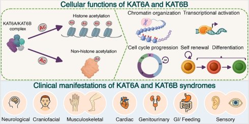
KAT6A and KAT6B are central acetyltransferases that acetylate histone H3 and non-histone proteins, thereby regulating the expression of many transcription factors and developmental genes, and modulating a wide array of key signaling pathways, such as Wnt/β-catenin and MAPK, as well as cellular pathways. These include chromatin organization, cell cycle regulation, proliferation, differentiation, maintenance of mesenchymal, hematopoietic and neural stem cells, as well as orchestration of multiple developmental programs, including hematopoietic, neurogenic, skeletal, and craniofacial development. Consistent with these key roles, heterozygous pathogenic variants in KAT6A and KAT6B result in neurodevelopmental syndromes. These syndromes are characterized by mild to severe intellectual disability, developmental delay including motor and speech delay, and a variety of malformations. This review provides a comprehensive summary of current evidence highlighting the impact of KAT6A and KAT6B on developmental programs and neurodevelopmental disorders resulting from pathogenic variants in KAT6A and KAT6B. Moreover, we describe the cellular and animal models of KAT6A and KAT6B syndromes, which are essential for studying and deciphering the molecular etiologies underlying the manifestation of the clinical phenotypes of these syndromes. An in-depth understanding of the molecular and cellular pathways impaired in KAT6A and KAT6B syndromes is essential for guiding future development of possible therapeutic interventions.
Keywords: KAT6A, KAT6B, lysine acetyltransferase, acetylation, epigenetics, gene expression, neurodevelopmental syndromes
Introduction
Epigenetic modifications are reversible chemical alterations of DNA or histones that regulate chromatin remodeling and gene expression [1-3]. Histone modifications include the extensively studied methylation and acetylation, as well as a multitude of histone acylations including propionylation, butyrylation, crotonylation, lactylation, succinylation, malonylation, glutarylation, 2-hydroxyisobutyrylation, and β-hydroxybutyrylation [3-5]. Accurate orchestration of histone modifications is essential for precise levels and spatiotemporal regulation of gene expression [6,7]. Proper cell proliferation, differentiation, and development rely on high levels of regulatory control over gene expression, which is crucial for various developmental processes, including skeletogenesis, craniofacial morphogenesis, hematopoiesis, and neurogenesis [7-10]. Accordingly, pathogenic variations in genes encoding histone-modifying enzymes that result in aberrant epigenetic regulation are frequently associated with neurodevelopmental disorders, which predominantly exhibit dominant inheritance [6,7,11-13]. These disorders are collectively called Mendelian disorders of epigenetic machinery (MDEM) or Mendelian disorders of chromatin machinery (MDCM) [11,14,15]. Among the histone-modifying enzymes are lysine acetyltransferases (KATs), including KAT6A and KAT6B, which catalyze ε-lysine acetylation on histone tails as well as on non-histone proteins [16]. Histone lysine acetylation promotes transcription by neutralizing the positive charges on histones, thereby weakening their interaction with negatively charged DNA and facilitating an open chromatin state. Similarly, post-translational acetylation of non-histone proteins neutralizes the positive charge of lysine residues, thereby resulting in modulation of protein conformation and function, stability, protein-protein or protein-DNA interaction and subcellular localization. Consequently, acetylation of histone and non-histone proteins impacts multiple cellular and physiological processes, including transcription, mitosis, autophagy, liquid-liquid phase separation, differentiation, and neural function [16].
KAT6A was first identified as part of a fusion transcript to CREB binding protein (CBP) in acute myeloid leukemia (AML) [17]. Its homolog KAT6B was subsequently identified a few years later [18]. In the past 3 decades, both KAT6A and KAT6B have been identified in chromosomal translocations and as aberrantly expressed in a multitude of cancer types, mostly overexpressed and operating as oncogenes [19,20]. While the role of KAT6A and KAT6B in malignancies is well-documented, pathogenic heterozygous KAT6A and KAT6B variants have subsequently been identified and linked to neurodevelopmental and intellectual disability syndromes, which have been studied in the past 15 years [19-21].
The role of KAT6A/KAT6B in cancer, as well as advances in the development of KAT6A/KAT6B inhibitors and their evaluation in preclinical and clinical trials, were recently described in detailed reviews [20,22,23]. In the current review, we focus on the roles of KAT6A and KAT6B in development and discuss findings from recent studies that explored how these central acetyltransferases regulate developmental programs, and specifically neurodevelopment, as well as the role of heterozygous pathogenic variations in KAT6A/KAT6B in neurodevelopmental disorders.
KAT6A and KAT6B: Structure and acyltransferase function
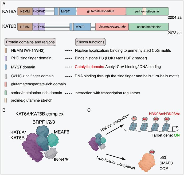
KAT6A (formerly named MOZ and MYST3) and KAT6B (aka MORF and MYST4) are two key members of the KAT family of lysine acetyltransferases, including KAT2A (GCN5), KAT2B (PCAF), KAT3A (CBP), KAT3B (p300), KAT5 (TIP60), KAT7 (HBO1, MYST2) and KAT8 (MOF, MYST1) [24]. The KAT family of lysine acetyltransferases is categorized into three principal families, GNAT, MYST, and p300/CBP, based on their conserved structural domains and function. KAT6A, KAT6B, and KAT7, which belong to the MYST family (along with KAT5 and KAT8), serve as alternating catalytic subunit components of a multi-subunit complex comprised of BRPF1/2/3, ING4/5, and MEAF6 (Figure 1A-B) [25-27]. This complex, when comprised of KAT6A or KAT6B, acylates histone H3 on lysine residues 9 and 23 (H3K9 and H3K23), as well as diverse non-histone proteins (Figure 1C). Among these proteins are p53, SMAD3, and COP1 (constitutive photomorphogenesis protein 1) [28-30]. Through these post-translational modifications, KAT6A and KAT6B regulate a broad range of key physiological and pathological processes, including transcription, cell cycle progression, differentiation (e.g., hematopoiesis, neurogenesis, skeletogenesis), DNA repair, maintenance of hematopoietic and neuronal stem cells, developmental programs, and tumorigenesis [20,27]. KAT6A and KAT6B possess identical domain architecture, as shown in Figure 1A, and high sequence homology, sharing 60% amino acid identity and 66% similarity [18]. They bind Acetyl-CoA through their conserved MYST domain [18,31]. The zinc finger domain and helix-turn-helix motifs within the MYST domain enable the binding of the MYST domain of KAT6A to DNA [32]. The conserved region in the N-terminal domain is called the NEMM (N-terminal part of Enok, MOZ or MORF) domain. The NEMM contains a region that is essential for the nuclear localization of the acetyltransferases, as well as two winged helix motifs (WH1 and WH2) and a double plant homeodomain (PHD) zinc finger domain, which interacts with histone H3 [33-35]. It was demonstrated that while both WH1 and WH2 motifs, located in the NEMM, bind to DNA, WH1 specifically enables binding of KAT6A/KAT6B to unmethylated CpG motifs and colocalizes with H3K18ac-rich CpG regions [36-38]. H3K18 is the acetylation substrate of the acetyltransferase p300, and notably, KAT6B was shown to interact with the TAZ domain of p300 via a hydrophobic motif within its WH1 domain [38]. This interaction suggests a functional cooperation of KAT6B and p300 in transcriptional regulation. KAT6A/KAT6B recruitment to the promoters of their target genes and subsequent acetylation activity relies on the DNA-binding capacity of the WH domain [37]. The unstructured C-terminal segment contains a glutamate/aspartate-rich region and serine/methionine-rich region. Within its serine/methionine-rich region, KAT6A contains a 55 amino acid stretch composed solely of proline and glutamine residues, which is absent in KAT6B [31,39]. In this respect, this intrinsically disordered region appears to contribute to liquid-liquid phase separation and the formation of biomolecular condensates of KAT6A in the nucleus of ovarian cancer cells [40]. The serine/methionine domain is essential for the interaction with transcription co-activators and the transcription activation capacity of KAT6A/KAT6B [31,35,41]. KAT6A/KAT6B recognize (i.e. 'read') and bind to specific histone acylation marks through their double PHD finger domain [42-44]. This interaction promotes their enzymatic activity (i.e. 'writing') and transcription at specific loci. Specifically, KAT6A recognizes unmodified H3R2 and H3K14ac, the latter of which is the acetylation substrate of KAT7 [42,45]. The association of the PHD finger domain with H3K14ac contributes to the localization of KAT6A to the promoter of HOXA9 in HEK-293 cells. In addition to acetylated H3K14, KAT6A also binds H3K14 harboring other acyl modifications such as crotonyl, butyryl, and propionyl groups, with a preference to crotonylated H3K14 [43]. Similarly, KAT6B recognizes H3K14 acylation, which promotes its role in H3K23 acetylation at specific loci [44,46]. While most studies describe the acetyltransferase function of KAT6A/KAT6B, both enzymes, as well as other members of the MYST family of lysine acetyltransferases, exert propionyl transferase activity as well [47,48].
KAT6A/KAT6B lysine acetyltransferases: structure and enzymatic function. A) Schematic representation of KAT6A/KAT6B's protein domain architecture, including representation of color-coded domains and regions: the N-terminal NEMM (contains two highly conserved winged-helix domains, WH1/WH2), double plant homeodomain (PHD) zinc finger domains, catalytic MYST domain (contains a C2HC zinc finger and helix-turn-helix motifs), and, in the C-terminal region, the acidic-rich and serine/methionine-rich domains. Domain coordinates were derived from UniProt (KAT6A (Q92794) (NEMM, 1-205; PHD-type 1, 206-265; PHD-type 2, 266-313; catalytic/MYST domain, 488-778; C2HC MYST-type, 537-562; acidic-rich domain, 787-1420; serine/methionine-rich domain 1421-1996; proline/glutamine stretch, 1647-1701). KAT6B (Q8WYB5) (NEMM, 1-212; PHD-type 1, 213-272; PHD-type 2, 273-320; catalytic/MYST domain, 715-1008; C2HC MYST-type, 748-773; acidic-rich domain, 1069-1529; serine/methionine-rich domain 1530-2073). A description of the functions of each domain is included. B) The 4-subunit KAT6A/KAT6B complex is composed of the catalytic subunit KAT6A/KAT6B and the scaffolding subunit BRPF1/2/3 that interacts with the ING4/5 and MEAF6 subunits. C) The KAT6A/KAT6B complexes acylate lysine residues K9 and K23 on histone H3 tails as well as non-histone proteins. KAT6A/KAT6B histone acetylation is associated with a transcriptionally active chromatin state.
Developmental roles of KAT6A and KAT6B
Embryonic and tissue development
According to the Human Protein Atlas [49] and the Harmonizome 3.0 [50] databases, both KAT6A and KAT6B are ubiquitously expressed in human adult tissues, including the heart, brain, lung, intestine, muscles, and hematopoietic system, underscoring their roles in diverse developmental and adult tissue pathways [17,18,20]. Both acetyltransferases play crucial roles in maintaining adult tissue homeostasis through regulation of stem cells and the immune system [20]. Mouse Kat6b is expressed as early as the embryonic stem (ES) cell stage and within the inner cell mass of blastocysts [51,52]. KAT6B modulates chromatin compaction and influences the interactions of Oct4 and Nanog with chromatin targets in ES cells [51]. Kat6a and Kat6b are both expressed from early mouse developmental stages, with transcripts detected in whole embryos at E8.5, E9.5, and E10.5, as well as in mouse embryonic fibroblasts (MEFs) and fetal hematopoietic progenitors [53]. Notably, Kat6a mRNA is expressed at higher levels than Kat6b mRNA in embryos at E8.5-E10.5 as well as in several embryonic tissues including the heart, pharyngeal arches (transient embryological structures in vertebrates that develop into the face, jaw, ears, and neck) [54], and in LSK cells (a population of hematopoietic stem and progenitor cells) [53].
Depending on the genetic background, homozygous Kat6a-deficient mice display embryonic lethality or die at birth [55,56]. KAT6A is a regulator of the expression of diverse homeodomain transcription factor (TF) genes, which serve as master regulators of fundamental developmental programs [19,20,27]. KAT6A was found to regulate the expression of HOX family genes, which regulate body segment identity [57-60], DLX genes, which modulate craniofacial development [61], as well as the T-box TFs TBX1 and TBX5, which are involved in heart development [62,63]. In line with the attenuated expression of Tbx1 and Tbx5 in Kat6a-deficient mice, these mice display heart defects including interrupted aortic arch and ventricular septal defects [62,63]. Unlike Kat6a-deficient mice, Kat6b deficiency in mice is not embryonic lethal. However, Kat6b-deficient mice display growth retardation when compared to control littermates [52,64] and do not survive past weaning [65,66].
Remarkably, a point mutation that abrogates the catalytic acetyltransferase activity of KAT6A leads to a milder phenotype in mice compared to the complete loss of the KAT6A protein [67]. This finding suggests that KAT6A possesses non-catalytic functions presumably mediated by interactions with other proteins. These mice survive to term, albeit exhibiting postnatal lethality characterized by reduced survival rates post-weaning. Mice harboring the point mutation in the catalytic domain exhibit only a few differentially expressed genes compared to the null phenotype at embryonic stage E9.5. KAT domain mutant mice display abnormal development of a thinner atlas vertebra of the axial skeleton and diminished fetal liver hematopoietic stem cell activity, albeit not complete elimination of hematopoietic stem cells as in the null phenotype. Moreover, unlike the null phenotype, they do not exhibit homeotic transformations or aortic arch defects, and only one out of five fetuses displayed ventricular septal defects.
A point mutation that abolishes the acetyltransferase activity of KAT6B did not lead to differentially expressed genes, cardiac abnormalities, or hematopoietic stem cell activity. However, the combined mutation of both the catalytic domains of KAT6A and KAT6B resulted in more severe phenotypes than the mutation in the KAT6A catalytic domain alone, yet less severe than that of the KAT6A-null mutation.
In a surprising and remarkable recent paper, it was demonstrated that Kat6b overexpression (4-fold upregulation) rescues embryonic lethality in homozygous null Kat6a mice, thereby restoring embryonic vitality [53]. Kat6b overexpression restored acetylation at histone H3 lysine 9 and 23 and reversed critical gene expression anomalies in Kat6a mutant mice. These findings imply that the target gene specificity of KAT6A can be substituted, at least in part, by the related paralog KAT6B, despite differences in amino acid sequence, as long as KAT6B is expressed at sufficiently high levels. The authors suggested that KAT6B overexpression in the absence of KAT6A yields a more favorable stoichiometry of complex subunits compared to endogenous levels of KAT6B, or KAT6B overexpression in the presence of KAT6A, enabling KAT6B to functionally substitute for KAT6A. Notably, KAT6B does not normally regulate critical processes which depend on KAT6A, as evidenced by distinct individual knockout phenotypes: Kat6a-null mice display embryonic lethality and hematopoietic stem cell loss, whereas Kat6b-deficient mice primarily display neurogenesis defects [52,53,55,56]. The clear phenotype observed in haploinsufficient Kat6a and Kat6b mouse models suggests that there is no compensation between KAT6A and KAT6B when they are expressed at endogenous levels [65,68]. Additionally, endogenous KAT6B expression levels were not upregulated following KAT6A deficiency to compensate for KAT6A loss in SH-SY5Y cells or neural progenitor cells (NPCs) differentiated from iPSCs derived from a KAT6A patient [69]. Collectively, these findings highlight the crucial role that these two central acetyltransferases play in development. Their roles in various developmental processes are discussed in greater detail in the subsequent sections.
Craniofacial and skeletal development
The zebrafish ortholog of KAT6A was found to be essential for the segment identity of pharyngeal arches two to four [58]. The impact of Kat6a deficiency on segment identity in zebrafish was attributed to the Kat6a-dependent regulation of hoxa2b and hoxb2a expression [58]. These genes function as "selector genes" which instruct this tissue to generate the support skeleton for the jaw instead of duplicating the lower jaw structure. Interestingly, in contrast to mice in which loss of a single Hoxa2 gene results in severe deformities, these genes in zebrafish function redundantly, and only when both genes are knocked down simultaneously severe developmental defects occur, specifically the transformation of second-arch cartilages into duplicate jaw structures. Similarly, Kat6a-deficient mouse embryos displayed homeotic transformation of anterior body segments that give rise to the neck and trunk region [57]. Kat6a deficiency in mice led to reduced expression of multiple Hox genes, which was accompanied by decreased H3K9 acetylation at these genes' loci [57,60]. Loss of function (LoF) of KAT6A in mice results in craniofacial anomalies in the jaws, including cleft palate [62]. Among the differentially expressed genes, 19 genes were related to cleft palate, including Dlx1, Dlx2, Dlx3 and Dlx5, as well as several genes regulated by DLX TFs [61]. KAT6A was shown to directly bind to the Dlx5 locus and maintain its H3K9 acetylation. In addition to the downregulation of multiple genes involved in craniofacial and specifically palate development, several bone collagen genes as well as genes involved in osteoblast differentiation, such as Runx2 and Nkx3-1, were upregulated in the pharyngeal arches and palatal shelves [61]. The upregulation of these genes could result from premature osteogenic lineage differentiation during palatal shelf development [61].
KAT6A was suggested to play a role in tooth formation via the regulation of odontoblast differentiation [70]. KAT6A overexpression increased the expression of the TFs Runx2 and Sp7, which are essential for commitment and differentiation, as well as genes encoding early and late markers of odontoblast maturation and mineralization, Alp, Col1a1, Ocn, Dspp, and Dmp1, in rat MDPC-23 dental pulp cells. The authors further demonstrated that KAT6A and RUNX2 are recruited to the promoters of Dspp and Dmp1 in MDPC-23 cells, and KAT6A overexpression synergistically increased Dspp promoter activity with RUNX2 in HEK-293T cells. Notably, both KAT6A and KAT6B interact with RUNX2 via their C-terminal serine/methionine-rich domain [41]. KAT6A also binds and promotes H3K9 acetylation on the chromatin of RUNX2 in HEK-293 cells, suggesting a role for KAT6A in regulating RUNX2 expression, in addition to functioning as its co-activator [36].
KAT6A expression is reduced in human aging bone marrow-derived mesenchymal stem cells (BM-MSCs), leading to impaired stemness characterized by decreased proliferation, self-renewal, and osteogenic differentiation [71]. BM-MSCs are essential for bone and cartilage formation, and decreased stemness of BM-MSCs has been associated with osteoporosis [72-74]. Using gain- and LoF experiments in young and old BM-MSCs, it was found that KAT6A promotes proliferation, colony formation, and osteogenic differentiation of BM-MSCs through the regulation of the Nrf2/ARE antioxidant signaling axis and inhibition of reactive oxygen species (ROS) accumulation [71]. KAT6A regulated the expression of the osteogenic-related genes RUNX2, OCN, and BMP2 following osteogenic differentiation of BM-MSCs.
Consistent with a broader role in bone remodeling, KAT6A is elevated in RANKL-induced osteoclast cell cultures and in the serum of patients with osteoporosis [75]. Knockdown of KAT6A in an in vitro model of osteoclast differentiation from mouse bone marrow-derived macrophages (BMDMs) and RAW264.7 cells revealed reduced capacity of osteoclast differentiation following stimulation [75]. Mechanistically, KAT6A binds the Tet1 promoter, increases H3K9 acetylation and RNA Pol II recruitment, resulting in upregulated Tet1, which promotes the transcription of TRPV4 (Transient Receptor Potential Cation Channel Subfamily V Member 4). TRPV4 is a nonselective calcium-permeable channel that is essential for osteoclast differentiation, function, and bone resorption [76].
Collectively, these findings indicate that KAT6A plays an important role in bone homeostasis by maintaining BM-MSCs, regulating the expression of key osteogenic genes, such as RUNX2 and DLX, and regulating osteoclastogenesis.
Interestingly, in addition to its functions during skeletal development, KAT6A has also been implicated in degenerative cartilage pathology. KAT6A was identified as a major driver of cartilage senescence in osteoarthritis [77]. In this respect, the long noncoding RNA ELDR mediates the formation of a protein complex consisting of KAT6A and hnRNPL, thereby altering chromatin accessibility. Specifically, this complex regulates histone modifications at the promoter region of IHH (Indian Hedgehog), a crucial regulator of osteoarthritis, thereby activating the Hedgehog signaling axis, resulting in chondrocyte senescence and cartilage degradation.
During mouse embryogenesis, Kat6b expression is upregulated within the developing craniofacial and dental structures, including the frontonasal and maxillary prominences at E11.5 and E12.5, eyelids at E15.5, and in tooth primordia [52,64,78]. Similar to Kat6a, Kat6b deficiency in mice results in craniofacial abnormalities, including microphthalmia (smaller eyes) and more ventrally positioned ears [52,64]. Mice homozygous for a Kat6b hypomorphic allele (90% Kat6b deficiency) exhibited irregular development of calvarial bones (i.e., the skullcap bones, including the frontal, parietal, and occipital bones) without an observed craniosynostosis [52]; the latter is defined as the premature fusion of the cranial sutures, hence causing an abnormal head shape. Additionally, mice homozygous for a Kat6b null allele presented shorter frontal bones and jaws [65].
The irregular craniofacial development in Kat6a- and Kat6b-deficient mice is consistent with craniofacial abnormalities presented by individuals harboring monoallelic pathogenic variants in KAT6A and KAT6B [19,20,79,80]. KAT6B pathogenic variants frequently lead to additional skeletal malformations, such as in the spine, patella, and digits [19,81]. In this respect, KAT6B was found to be epigenetically silenced in patients with congenital scoliosis (CS) [82]. CS is characterized by perturbations in spine development. KAT6B downregulation in chondrocytes from CS patients led to their reduced proliferation, presumably due to the observed decreased RUNX2 expression. It is well established that RUNX2 is a master TF that regulates the entire skeletal developmental program [83,84]. In this respect, RUNX2 is crucial for the differentiation of osteoblasts and the maturation of chondrocytes. It further plays a complex and significant role in their proliferation. Hence, RUNX2 is important during cartilage and bone formation. Kat6b-deficient mice exhibited an impaired femur growth plate, suggesting a role of KAT6B in chondrocyte differentiation [64]. Consistently, KAT6B was highly expressed in the undifferentiated mesenchyme of the skull and in the mesoderm surrounding cartilage of the limbs [52,64]. Kat6b deficiency in mice led to increased chondrocyte hypertrophy and premature ossification, resulting in increased bone density in the skull, shortened skeletal elements, and thickened bone [85]. Kat6b deficiency did not alter chondrocyte proliferation in growth plate regions. Chondrocyte hypertrophy is the process of an increase in the chondrocyte volume prior to a shift from a collagen-rich to a calcified matrix in the remodeling of cartilage into bone tissue [86]. The phenotype observed in Kat6b-deficient mice is consistent with skull malformations and craniosynostosis observed in some individuals harboring pathogenic heterozygous KAT6B variants [81,87,88]. In line with the observed premature ossification, Kat6b-deficient MEFs that were induced to differentiate into osteoblast-like cells (pre-osteoblasts), displayed increased expression of genes associated with osteoblast development, compared to control MEFs [85]. Among the upregulated genes were several genes whose gain-of-function is associated with craniosynostosis in humans. Consistent with the above findings, Runx2 was upregulated following osteogenic induction. Conversely, unlike wild-type (WT) pre-osteoblasts, Sox9 was not upregulated in Kat6b-deficient pre-osteoblasts. Notably, SOX9 and RUNX2 are antagonistic regulators of chondrocyte fate during growth plate maturation and ossification [89]. Additionally, SOX9 and RUNX2 can physically interact and reciprocally inhibit each other's transactivation [90,91]. SOX9 is an essential key regulator of chondrocyte lineage commitment and is downregulated during chondrocyte dedifferentiation/osteoblastic redifferentiation [86,89,92]. SOX9 maintains chondrocyte identity and prevents premature hypertrophy, dedifferentiation, and transition towards an osteoblast fate, whereas RUNX2 promotes chondrocyte hypertrophy and osteoblast differentiation. [89,93].
Maintenance of hematopoietic stem cells and the development of the immune system
Both KAT6A and KAT6B acetyltransferases were found to function in the maintenance of hematopoietic stem cells (HSCs) [55,56,94-98]. Deletion of Kat6a or a mutation that results in catalytic inactivation of KAT6A leads to a reduced number of fetal and adult HSCs and loss of quiescent adult HSCs [55,56,99,100]. KAT6A suppresses senescence via inhibition of the INK4A-ARF pathway in hematopoietic and neural progenitors, as well as in fibroblasts [97,101].
While most studies described the significant role of KAT6B in relation to neural stem cells, KAT6B was also found to regulate hematopoietic stem cells [95]. In this respect, KAT6A haploinsufficiency reduced H3K23ac, whereas KAT6B haploinsufficiency diminished H3K9ac in fetal hematopoietic stem cells and progenitor cell populations. Similar to the loss of KAT6A, KAT6B deficiency led to a reduced number of HSCs as well as a reduced number of quiescent cells and a greater number of cells in G1 phase of the cell cycle. While KAT6A exerted a predominant regulatory impact, both KAT6A and KAT6B cooperatively regulated the transcription of hematopoiesis-associated genes including Pbx1, Meis1, Erg, Flt3, and genes belonging to the HOXA and GATA families [95]. HSCs harboring combined monoallelic loss of both Kat6a and Kat6b (Kat6a+/-Kat6b+/-) exhibited more severe transcriptional deficits than loss of either alone. Notably, among the downregulated genes in Kat6a+/-Kat6b+/- HSCs were Pbx1, Tie2 and Mpl, which are essential to maintain the quiescence of HSCs, as well as Meis1, Hoxa9, and Erg, which are implicated in self-renewal of HSCs [95]. These findings support cooperative functions in vivo. However, chromatin co-occupancy data for KAT6A and KAT6B are currently limited to leukemia models [102], leaving normal tissues uncharacterized regarding direct target-gene overlap. While global histone marks (like H3K23ac or H3K9ac) were mapped using the CUT&TAG (Cleavage under targets and tagmentation) technique in normal mouse primary tissues such as MEFs and neural stem and progenitor cells (NSPCs) [53,66,103], direct binding profiles for KAT6A/KAT6B were presumably limited by the lack of high-quality, reliable and specific antibodies against KAT6A and KAT6B, which often necessitates the use of a tagged fusion protein [66,68].
Interestingly, the above study revealed 126 genes co-occupied by both KAT6A and KAT6B in human AML MOLM-13 cells [102]. Among these genes were HOXA-cluster genes and MEIS1, suggesting that these genes are directly regulated by both KAT6A and KAT6B. Moreover, this study revealed that KAT6B loss had modest effects on leukemia cell growth compared to KAT6A KO, further suggesting the functional dominance of KAT6A over KAT6B in hematopoietic cells [102]. Studies in AML cell lines have demonstrated that KAT6A cooperates with the nuclear chromatin reader protein ENL (eleven-nineteen leukemia) [104,105]. The acetyl-lysine reader ENL binds H3K9ac catalyzed by KAT6A at gene promoters and cooperates with a network of chromatin factors to drive transcriptional elongation. While this cooperative writing-reading function was identified in malignant cells, whether this epigenetic crosstalk occurs under physiological conditions remains to be elucidated. Notably, KAT6A colocalized with ENL at promoter-proximal regions of HOXA5 and HOXA9 in HEK293T cells [105], suggesting that this functional interaction may occur under physiological conditions as well.
In addition to their role in the maintenance of HSCs, both KAT6A and KAT6B regulate lineage differentiation. KAT6A deficiency and downregulation of KAT6B reduced erythroid differentiation capacity, resulting in enhanced myeloid lineage differentiation [55,94]. KAT6A is crucial for the differentiation of monocytes into macrophages and is further implicated in their polarization into M1 macrophages [106,107]. M1 macrophages are essential for proinflammatory responses, whereas M2 macrophages have a role in anti-inflammatory responses and promote tissue repair [108]. In this respect, Kat6a was upregulated in mouse macrophages stimulated to induce M1 polarization [107], in macrophages that participate in inflammatory processes such as periodontitis [109] and sepsis-induced acute lung injury [110] in mouse models, as well as in peripheral blood samples of sepsis patients [110]. Conversely, Kat6a was downregulated in IL-4-stimulated (to induce M2 polarization) primary mouse BMDMs [106]. Kat6a downregulation in stimulated (to induce M1 polarization) mouse macrophage-like cell line and BMDMs led to impaired phosphorylation of AKT, ERK, p38, and JNK, which are involved in signaling pathways that are essential for the polarization of M1 macrophages [106,111]. Additionally, Kat6a downregulation resulted in reduced expression of pro-inflammatory cytokines, including IL-1β, tumor necrosis factor (TNF) α, and IL-6 [106,109,110]. A recent study has further demonstrated that inhibition of KAT6A in mouse peritoneal macrophages and BMDMs, using the KAT6A/KAT6B inhibitor WM1119, as well as KAT6A knockdown in M1-induced RAW264.7 macrophages, reduced multiple glycolytic genes, including Glut1, Hk2, Pkm, Eno1, and Ldha [110]. Consequently, glucose uptake and glycolysis were reduced, whereas mitochondrial respiration was increased. KAT6A inhibition led to the reduced expression of both HIF1α and c-Myc, which regulate the expression of genes encoding glycolytic enzymes and pro-inflammatory factors [110,112,113]. Moreover, KAT6A inhibition was associated with diminished PI3K-AKT-mTORC1 pathway, a central signaling axis supporting macrophage activation and M1 polarization. The PI3K-AKT-mTORC1 axis is associated with glucose uptake and glycolysis as well as regulation of the TFs MYC and HIF-1α [114]. In this respect, KAT6A drives MYC expression in AML cells, thus promoting tumorigenesis [104].
In contrast to KAT6A, KAT6B is downregulated following the activation of M1 macrophages, suggesting that KAT6A and KAT6B may serve different roles in the context of M1 macrophage polarization [107].
Apart from KAT6A's role in the differentiation of cells of the myeloid lineage, KAT6A was found to regulate processes in cells of the lymphoid lineage, such as B cells and T cells. B cell progenitor proliferation is dependent on KAT6A, which was shown in mice to directly bind the Meis1 locus and regulate the expression of TFs that are critical for hematopoietic cell proliferation, such as Hoxa7, Hoxa9, Pbx1, and Meis1 [115].
Consistent with its function in hematopoietic self-renewal and differentiation, KAT6A binds to and co-activates TFs implicated in hematopoiesis, such as Runt-related transcription factor 1 (RUNX1 aka AML1) and PU.1, which are critical TFs for HSC maintenance, lineage specification, and differentiation [35,55,116-118].
KAT6A is also essential for the immune response by regulating crucial processes in memory B- and T- cell formation and function [119-121]. Specifically, KAT6A-deficient B cells displayed increased differentiation into IgM and low-affinity IgG1 memory B cells during primary response and decreased secondary high-affinity antibody-secreting cells [121]. By directly acetylating H3K9 in the promoter and E8I enhancer at the Cd8 locus, KAT6A regulates its expression in CD8+ T cells [119]. Consequently, KAT6A deficiency reduced CD8 coreceptor expression, which was associated with diminished T cell receptor signaling and effector-like memory T cells. KAT6A is also essential for the proliferation of CD4+ T cells and the differentiation of a subset population of pro-inflammatory Th1 and Th17 cells [120]. KAT6A exerts its role in CD4+ T cells via the regulation of expression of cell cycle- and glucose metabolism-related genes. KAT6A regulation of glucose metabolic reprogramming was dependent on the binding of MYC and HIF-1α TFs to these genes.
These data lend support to the importance of both KAT6A and KAT6B in the maintenance of hematopoietic stem cells as well as in the differentiation of several hematopoietic cell lineages.
Maintenance of neural stem cells and neurodevelopment
In addition to their role in the maintenance of hematopoietic stem cells, KAT6A and KAT6B also support the maintenance of neural stem cells (NSCs) [66,97,98,122]. Reduced proliferation and increased senescence of neural cells were identified both in vivo in the brain of mouse embryos harboring catalytically inactive KAT6A and ex vivo in cells isolated from the telencephalon of these mice to generate neurospheres [97]. Importantly however, a recent study revealed that while homozygous loss of Kat6a expression in mouse embryonic NSPCs from the dorsal telencephalon led to reduced proliferation, haploinsufficiency of Kat6a impacted the differentiation of NSPCs but not their proliferation [103]. Complete loss of Kat6a expression resulted in a moderate reduction in global H3K23ac in NSPCs. In-depth chromatin profiling revealed a Kat6a dose-dependent reduction in H3K9ac and H3K23ac in promoters, enhancers, and gene bodies in differentiating NSPCs. Proliferating NSPCs exhibited a similar trend of reduced H3K23ac, whereas H3K9ac was reduced only in Kat6a-deficient cells. KAT6A deficiency in differentiating NSPCs led to a decreased expression of multiple genes associated with nervous system development. The list of downregulated genes includes SOX family genes and TF-encoding genes that were previously shown to be downregulated following KAT6A deficiency in other cell types; these include Myc [104,110], DLX family members [61], and MEIS genes [104,115]. Interestingly, KAT6A deficiency was associated with diminished recruitment of the methyltransferase MLL1 (Mixed-Lineage Leukemia protein-1, KMT2A) to its target genes, accompanied by reduced H3K4 methylation at these loci. These alterations were positively correlated with observed changes in gene expression levels. Importantly, MLL1 is a central epigenetic regulator, acting as a transcriptional activator that is essential for hematopoiesis, neural progenitor proliferation and neuronal differentiation [123-125]. Consistently, KAT6A was previously shown to impact MLL1 recruitment to HOX gene loci in mouse embryos [57]; moreover, KAT6A and MLL1 cooperatively regulate the expression of HOX genes in human cord blood CD34+ cells [126]. Evidence for the cooperative regulation of gene expression by KAT6A and MLL1 was additionally demonstrated in B-cell progenitors [115] and HEK-293T cells, where they colocalized at the promoters of many transcriptionally active genes, including MYC and HOXA9 [127], in REH acute lymphoblastic leukemia (ALL) cells [127], AML MOLM-13 cells [102], and in gastrointestinal stromal tumor cells [128].
Kat6b-deficient mouse embryonic stem cells (mESC) exhibit reduced capacity of neuronal differentiation [51,129]. Recently it was shown that KAT6A and KAT6B are essential for the activation of genes at bivalent chromatin during an in vitro neuronal differentiation from ESCs [129]. Bivalent nucleosomes are asymmetric for the repressive histone mark H3K27me3 and the activating histone mark H3K4me3 [130]. A possible role for these bivalent domains in ESCs is to poise genes for timely activation or repression upon cell-lineage specification and differentiation. Histone bivalency also occurs in NPCs and in neurons, albeit to a lesser extent [130,131]. KAT6B and its complex subunits BRPF1/2 and ING5 specifically bind asymmetric bivalent nucleosomes in ESCs [129]. Moreover, KAT6B preferentially binds bivalent over monovalent H3K4me3 at promoter sites. The recruitment of the KAT6B complex to bivalent nucleosomes is presumably mediated by the ability of the MYST domain of KAT6B to act as an H3K27me3 reader and the recognition of the H3K4me3 mark by the PHD finger domain of ING5 [129,132]. H3K23ac was markedly reduced in Kat6b-deficient mESC and to a lesser extent in Kat6a-deficient mESC [129]. However, KAT6A- or KAT6B deficiency did not affect the proliferation and maintenance of mESC, nor the expression of genes in ESCs. Sequential differentiation of mESC towards glutamatergic neurons revealed that KAT6A and KAT6B are essential for glutamatergic neuronal differentiation. The expression of neural progenitor as well as early and mature neuronal marker genes was preferentially downregulated in genes associated with bivalency at their promoters. Loss of KAT6A/KAT6B preferentially affected bivalent genes that are activated during neuronal differentiation, indicating that these acetyltransferases preserve a poised chromatin state that is required for proper developmental gene induction.
KAT6B expression increases during cerebral cortex development, and KAT6B was found to be crucial for neurogenesis and the development of the cerebral cortex [52,66]. Loss of KAT6B in embryonic NSPCs derived from the dorsal telencephalon of mice impaired neuronal differentiation and neurite outgrowth, in addition to reducing proliferation and self-renewal of NSPCs in vitro [66]. Conversely, Kat6b overexpression promoted neurite outgrowth in cortical neurons [133]. Consistently, Kat6b-deficient mouse embryos displayed a smaller cortical plate and adult cortex with fewer layer V pyramidal neurons and GAD67-positive interneurons [52]. Apart from embryonic neurogenesis, KAT6B displays a role in adult neurogenesis too. KAT6B gene expression is high in the neurogenic subventricular zone of adult mice, and Kat6b deficiency in mice resulted in reduced self-renewal of NPCs, a reduced number of neuroblasts in the subventricular zone, as well as diminished capacity for neuronal differentiation in vitro, along with smaller olfactory bulbs [122]. Conversely, overexpression of Kat6b in mice led to enhanced self-renewal of subventricular zone-derived NSPCs and skewed differentiation towards a neuronal rather than an astrocytic fate in vitro and in vivo [133].
The molecular function and gene targets of KAT6B in neurogenesis have only recently begun to be deciphered. Using Kat6b-deficient and -overexpressing mice, KAT6B was found to catalyze global H3K9ac in embryonic NSPCs and global H3K9ac and H3K23ac in the dorsal telencephalon, at E12.5 [66,133]. H3K23ac was reduced at promoters, gene bodies, and active enhancers at specific genomic sites in Kat6b-deficient NSPCs. Transcriptomic profiling of Kat6b-deficient NSPCs revealed a reduction in the expression of nervous system development-associated genes and neural precursor proliferation genes, which was positively correlated with a reduction in H3K9ac. This gene list included early regulators of neuroepithelium, brain development, and NSC function, including Sox1, Sox2, Pax6, and Id2, and genes related to neurite elongation [66]. Loss of KAT6B also led to downregulation of brain and neuronal development genes in the dorsal telencephalon, which was established to give rise to the cerebral cortex, at E12.5. Among the downregulated genes in both NSPCs, E12.5 dorsal telencephalon, and E15.5 cortex were many SOX family genes, which are implicated in neural precursor proliferation and brain development [134]. KAT6B was found to bind the genomic loci of SOX2 and PAX6 and directly regulate their gene expression.
Congruently, many upregulated genes in Kat6b-overexpressing NSPCs and dorsal telencephalon were associated with nervous system, neuronal development and maturation genes [133]. Interestingly, fewer genes were detected as differentially expressed in more mature areas, i.e., the cortex at E15.5, when KAT6B was deficient or overexpressed, suggesting a possible role in early developmental stages of neurogenesis or an increased cell type complexity, which masks differential expression of genes in specific cell types [66].
Collectively, these studies reveal that KAT6B is a key epigenetic regulator of neurodevelopment, controlling neural stem/progenitor proliferation, neuronal differentiation, and cortex formation. While KAT6A was shown to be essential for NSPCs proliferation and early differentiation, as stated above, its role during the early stages of neurogenesis has just begun to be elucidated and is further discussed later in this review.
A critical goal in understanding KAT6A and KAT6B syndromes involves resolving KAT6A/KAT6B cell-type-specific functions within the developing brain. Bulk assays used in the studies described above mask cell-type-specific perturbations, particularly given the complex cellular heterogeneity of the developing brain [135]. Future studies leveraging single-cell RNA sequencing (scRNA-seq) and scATAC-seq (single-cell assay for transposase-accessible chromatin with high-throughput sequencing) could potentially determine whether impaired function of KAT6A/KAT6B impacts specific cell types, progenitor subtypes, and neural lineages. Importantly, the utilization of a combined approach of several omics, as was done with dermal fibroblasts obtained from KAT6A patients [136] and NSPCs obtained from Kat6b-deficient mice [66], is beneficial in order to comprehensively profile the epigenome and transcriptome changes caused by KAT6A and KAT6B deficiency in neurodevelopment. This multi-omics approach, including the utilization of scATAC-seq and CUT&RUN (Cleavage under targets and release using nuclease) or CUT&TAG followed by sequencing, combined with scRNA-seq, could profile chromatin accessibility and link epigenomic alterations with transcriptional changes at specific loci within specific cell types.
KAT6A/KAT6B-associated neurodevelopmental disorders
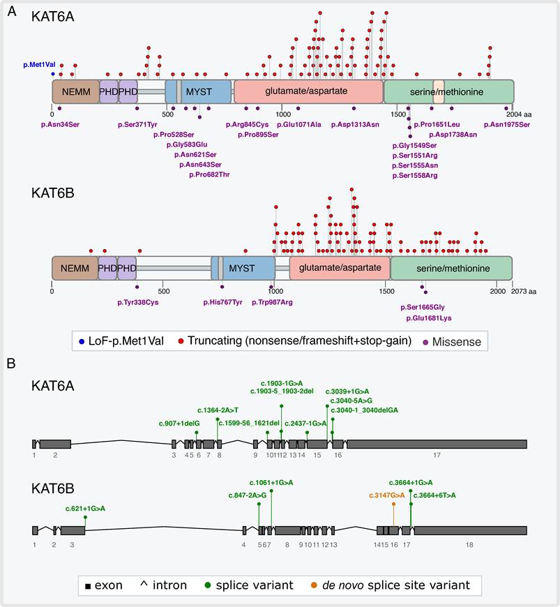
The established roles of KAT6A and KAT6B as pivotal epigenetic modifiers, particularly in regulating NSC homeostasis and neurodevelopment, underscore why their dysregulation has been linked to serious neurodevelopmental disorders. While pathogenic KAT6A variants are associated with KAT6A syndrome, aka Arboleda-Tham syndrome (ARTHS, OMIM#616268) [137-139], pathogenic KAT6B variants are associated with Say-Barber-Biesecker-Young-Simpson variant of Ohdo syndrome (SBBYSS, OMIM #603736) [78], Genitopatellar syndrome (GTPTS, OMIM #606170) [140,141], Noonan syndrome-like phenotype [64] and intermediate or combined phenotypes [81,142,143], which are now collectively referred to as “KAT6B disorders” [80,81,88]. Pathogenic KAT6A and KAT6B variants are predominantly heterozygous de novo truncating variants [79,81,144,145], although a few inherited variants were reported as well (Figure 2; Supplementary Tables S1, S2) [81,144,146-151]. Missense variants are less common and are often located within functionally critical domains, such as the MYST acetyltransferase domain or the acidic and serine/methionine-rich domains, where they are predicted to impair enzymatic activity or protein-protein interactions (Figure 2A) [19,79]. Several splice variants were reported as well (Figure 2B) [80,81,139,144,148,152-155]. The pathogenic variants of KAT6A that were described to date occur throughout the gene, although there is an evident clustering of truncating variants (64%, 61/95 truncating variants) in the glutamate/aspartate region of KAT6A, which is encoded by the last exons (exon 15 through the proximal region of exon 17) (Figure 2A; Supplementary Table S1). As for KAT6B, pathogenic variants that were previously reported predominantly reside in the glutamate/aspartate and serine/methionine regions, which are encoded by exons 16-18 (Figure 2A; Supplementary Table S2). Interestingly, all GTPTS-associated variants cluster within the distal part of exon 17 and the proximal region of exon 18 (reported variants mapped between amino acids 1194-1515 in the acidic domain; Supplementary Table S2) [81,88,156].
Distribution of pathogenic variants associated with KAT6A/KAT6B neurodevelopmental syndromes. A) Pathogenic variants associated with KAT6A- and KAT6B-related neurodevelopmental disorders are plotted according to their relative position along the schematic representation of the protein domains. A loss of function variant (p.Met1Val) abolishing KAT6A's start-codon is indicated by a blue lollipop and label. Truncating variants (nonsense and frameshift variants that lead to a premature stop codon) are shown above the protein (red lollipops); missense variants are shown below the protein (purple lollipops and labels). B) Splice site variants (green lollipops and labels) are plotted along the exon-intron gene structure (exons/introns are scaled according to their relative size). Recurrent de novo alterations of the synonymous variant c.3147G>A (orange lollipop and label) (n=8) indicate the presence of a mutation hotspot, which is predicted to result in a protein truncation (p.Ala1008Argfs*62) through an aberrant, newly created cryptic splice site [185-189]. Pathogenic KAT6A/KAT6B variants that were previously reported for KAT6A/KAT6B syndromes are summarized in Supplementary Tables S1 and S2. Recurrent variants are plotted in the figure only once to maintain figure clarity and to avoid redundancy of pathogenic variants in individuals that were described more than once in the literature.
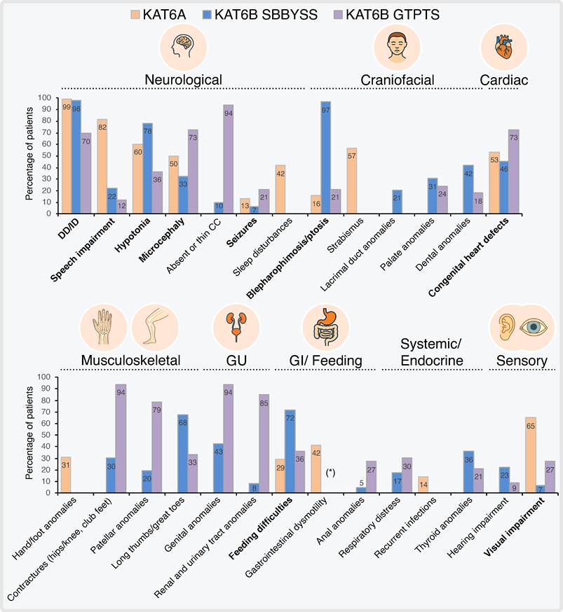
Common phenotypic features of KAT6-associated disorders include varying degrees of intellectual disability (ID), developmental delay (DD), and multisystemic anomalies (Figure 3) [80,81,144,157]. Additional common features include facial dysmorphisms, speech delay, hypotonia, seizures, microcephaly, feeding difficulties, as well as congenital heart defects. The most consistent facial features in KAT6A syndrome include a broad nasal tip and a thin, tented upper lip. Other characteristic features may include strabismus, bitemporal narrowing, a prominent nasal bridge and a flat philtrum. Some patients present epicanthic folds and low-set and posteriorly rotated ears, which are occasionally folded, and micro/retrognathia [144,157]. The hallmark of SBBYSS is the "mask-like" facies, blepharophimosis and ptosis. Lacrimal duct anomalies (e.g., dacryostenosis) are also more specific to this subtype of KAT6B syndrome [80,81]. Common KAT6B spectrum features include a nose with a bulbous end or a broad nasal bridge, low-set and posteriorly rotated ears, prominent cheeks, downslanting palpebral fissures, long philtrum, thin upper lip, thin lip vermilion, and micro/retrognathia [81,158]. While KAT6A and KAT6B syndromes exhibit some phenotypic overlap, each presents additional distinct clinical manifestations that further diverge between patients with GTPTS and with SBBYSS (Figure 3). Some individuals with KAT6A syndrome display digestive problems, sleep disturbances, motor disorders, visual problems, and autism features [144,152]. Common features of individuals with KAT6B syndrome include craniofacial and skeletal anomalies, such as a cleft palate; with some features that are more common in GTPTS patients, including patellar hypoplasia, contractures of the hips and knees, as well as genital anomalies [80,81,88,158]. Long thumbs or long great toes are seen particularly in the SBBYSS subtype. Similar to KAT6A syndrome patients, gastrointestinal complications, including gastroesophageal reflux and constipation, were reported for patients with KAT6B syndrome [79,81,88]. Sleep disturbances are not frequently reported in patients with KAT6B syndrome, but several patients were reported to have obstructive sleep apnea [88,143].
Phenotypic spectrum and prevalence of clinical features associated with KAT6A, KAT6B-SBBYSS, and KAT6B-GTPTS syndromes. The bar graph depicts the percentage of individuals presenting with major clinical features across KAT6A and KAT6B syndromes (including the SBBYSS and GTPTS subtypes), highlighting both shared and syndrome-specific phenotypic profiles. Shared clinical features across KAT6A and both KAT6B subtypes are indicated in bold. Clinical features are categorized into organ system domains: neurological, craniofacial, cardiac, musculoskeletal, genitourinary (GU), gastrointestinal (GI)/feeding, systemic/endocrine, and sensory. Percentages for each clinical feature are indicated within the bars. CC denotes corpus callosum. DD/ID denotes developmental delay/intellectual disability. (*) Gastrointestinal complications, including gastroesophageal reflux and constipation, were also reported in patients with KAT6B syndromes [81,88,158]. Data for the figure were collated from previous publications [79-81,157].
Magnetic resonance imaging (MRI) of the brain did not reveal structural anomalies in most KAT6A syndrome patients [144,157,159]. However, reported variations include microcephaly and infrequent malformations such as craniosynostosis, thinning of the corpus callosum, cisterna magna enlargement, Chiari malformation (i.e., the pushing of the cerebellum downwards into the spinal canal), and white matter hyperintensities. Conversely, KAT6B syndrome patients were more frequently reported to have structural malformations [81]. Approximately one-third of SBBYSS patients were found to bear microcephaly, and a few displayed agenesis or hypoplasia of the corpus callosum [80,158]. Structural anomalies reported for the majority of GTPTS patients included agenesis or hypoplasia of the corpus callosum and microcephaly. Some GTPTS patients were also reported to exhibit cortical malformations, hydrocephaly, or ventriculomegaly [158].
Several studies addressed the evaluation of the cognitive and behavioral functions of KAT6A syndrome patients in recent years [152,160-163], while one study also compared the cognitive profile of 13 KAT6B syndrome patients to that of 15 KAT6A syndrome patients [160]. These studies reported that both KAT6A and KAT6B patients exhibit difficulties in adaptive functioning as well as impaired non-verbal cognition and receptive language skills. KAT6A syndrome patients were also found to display impaired expressive language skills, while this skill remains to be systematically evaluated in KAT6B syndrome patients [12,152,160]. Notably, while both groups shared autism-related behaviors with low rates of externalizing behavior problems, only KAT6B syndrome patients exhibited reduced social motivation [160].
Two studies investigating the impact of distal versus proximal truncating variants in KAT6A on the severity of the phenotype reported a correlation between the position of the pathogenic variant and the level of ID [144,152]. Individuals carrying truncating variants in the last two exons exhibited a shift towards a more severe degree of ID than those with variants in the first 15 exons, including lower standard scores of adaptive behaviors, such as communication, daily living skills, and socialization [152]. However, two other studies did not find a correlation between early versus late truncating variants and the neuropsychological phenotype, due to the small number of participants [161,163]. Nevertheless, the authors suggested a trend towards relatively higher cognitive functioning in individuals carrying missense variants compared to those harboring truncating variants. Notably, specific missense variants in KAT6A and KAT6B were associated with milder phenotypes compared to truncating variants [19]. Across major cohorts, missense variants exhibit a reduced prevalence or complete absence of congenital heart defects [79,144]. A point mutation that abrogates the catalytic acetyltransferase activity of KAT6A was shown to lead to a milder phenotype in mice compared to KAT6A null mice, revealing that KAT6A possesses non-catalytic functions too [67]. Similarly, missense variants within the MYST domain of human KAT6A may selectively impair its catalytic activity, while preserving its non-catalytic functions. Additionally, KAT6A and KAT6B physically bind to the N-terminal region of the BRPF1/2/3 scaffolding subunit through their catalytic MYST domain [25]. BRPF1/2/3 acts as the structural core of the complex by binding both KAT6A/KAT6B and the ING4/5 and MEAF6 subunits. Consequently, missense variants located in the MYST domain may disrupt both KAT6A/KAT6B's catalytic acetyltransferase activity and BRPF1-mediated complex assembly. Conversely, missense variants in non-catalytic domains are more likely to disrupt scaffolding functions or specific protein-protein interactions. Missense variants in KAT6A or KAT6B located outside the MYST domain, such as those within the N-terminal NEMM domain, the double PHD fingers, or the C-terminal acidic and serine/methionine-rich regions, can selectively impair chromatin recruitment, histone/DNA binding, transactivation, and TF interactions [27,144]. Preserving partial functional integrity might account for the milder phenotypes observed with certain missense KAT6A variants. Clearly, additional studies with a larger sample size are warranted to determine whether there is a statistically significant correlation between the variant type and the resulting phenotype [161,163].
In vitro and in vivo modeling of KAT6A and KAT6B syndromes
In order to decipher the molecular and cellular mechanisms underlying KAT6-associated disorders, it is imperative to implement disease models, such as transgenic mice and patient-derived induced pluripotent stem cells (iPSCs). The advantages and limitations of each in vitro and in vivo model are summarized in Table 1. While the roles of KAT6A/KAT6B in development, and neurodevelopment in particular, could be explored in KAT6A/KAT6B-deficient or -overexpressing animal and cell models [51,52,57,66,97,103,122,133], KAT6A/KAT6B disorders result from pathogenic heterozygous mutations. Hence, these cells presumably express at least 50% WT transcript, and animal and cell models of these disorders should be heterozygous for the pathogenic KAT6A/KAT6B variants or present haploinsufficiency. By recapitulating the environment of cells harboring pathogenic KAT6A/KAT6B variants, these models could enable the systematic identification of molecular perturbations and dysfunctional regulatory networks and possibly the validation of potential therapeutic targets. In this respect, two recent studies explored the impact of Kat6a haploinsufficiency in mice on neurogenesis, cognitive function, and behavior [68,164]. The researchers used mice harboring a deletion of exon 4 or exons 4-8 in one Kat6a allele, which resulted in a premature stop codon, mirroring the genotype of KAT6A in patients harboring early truncating pathogenic variants [68,164], as well as mice with KAT6A haploinsufficiency restricted to the neocortex and hippocampus (Kat6aNEX- cKO mice) [164]. In contrast to the embryonic or perinatal lethality and early developmental arrest observed in complete Kat6a deficiency models, Kat6a-haploinsufficient mice survived to adulthood, although they displayed smaller body size [68,164]. Kat6a-haploinsufficient mice displayed impaired cognitive function, a deficit in memory, and reduced anxiety levels. A difference between these two studies was that while one study observed normal social behavior of Kat6a-haploinsufficient mice [164], the other study reported a slightly reduced preference for interaction [68].
Summary of advantages and limitations of in vitro and in vivo modeling of KAT6A and KAT6B syndromes.
| Ref. for KAT6 syndromes modeling | Limitations | Advantages | Key Features | Model Type |
|---|---|---|---|---|
| [64,65,68,88] | -Lacks patient-specific physiological, genomic, and chromatin architecture background. -Does not capture complex neurodevelopmental or tissue-specific pathology. -Overexpression can lead to non-physiological protein stoichiometry. | -Easy to culture, expand, and transfect efficiently. -Ideal for rapid biochemical assaying (e.g., measuring H3K9/H3K23 acetylation). -Enables straightforward structural and domain-function mechanistic studies (e.g., testing BRPF1 complex interactions) | -Heterologous mammalian in vitro expression system. -Enables transient or stable transfection/CRISPR gene editing to express wild-type or mutant KAT6A/KAT6B constructs. -Standard tool for investigating enzyme biochemistry and protein-protein interactions. | HEK293 cell line |
| [48,64,69,136,190] | -High patient-to-patient variability and line-to-line heterogeneity. -iPSC reprogramming and neuronal differentiation protocols are labor-intensive and costly. -Cannot replicate systemic whole-organism physiology or behavioral deficits. | -Highly clinically relevant human physiological model. -Useful for identifying disease-relevant downstream target genes and epigenetic biomarkers (e.g., histone acetylation status in blood/cells). -Provides a translationally relevant platform for testing therapeutic interventions. | -Derived directly from individuals with KAT6A or KAT6B neurodevelopmental syndromes. -Preserves patient-specific heterozygous variants (nonsense, frameshift, or missense) and background genome. -Can be differentiated into target cell types (e.g., neural progenitor cells, cortical neurons). | Patient-derived cells (e.g., dermal fibroblasts, iPSCs, peripheral blood cells) |
| [65,68,103,164] | -Interspecies differences between mouse and human genetic/developmental networks. -Resource-intensive, longer experimental timelines, and high maintenance costs. | -Captures full systemic phenotypes, including neurodevelopment, craniofacial features, heart, and hematopoiesis defects. -Allows evaluation of motor and cognitive skills, as well as social behaviors. -Essential for testing preclinical systemic efficacy, pharmacokinetics and pharmacodynamics of potential therapeutic compounds. | -Whole organism genetic models (e.g., haploinsufficient Kat6a+/- or Kat6b+/- mice). -Models complex gene-dosage effects (haploinsufficiency vs. full KO), systemic development, and neurocircuitry. | In vivo models (e.g., Kat6a/Kat6b transgenic mouse lines) |
Similar to the study on KAT6A, the impact of Kat6b haploinsufficiency on cognitive function and behavior was also explored in mice [65]. Unlike the homozygous Kat6b KO mouse models, which failed to survive past weaning, Kat6b-haploinsufficient mice survived to adulthood [65]. Kat6b-haploinsufficient mice exhibited impaired learning and memory as well. These mice also displayed reduced social recognition and interaction, resembling KAT6B syndrome patients [160]. Interestingly, KAT6B overexpression in mice resulted in aggression, anxiety, and seizures [133].
Similar to the majority of KAT6A syndrome patients, who were not reported to have structural anomalies of the brain, Kat6a-haploinsufficient mice had normal morphology of the brain, cerebral cortex, cortical layers, and ventricles [68]. Similarly, Kat6aNEX-cKO mice exhibited normal cortical morphology and hippocampal cell density [164].
The study by Liu et al. [164] revealed that Kat6a-haploinsufficient CA3 hippocampal neurons display decreased spine density and impaired synaptic activity. The impaired synaptic activity and cognitive function of the mice were mainly attributed to a reduction in Rspo2, whose expression is directly regulated by KAT6A, and consequently, RSPO2-mediated Wnt signaling was downregulated [164].
Consistent with the observations in fetal hematopoietic stem cells, Kat6a haploinsufficiency caused a reduction of H3K23ac in the brain and peripheral white blood cells of mice, as well as in human HEK-293T cells carrying several pathogenic heterozygous KAT6A variants [68]. In contrast, Kat6b haploinsufficiency induced a reduction of H3K9ac in the cortex of mice at 2 weeks of age (but not at 3 weeks of age) and in peripheral white blood cells of the mice [65]. These findings suggest that although both KAT6A and KAT6B were shown to acetylate histone H3K9 and H3K23 [19], each of them acetylates a preferential H3 lysine residue.
In line with the reduced H3K9ac levels resulting from Kat6b haploinsufficiency, Kat6b-deficiency in mouse embryonic dorsal telencephalon and adult cerebral cortex, as well as a lymphoblastoid cell line derived from a patient with a KAT6B variant, implicated dysregulation of the MAPK signaling pathway [64]. Specifically, expression analyses in the Kat6b-deficient mouse model revealed altered expression of genes related to the MAPK pathway. Moreover, the patient-derived cells exhibited hyperactivation of this pathway, specifically characterized by increased phosphorylation of MEK1/2, ERK1/2, and AKT. Clinically, this individual presented a Noonan syndrome-like phenotype. Noonan syndrome (OMIM #163950) shares several features with KAT6B-related disorders, including cognitive impairment, developmental delay, facial dysmorphism, cardiac defects, genitourinary anomalies, and skeletal abnormalities [165]. Notably, similar to the findings observed in the KAT6B patient-derived cells, Noonan syndrome is caused by pathogenic variants in the RAS/MAPK pathway genes that increase MEK1/2 and ERK phosphorylation. These results suggest a functional relationship between KAT6B deficiency and dysregulated MAPK/AKT signaling.
A recent study utilized KAT6A patient-derived iPSCs differentiated into NPCs and cortical neurons to model the KAT6A disorder [69]. This study revealed a significantly altered transcriptome in KAT6A-mutant NPCs and cortical neurons compared to their healthy counterparts. Interestingly, a great number of the dysregulated genes were found to be associated with autism spectrum disorder (ASD). KAT6A-mutant cortical neurons exhibited premature elevation of many key neuronal development genes at an early stage of the differentiation. This altered transcriptome apparently led to a precocious maturation, as demonstrated by the hyperexcitability and increased spatial coverage, dendritic arbor complexity, neurite width, and soma size of the KAT6A-mutant neurons at an early stage of the differentiation. Notably, these neurons exhibited a markedly lower percentage of GABA (gamma-aminobutyric acid)-positive neurons. KAT6A-mutant cortical neurons that were differentiated for a longer time became hypoexcitable compared to control neurons, apparently due to downregulation of key ion channel genes [69]. In line with these observations, Kat6a-haploinsufficient mice displayed upregulation of neuronal development and maturation genes, including axon guidance and synapse organization-related genes, in the developing dorsal telencephalon at E12.5, compared to control mice [68]. Strikingly, a similar phenotype of early maturation, hyperexcitability, and a reduced percentage of GABA-positive neurons was previously also found in neurons differentiated from iPSCs obtained from patients with ASD- and ID-related variants [69,166-168]. Collectively, these findings suggest that KAT6A downregulation impacts the timely maturation of neurons, leading to an early maturation of cortical neurons.
Evaluation of potential treatments in mouse models of KAT6A and KAT6B syndromes
To date, there is no direct treatment for KAT6A or KAT6B syndromes; patients therefore receive symptomatic and supportive care. These treatments include physical, feeding, speech and language therapies, as well as treatment against sleep disturbances and seizures [144,147,157,158,161,163,169]. Histone acetylation deficiency could potentially be treated by inhibition of histone deacetylases (HDAC), which are the epigenetic erasers of acetyl groups [170]. In this respect, treatment with HDAC inhibitors (HDACi) like valproic acid (VPA) was found to be neuroprotective in mouse models of brain injury and neurodegenerative diseases [170].
VPA is a well-established HDACi used as an FDA-approved treatment for various types of epilepsy and as a mood stabilizer, especially in the manic phase of bipolar disorder [171,172]. However, a major limitation of broad epigenetic drugs (such as HDACi like VPA) is their lack of organ and locus-specificity. Since they broadly alter global histone acetylation, they carry high risks of systemic toxicity [173]. Indeed, the clinical utility of VPA has been limited by substantial side effects, including neurotoxicity, especially when taken at certain developmental stages of a child's brain, hepatotoxicity and teratogenicity [173]. VPA's neurotoxic effects result from its ability to increase ROS production, cause mitochondrial dysfunction, alter epigenetics, influence signaling pathways and neurotrophic cascades, as well as shift the balance between excitatory/inhibitory neurotransmission. Additionally, HDACi administration causes gastrointestinal disturbances, immune dysregulation, and off-target effects [174]. Thus, VPA is currently serving as a scaffold for drug development of analogs aimed at augmenting treatment efficacy while diminishing untoward toxicity [175]. Notably, a precise balance of histone acetylation and deacetylation is crucial in neurodevelopment [176]. Similar to acetyltransferases, HDACs play essential roles during neurodevelopment, synaptic transmission, plasticity, and cognition [177,178]. Consequently, aberrant expression of HDACs leads to neurodevelopmental syndromes [179-181]. Mechanistically, treatment with HDACi such as VPA during specific postnatal periods significantly decreases neural stem cell progeny production in the dentate gyrus, rostral migratory stream, and olfactory bulb [182].
Another treatment approach is to increase the levels of Acetyl-CoA, the canonical acetyl donor substrate of acetyltransferases, in order to enhance histone acetylation. Following this rationale, Kat6b-haploinsufficient mice were treated with VPA, or with acetyl-carnitine (ALCAR), which is an acetyl donor [65]. Both VPA and ALCAR treatments resulted in increased acetylation levels of H3K9, H3K14 and H3K23 in the adult cortex. Notably, vitro treatment with VPA as well as with ALCAR partially restored gene expression in cortical neurons, which were isolated from Kat6b-haploinsufficient mice. While both VPA and ALCAR treatment partially reversed sociability deficits, ALCAR treatment further rescued learning and memory deficits in Kat6b-haploinsufficient mice. Kat6b-deficient mice displayed reduced GABAergic neurons [52], and the improved sociability could possibly be attributed to the reported ability of VPA to increase GABA levels, which is essential for social behavior [65,183]. Notably, while VPA treatment elicited some beneficial effects, it also exerted adverse effects on the motor skills of these mice. Similarly, in addition to cognitive and language delays, developmental motor delays are reported in children who were exposed to VPA during prenatal development [173].
Similar to the treatment of Kat6b-haploinsufficient mice, supplementation of ALCAR was found to restore H3K23ac levels in the brain of Kat6a-haploinsufficient mice and significantly improved their learning and memory, as well as reduced their hyperactivity [68]. However, in contrast to Kat6b-haploinsufficient mice, ALCAR treatment did not improve the sociability of Kat6a-haploinsufficient mice. It should be noted that KAT6A syndrome patients exhibit social motivation, and another Kat6a-haploinsufficient mouse model did not exhibit impaired sociability, as stated above [160,164].
Importantly, given that ALCAR and VPA exert pleiotropic effects, they could potentially ameliorate the phenotype of Kat6a- and Kat6b-haploinsufficient mice through compensatory mechanisms, rather than through the mere restoration of normal acetylation levels at specific gene loci [65,68]. Such mechanisms include the elevation of the neurotransmitters GABA, acetylcholine, noradrenaline and serotonin, as well as elevation of a glutamatergic receptor. Elevation of acetylcholine could result in the improvement of learning and memory [68,184].
In summary, the above findings reveal that treatment with an acetyl donor had beneficial outcomes in mouse models of KAT6A and KAT6B syndromes. However, additional studies are warranted to carefully examine and assess this treatment option for KAT6A and KAT6B patients.
While the efficacy and safety of treatment with epigenetic modulators constitute major concerns [174], utilizing in vitro and in vivo models of KAT6A and KAT6B syndromes aims to uncover specific downstream molecular perturbations. Pinpointing these molecular pathways could reveal novel targets for therapeutic interventions. Moreover, this downstream approach could be beneficial and circumvents the risks of gene overexpression, which has been shown to cause adverse effects with Kat6b overexpression [133].
Furthermore, establishing a precise therapeutic window is essential. As stated above, early postnatal treatments yielded favorable therapeutic effects in mouse models of KAT6A and KAT6B syndromes, ameliorating specific phenotypes such as hyperactivity and improving learning and memory [65,68]. Future studies must determine whether these neurobehavioral rescues are restricted to a specific window, during early critical periods of neurodevelopment in early childhood, and whether a specific intervention remains beneficial in adulthood.
Concluding remarks and future directions
KAT6A and KAT6B lysine acetyltransferases are essential for a multitude of key signaling cascades, cellular functions and developmental pathways, as shown in Figures 4 and 5. These include chromatin landscape, proliferation, differentiation, support of mesenchymal, hematopoietic, and neural stem cell maintenance, metabolic pathways such as glycolysis, and orchestration of developmental programs. Abnormal acetylation of histone and non-histone proteins disrupts chromatin dynamics and leads to imbalanced gene expression programs and aberrant regulation of various key cellular processes. Collectively, the role of KAT6A/KAT6B in a wide range of central developmental pathways, including neurogenesis, provides a mechanistic framework for understanding how their aberrant expression leads to neurodevelopmental syndromes. However, the pathogenic molecular mechanisms have only begun to be deciphered by using cellular and animal models of KAT6A and KAT6B syndromes in recent years. Additional studies are warranted to further uncover the aberrantly expressed genes and defects in molecular and cellular pathways that lead to the clinical phenotypes of these syndromes. Elucidating the molecular and cellular pathways impaired by aberrant expression and function of KAT6A and KAT6B in these disorders is critical for the possible rational development of targeted therapeutic interventions.
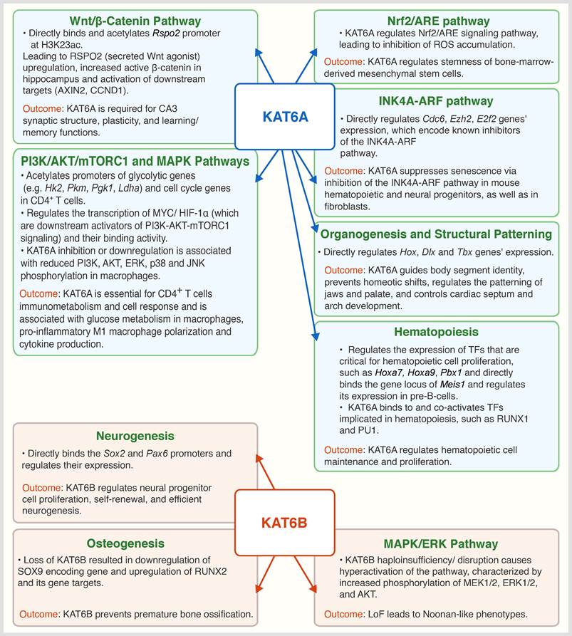
KAT6A and KAT6B orchestrate key physiological molecular pathways. A schematic overview of KAT6A- and KAT6B-dependent regulation across major molecular pathways (signal transduction, metabolic, and gene regulatory/epigenetic pathways), presenting epigenetic targets, downstream signaling cascades, and physiological outcomes. Data for KAT6A were collated from references: [164] (Wnt/β-catenin signaling), [106,110,120] (PI3K-AKT-mTORC1/ glycolysis in CD4+ T cells and macrophages), [71,109] (inflammatory and stress axes), expression of Hox [57-60], Dlx [61] and Tbx genes [62,63], [97,101] (INK4A-ARF pathway), [55,115,116] (hematopoiesis). Data for KAT6B were derived from references: [66] (neurogenesis), [85] (bone ossification), [64] (MAPK/ERK pathway).
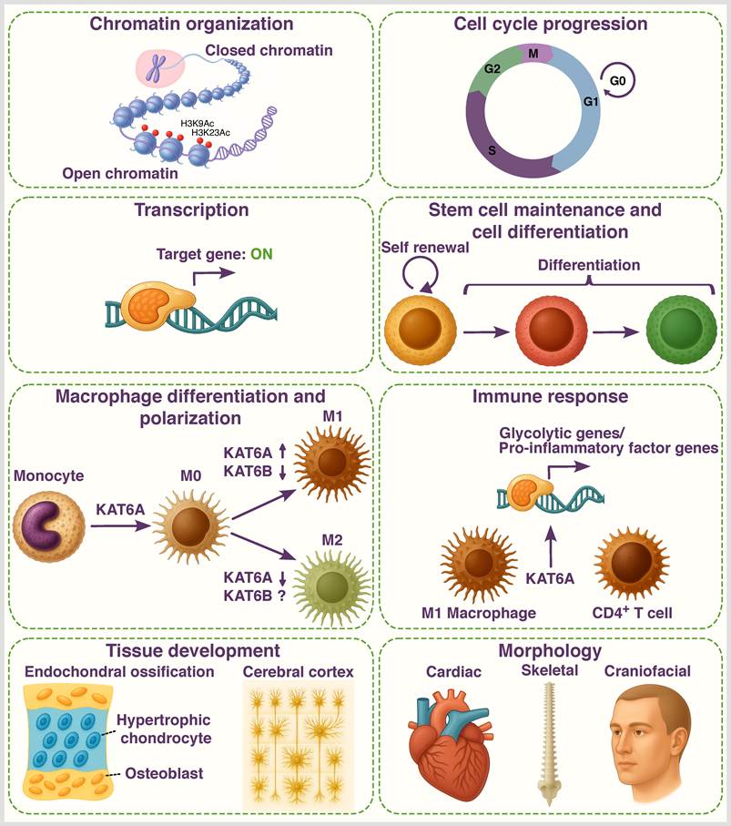
KAT6A and KAT6B acetyltransferases regulate a broad spectrum of essential cellular and developmental processes. At the cellular level, they regulate chromatin organization, transcription regulation, cell cycle progression, stem cell maintenance including mesenchymal, hematopoietic, and neural stem cells, and lineage differentiation. Moreover, KAT6A, in particular, has been linked to modulating immune responses, including macrophage differentiation and polarization, immune cell function and metabolic programming. It plays an important role in the formation and function of memory B- and T cells, and promotes glycolytic gene expression programs in activated CD4+ T cell sub-populations, as well as glycolytic and pro-inflammatory transcriptional programs in M1 macrophages. At the tissue and organ level, KAT6A and KAT6B are key regulators of multiple developmental processes, including hematopoiesis, neurogenesis, skeletogenesis, body segment identity, cerebral cortex formation, as well as craniofacial and cardiac development.
Abbreviations
ALCAR: acetyl-carnitine; ALL: Acute lymphoblastic leukemia; AML: Acute myeloid leukemia; ARTHS: Arboleda-Tham syndrome; ASD: autism spectrum disorder; BMDMs: bone marrow-derived macrophages; BM-MSCs: bone marrow-derived mesenchymal stem cells; CBP: CREB binding protein; CS: congenital scoliosis; CUT&RUN: cleavage under targets and release using nuclease; CUT&TAG: cleavage under targets and tagmentation; DD: developmental delay; ES: embryonic stem; GABA: gamma-aminobutyric acid; GTPTS: Genitopatellar syndrome; HDAC: histone deacetylase; HDACi: histone deacetylase inhibitors; HSCs: hematopoietic stem cells; ID: intellectual disability; iPSCs: induced pluripotent stem cells; KATs: lysine acetyltransferases; LoF: loss of function; MDCM: Mendelian disorders of chromatin machinery; MDEM: Mendelian disorders of the epigenetic machinery; MEFs: mouse embryonic fibroblasts; mESC: mouse embryonic stem cells; MLL1: Mixed-Lineage Leukemia protein-1; MRI: Magnetic resonance imaging; NEMM: N-terminal part of Enok, MOZ or MORF; NPCs: neural progenitor cells; NSCs: neural stem cells; NSPCs: neural stem and progenitor cells; PHD: plant homeodomain; ROS: reactive oxygen species; RUNX1: Runt-related transcription factor 1; SBBYSS: Say-Barber-Biesecker-Young-Simpson syndrome; scATAC-seq: single-cell assay for transposase-accessible chromatin with high-throughput sequencing; scRNA-seq: single-cell RNA sequencing; TF: transcription factor; TNF: tumor necrosis factor; VPA: valproic acid; WH: winged helix; WT: wild-type.
Supplementary Material
Supplementary tables.

Acknowledgements
This study was supported by the Ginsburg Fund at the Technion-Israel Institute of Technology, Haifa, Israel.
Microsoft 365 Copilot was used to generate individual images within the Figures.
Competing Interests
The authors have declared that no competing interest exists.
References
1. Allis CD, Jenuwein T. The molecular hallmarks of epigenetic control. Nat Rev Genet. 2016;17:487-500
2. Cavalli G, Heard E. Advances in epigenetics link genetics to the environment and disease. Nature. 2019;571:489-99
3. Kong DA, Wang H, Wang M, Chen S, Cao D, Liu X. et al. The histone acylation network: emerging therapeutic targets for remodeling the epigenetic landscape in pancreatic ductal adenocarcinoma. Cell Oncol. 2026;49:63
4. Zhao S, Zhang X, Li H. Beyond histone acetylation-writing and erasing histone acylations. Curr Opin Struct Biol. 2018;53:169-77
5. Nitsch S, Shahidian LZ, Schneider R. Histone acylations and chromatin dynamics: concepts, challenges, and links to metabolism. EMBO Rep. 2021;22:e52774
6. Al Ojaimi M, Banimortada BJ, Alragheb A, Hajir RS, Alves C, Walid D. et al. Molecular and clinical aspects of histone-related disorders. Hum Genomics. 2025;19:47
7. Park J, Lee K, Kim K, Yi SJ. The role of histone modifications: from neurodevelopment to neurodiseases. Signal Transduct Target Ther. 2022;7:1-23
8. Yi SJ, Lee H, Lee J, Lee K, Kim J, Kim Y. et al. Bone remodeling: Histone modifications as fate determinants of bone cell differentiation. Int J Mol Sci. 2019;20:3147
9. Selleri L, Rijli FM. Shaping faces: genetic and epigenetic control of craniofacial morphogenesis. Nat Rev Genet. 2023;24:610-26
10. Meng Y, Nerlov C. Epigenetic regulation of hematopoietic stem cell fate. Trends Cell Biol. 2025;35:217-29
11. Ng R, Kalinousky A, Harris J. Epigenetics of cognition and behavior: insights from Mendelian disorders of epigenetic machinery. J Neurodev Disord. 2023;15:1-11
12. St John M, Tripathi T, Morgan AT, Amor DJ. To speak may draw on epigenetic writing and reading: Unravelling the complexity of speech and language outcomes across chromatin-related neurodevelopmental disorders. Neurosci Biobehav Rev. 2023;152:105293
13. Nothof SA, Magdinier F, Van-Gils J. Chromatin Structure and Dynamics: Focus on Neuronal Differentiation and Pathological Implication. Genes (Basel). 2022;13:639
14. Donoghue S, Wright J, Voss AK, Lockhart PJ, Amor DJ. The Mendelian disorders of chromatin machinery: Harnessing metabolic pathways and therapies for treatment. Mol Genet Metab. 2024;142:108360
15. Karlsson MG, Harris J. Epigenetics in neurodevelopmental disabilities. Capute and Accardo's Neurodevelopmental Disabilities in Infancy and Childhood. Academic Press. 2024 p. 55-70
16. Shvedunova M, Akhtar A. Modulation of cellular processes by histone and non-histone protein acetylation. Nat Rev Mol Cell Biol. 2022;23:329-49
17. Borrow J, Stanton VP, Andresen JM, Becher R, Behm FG, Chaganti RSK. et al. The translocation t(8;16)(p11;p13) of acute myeloid leukaemia fuses a putative acetyltransferase to the CREB-binding protein. Nat Genet. 1996;14:33-41
18. Champagne N, Bertos NR, Pelletier N, Wang AH, Vezmar M, Yang Y. et al. Identification of a Human Histone Acetyltransferase Related to Monocytic Leukemia Zinc Finger Protein. J Biol Chem. 1999;274:28528-28536
19. Wiesel-Motiuk N, Assaraf YG. The key roles of the lysine acetyltransferases KAT6A and KAT6B in physiology and pathology. Drug Resist Updat. 2020;53:100729
20. Tan Y, Wang J, Ma F. Lysine Acetyltransferase 6 in Health and Disease. MedComm. 2025;6:e70520
21. Tripathi T, St John M, Wright J, Esber N, Amor DJ. Research Themes in KAT6A Syndrome: A Scoping Review. DNA. 2025;5:21
22. Xi Z, Wang J, Ge X, Zhang H, Ma Y. Dancing with KAT6A: Current advances and therapeutic potential in oncology of KAT6A inhibitors. Bioorg Chem. 2026;170:109533
23. Mukohara T. KAT6 inhibitors under investigation for solid tumors: the preclinical and early phase progress. Expert Opin Investig Drugs. 2026;35:215-21
24. Lin R, Zhang Y, Li H, Liang F. The role of histone acetyltransferases in tumorigenesis and their therapeutic potential: A review. Biochem Biophys Res Commun. 2026;795:153097
25. Ullah M, Pelletier N, Xiao L, Zhao SP, Wang K, Degerny C. et al. Molecular Architecture of Quartet MOZ/MORF Histone Acetyltransferase Complexes. Mol Cell Biol. 2008;28:6828-43
26. Doyon Y, Cayrou C, Ullah M, Landry AJ, Côté V, Selleck W. et al. ING tumor suppressor proteins are critical regulators of chromatin acetylation required for genome expression and perpetuation. Mol Cell. 2006;21:51-64
27. Viita T, Côté J. The MOZ-BRPF1 acetyltransferase complex in epigenetic crosstalk linked to gene regulation, development, and human diseases. Front Cell Dev Biol. 2023;10:1115903
28. Rokudai S, Laptenko O, Arnal SM, Taya Y, Kitabayashi I, Prives C. MOZ increases p53 acetylation and premature senescence through its complex formation with PML. Proc Natl Acad Sci U S A. 2013;110:3895-900
29. Yu B, Luo F, Sun B, Liu W, Shi Q, Cheng SY. et al. KAT6A Acetylation of SMAD3 Regulates Myeloid-Derived Suppressor Cell Recruitment, Metastasis, and Immunotherapy in Triple-Negative Breast Cancer. Adv Sci. 2021;8:2100014
30. Liu W, Zhan Z, Zhang MM, Sun B, Shi Q, Luo F. et al. KAT6A, a novel regulator of β-catenin, promotes tumorigenicity and chemoresistance in ovarian cancer by acetylating COP1. Theranostics. 2021;11:6278-92
31. Champagne N, Pelletier N, Yang X-J. The monocytic leukemia zinc finger protein MOZ is a histone acetyltransferase. Oncogene. 2001;20:404-9
32. Holbert MA, Sikorski T, Carten J, Snowflack D, Hodawadekar S, Marmorstein R. The human monocytic leukemia zinc finger histone acetyltransferase domain contains DNA-binding activity implicated in chromatin targeting. J Biol Chem. 2007;282:36603-13
33. Ali M, Yan K, Lalonde ME, Degerny C, Rothbart SB, Strahl BD. et al. Tandem PHD fingers of MORF/MOZ acetyltransferases display selectivity for acetylated histone H3 and are required for the association with chromatin. J Mol Biol. 2012;424:328-38
34. Dreveny I, Deeves SE, Fulton J, Yue B, Messmer M, Bhattacharya A. et al. The double PHD finger domain of MOZ/MYST3 induces α-helical structure of the histone H3 tail to facilitate acetylation and methylation sampling and modification. Nucleic Acids Res. 2014;42:822-35
35. Kitabayashi I, Aikawa Y, Nguyen LA, Yokoyama A, Ohki M. Activation of AML1-mediated transcription by MOZ and inhibition by the MOZ-CBP fusion protein. EMBO J. 2001;20:7184-96
36. Weber LM, Jia Y, Stielow B, Gisselbrecht SS, Cao Y, Ren Y. et al. The histone acetyltransferase KAT6A is recruited to unmethylated CpG islands via a DNA binding winged helix domain. Nucleic Acids Res. 2023;51:574-94
37. Becht DC, Klein BJ, Kanai A, Jang SM, Cox KL, Zhou BR. et al. MORF and MOZ acetyltransferases target unmethylated CpG islands through the winged helix domain. Nat Commun. 2023;14:697
38. Becht DC, Kanai A, Biswas S, Halawa M, Zeng L, Cox KL. et al. The winged helix domain of MORF binds CpG islands and the TAZ2 domain of p300. iScience. 2024;27:109367
39. Yang XJ, Ullah M. MOZ and MORF, two large MYSTic HATs in normal and cancer stem cells. Oncogene. 2007;26:5408-19
40. Zhan Z, Zhang J, Liang H, Wang C, Hong L, Liu W. KAT6A Condensates Impair PARP1 Trapping of PARP Inhibitors in Ovarian Cancer. Adv Sci. 2024;11:2400140
41. Pelletier N, Champagne N, Stifani S, Yang XJ. MOZ and MORF histone acetyltransferases interact with the Runt-domain transcription factor Runx2. Oncogene. 2002;21:2729-40
42. Qiu Y, Liu L, Zhao C, Han C, Li F, Zhang J. et al. Combinatorial readout of unmodified H3R2 and acetylated H3K14 by the tandem PHD finger of MOZ reveals a regulatory mechanism for HOXA9 transcription. Genes Dev. 2012;26:1376-91
43. Xiong X, Panchenko T, Yang S, Zhao S, Yan P, Zhang W. et al. Selective recognition of histone crotonylation by double PHD fingers of MOZ and DPF2. Nat Chem Biol. 2016;12:1111-8
44. Klein BJ, Jang SM, Lachance C, Mi W, Lyu J, Sakuraba S. et al. Histone H3K23-specific acetylation by MORF is coupled to H3K14 acylation. Nat Commun. 2019;10:4724
45. Yokoyama A, Niida H, Kutateladze TG, Côté J. HBO1, a MYSTerious KAT and its links to cancer. Biochim Biophys Acta - Gene Regul Mech. 2024;1867:195045
46. Klein BJ, Simithy J, Wang X, Ahn JW, Andrews FH, Zhang Y. et al. Recognition of Histone H3K14 Acylation by MORF. Structure. 2017;25:650-4
47. Han Z, Wu H, Kim S, Yang X, Li Q, Huang H. et al. Revealing the protein propionylation activity of the histone acetyltransferase MOF (males absent on the first). J Biol Chem. 2018;293:3410-20
48. Yan K, Rousseau J, Machol K, Cross LA, Agre KE, Gibson CF. et al. Deficient histone H3 propionylation by BRPF1-KAT6 complexes in neurodevelopmental disorders and cancer. Sci Adv. 2020;6:1-17
49. Thul PJ, Lindskog C. The human protein atlas: A spatial map of the human proteome. Protein Sci. 2018;27:233-44
50. Diamant I, Clarke DJB, Evangelista JE, Lingam N, Ma'Ayan A. Harmonizome 3.0: Integrated knowledge about genes and proteins from diverse multi-omics resources. Nucleic Acids Res. 2025;53:D1016-28
51. Cosentino MS, Oses C, Vázquez Echegaray C, Solari C, Waisman A, Álvarez Y. et al. Kat6b Modulates Oct4 and Nanog Binding to Chromatin in Embryonic Stem Cells and Is Required for Efficient Neural Differentiation. J Mol Biol. 2019;431:1148-59
52. Thomas T, Voss AK, Chowdhury K, Gruss P. Querkopf, a MYST family histone acetyltransferase, is required for normal cerebral cortex development. Development. 2000;127:2537-48
53. Bergamasco MI, Yang Y, Garnham AL, Sheikh BN, Smyth GK, Voss AK. et al. KAT6B overexpression rescues embryonic lethality in homozygous null KAT6A mice restoring vitality and normal lifespan. Nat Commun. 2025;16:1-16
54. Toro-Tobon S, Paredes-Gutierrez J, Mantilla-Rivas E, Ahmad L, Rogers GF. Pharyngeal Arches, Chapter 1: Normal Development and Derivatives. J Craniofac Surg. 2023;34:2237
55. Katsumoto T, Aikawa Y, Iwama A, Ueda S, Ichikawa H, Ochiya T. et al. MOZ is essential for maintenance of hematopoietic stem cells. Genes Dev. 2006;20:1321-30
56. Thomas T, Corcoran LM, Gugasyan R, Dixon MP, Brodnicki T, Nutt SL. et al. Monocytic leukemia zinc finger protein is essential for the development of long-term reconstituting hematopoietic stem cells. Genes Dev. 2006;20:1175-86
57. Voss AK, Collin C, Dixon MP, Thomas T. Moz and Retinoic Acid Coordinately Regulate H3K9 Acetylation, Hox Gene Expression, and Segment Identity. Dev Cell. 2009;17:674-86
58. Miller CT, Maves L, Kimmel CB. moz regulates Hox expression and pharyngeal segmental identity in zebrafish. Development. 2004;131:2443-61
59. Crump JG, Swartz ME, Eberhart JK, Kimmel CB. Moz-dependent Hox expression controls segment-specific fate maps of skeletal precursors in the face. Development. 2006;133:2661-9
60. Sheikh BN, Downer NL, Phipson B, Vanyai HK, Kueh AJ, McCarthy DJ. et al. MOZ and BMI1 play opposing roles during Hox gene activation in ES cells and in body segment identity specification in vivo. Proc Natl Acad Sci USA. 2015;112:5437-42
61. Vanyai HK, Garnham A, May RE, McRae HM, Collin C, Wilcox S. et al. MOZ directs the distal-less homeobox gene expression program during craniofacial development. Development. 2019;146:dev175042
62. Voss AK, Vanyai HK, Collin C, Dixon MP, McLennan TJ, Sheikh BN. et al. MOZ Regulates the Tbx1 Locus, and Moz Mutation Partially Phenocopies DiGeorge Syndrome. Dev Cell. 2012;23:652-63
63. Vanyai HK, Thomas T, Voss AK. Mesodermal expression of Moz is necessary for cardiac septum development. Dev Biol. 2015;403:22-9
64. Kraft M, Cirstea IC, Voss AK, Thomas T, Goehring I, Sheikh BN. et al. Disruption of the histone acetyltransferase MYST4 leads to a noonan syndrome-like phenotype and hyperactivated MAPK signaling in humans and mice. J Clin Invest. 2011;121:3479-91
65. Bergamasco MI, Vanyai HK, Garnham AL, Geoghegan ND, Vogel AP, Eccles S. et al. Increasing histone acetylation improves sociability and restores learning and memory in KAT6B-haploinsufficient mice. J Clin Invest. 2024;134:e167672
66. Bergamasco MI, Abeysekera W, Garnham AL, Hu Y, Li-Wai-suen CSN, Sheikh BN. et al. KAT6B is required for histone 3 lysine 9 acetylation and SOX gene expression in the developing brain. Life Sci Alliance. 2024;8:e202402969
67. Thomas T, Malelang S, Yang Y, Bergamasco MI, Kueh AJ, Garnham AL. et al. The roles of the acetyltransferase domains of the chromatin regulators KAT6A and KAT6B in vivo. Development. 2026;153:dev205559
68. Eccles S, Vanyai HK, Bergamasco MI, Malelang S, Pehlivanoglu H, Garnham AL. et al. Acetyl-carnitine improves hyperactivity and learning deficits in KAT6A haploinsufficient mice. Life Sci Alliance. 2026;9:e202503549
69. Wiesel-Motiuk N, Choudhary A, Hussein Y, Tripathi U, Rike WA, Shemen A. et al. KAT6A-Mutant iPSC-Derived Cortical Neurons Display Precocious Maturation, Dysregulated Electrophysiology, and Altered Morphology. Int J Biol Sci. 2026;22:8448-75
70. Heair HM, Kemper AG, Roy B, Lopes HB, Rashid H, Clarke JC. et al. MicroRNA-665 Regulates Dentinogenesis through MicroRNA-Mediated Silencing and Epigenetic Mechanisms. Mol Cell Biol. 2015;35:3116-30
71. Fei D, Wang Y, Zhai Q, Zhang X, Zhang Y, Wang Y. et al. KAT6A regulates stemness of aging bone marrow-derived mesenchymal stem cells through Nrf2/ARE signaling pathway. Stem Cell Res Ther. 2021;12:104
72. Levi G, Narboux-Nême N, Cohen-Solal M. DLX Genes in the Development and Maintenance of the Vertebrate Skeleton: Implications for Human Pathologies. Cells. 2022;11:3277
73. Wang L, Ruan M, Bu Q, Zhao C. Signaling Pathways Driving MSC Osteogenesis: Mechanisms, Regulation, and Translational Applications. Int J Mol Sci. 2025;26:1311
74. Li C, Li B, Zhang J, Liu K, Du G, Guo C. et al. Bone Marrow Mesenchymal Stem Cell Senescence in the Development of Osteoporosis: Mechanisms, Interventions, and Future Directions. Curr Mol Med. 2026;26:674-86
75. Cao B, Dai X, Xiao L. KAT6A regulates osteoclast differentiation and bone resorption through TET1-mediated TRPV4 expression. Naunyn Schmiedebergs Arch Pharmacol. 2026;399:5399-412
76. Liu N, Lu W, Dai X, Qu X, Zhu C. The role of TRPV channels in osteoporosis. Mol Biol Rep. 2022;49:577-85
77. Ji M liang, Li Z, Hu XY, Zhang W tuo, Zhang H xiang, Lu J. Dynamic chromatin accessibility tuning by the long noncoding RNA ELDR accelerates chondrocyte senescence and osteoarthritis. Am J Hum Genet. 2023;110:606-24
78. Clayton-Smith J, O'Sullivan J, Daly S, Bhaskar S, Day R, Anderson B. et al. Whole-exome-sequencing identifies mutations in histone acetyltransferase gene KAT6B in individuals with the say-barber-biesecker variant of Ohdo syndrome. Am J Hum Genet. 2011;89:675-81
79. Bayat S, Gholami M, Khodadadi H, Ghazavi M, Nasiri J, Kheirollahi M. Comprehensive review and outline of genotypes and phenotypes of Arboleda-Tham syndrome spectrum: insights from novel variants. Mol Biol Rep. 2025;52:1-8
80. Maglione V, Pizzuti A, Mastromoro G, Cresta E, Favata P, Digilio MC. et al. Phenotypic Characterization of Seven Pediatric Patients Diagnosed With KAT6B-Related Disorders: Case Series and Review of the Literature. Am J Med Genet Part A. 2025;197:e64100
81. Zhang LX, Lemire G, Gonzaga-Jauregui C, Molidperee S, Galaz-Montoya C, Liu DS. et al. Further delineation of the clinical spectrum of KAT6B disorders and allelic series of pathogenic variants. Genet Med. 2020;22:1338-47
82. Wu Y, Zhang H, Tang M, Guo C, Deng A, Li J. et al. High methylation of lysine acetyltransferase 6B is associated with the Cobb angle in patients with congenital scoliosis. J Transl Med. 2020;18:210
83. Komori T. Molecular Mechanism of Runx2-Dependent Bone Development. Mol Cells. 2020;43:168-75
84. Azarkina K, Gromova E, Malashicheva A. “A Friend Among Strangers” or the Ambiguous Roles of Runx2. Biomolecules. 2024;14:1392
85. Bergamasco MI, Ogier JM, Garnham AL, Whitehead L, Rogers K, Smyth GK. et al. Loss of KAT6B causes premature ossification and promotes osteoblast differentiation during development. Dev Biol. 2025;520:141-54
86. Hallett SA, Ono W, Ono N. The hypertrophic chondrocyte: To be or not to be. Histol Histopathol. 2021;36:1021
87. Bashir RA, Dixit A, Goedhart C, Parboosingh JS, Innes AM, Ferreira P. et al. Lin-Gettig syndrome: Craniosynostosis expands the spectrum of the KAT6B related disorders. Am J Med Genet Part A. 2017;173:2596-604
88. Yabumoto M, Kianmahd J, Singh M, Palafox MF, Wei A, Elliott K. et al. Novel variants in KAT6B spectrum of disorders expand our knowledge of clinical manifestations and molecular mechanisms. Mol Genet Genomic Med. 2021;9:e1809
89. Haseeb A, Kc R, Angelozzi M, de Charleroy C, Rux D, Tower RJ. et al. SOX9 keeps growth plates and articular cartilage healthy by inhibiting chondrocyte dedifferentiation/ osteoblastic redifferentiation. Proc Natl Acad Sci U S A. 2021;118:e2019152118
90. Zhou G, Zheng Q, Engin F, Munivez E, Chen Y, Sebald E. et al. Dominance of SOX9 function over RUNX2 during skeletogenesis. Proc Natl Acad Sci U S A. 2006;103:19004-9
91. Carbonare LD, Bertacco J, Minoia A, Cominacini M, Bhandary L, Elia R. et al. Modulation of miR-204 Expression during Chondrogenesis. Int J Mol Sci. 2022;23:2130
92. Lui JC, Yue S, Lee A, Kikani B, Temnycky A, Barnes KM. et al. Persistent Sox9 expression in hypertrophic chondrocytes suppresses transdifferentiation into osteoblasts. Bone. 2019;125:169-77
93. Chen H, Ghori-Javed FY, Rashid H, Adhami MD, Serra R, Gutierrez SE. et al. Runx2 regulates endochondral ossification through control of chondrocyte proliferation and differentiation. J Bone Miner Res. 2014;29:2653-65
94. Khokhar ES, Borikar S, Eudy E, Stearns T, Young K, Trowbridge JJ. Aging-Associated Decrease in the Histone Acetyltransferase KAT6B is Linked to Altered Hematopoietic Stem Cell Differentiation. Exp Hematol. 2020;82:43-52
95. Bergamasco MI, Ranathunga N, Abeysekera W, Li-Wai-Suen CSN, Garnham AL, Willis SN. et al. The histone acetyltransferase KAT6B is required for hematopoietic stem cell development and function. Stem Cell Reports. 2024;19:469-85
96. Sheikh BN, Yang Y, Schreuder J, Nilsson SK, Bilardi R, Carotta S. et al. MOZ (KAT6A) is essential for the maintenance of classically defined adult hematopoietic stem cells. Blood. 2016;128:2307-18
97. Perez-Campo FM, Costa G, Lie-a-Ling M, Stifani S, Kouskoff V, Lacaud G. MOZ-Mediated Repression of p16 INK4a Is Critical for the Self-Renewal of Neural and Hematopoietic Stem Cells. Stem Cells. 2014;32:1591-601
98. Sheikh BN, Dixon MP, Thomas T, Voss AK. Querkopf is a key marker of self-renewal and multipotency of adult neural stem cells. Development. 2012;139:295-309
99. Sheikh BN, Metcalf D, Voss AK, Thomas T. MOZ and BMI1 act synergistically to maintain hematopoietic stem cells. Exp Hematol. 2017;47:83-97.e8
100. Perez-Campo FM, Borrow J, Kouskoff V, Lacaud G. The histone acetyl transferase activity of monocytic leukemia zinc finger is critical for the proliferation of hematopoietic precursors. Blood. 2009;113:4866-74
101. Sheikh BN, Phipson B, El-Saafin F, Vanyai HK, Downer NL, Bird MJ. et al. MOZ (MYST3, KAT6A) inhibits senescence via the INK4A-ARF pathway. Oncogene. 2015;34:5807-20
102. Gordon SJV, Perner F, Macpherson L, Fennell KA, Wenge D V, Bourgeois W. et al. Catalytic Inhibition of KAT6/KAT7 Enhances the Efficacy and Overcomes Primary and Acquired Resistance to Menin Inhibitors in MLL Leukemia. Cancer Discov. 2025;15:2117-38
103. Voss AK, Eccles S, Wichmann J, Abeysekera W, Bergamasco MI, Garnham AL. et al. KAT6A is essential for developmental control gene expression in neural stem and progenitor cells. PLoS Genet. 2026;22:e1012133
104. Yan F, Li J, Milosevic J, Petroni R, Liu S, Shi Z. et al. KAT6A and ENL Form an Epigenetic Transcriptional Control Module to Drive Critical Leukemogenic Gene-Expression Programs. Cancer Discov. 2022;12:792-811
105. Komata Y, Kanai A, Maeda T, Inaba T, Yokoyama A. MOZ/ENL complex is a recruiting factor of leukemic AF10 fusion proteins. Nat Commun. 2023;14:1979
106. Jiang M, Zhang J, Qian L, Miao Y, Song W, Liu H. et al. MOZ Forms an Autoregulatory Feedback Loop with miR-223 in AML and Monocyte/Macrophage Development. iScience. 2019;11:189-204
107. Shukla S, Levine C, Sripathi RP, Elson G, Lutz CS, Leibovich SJ. The kat in the HAT: The histone acetyl transferase Kat6b (MYST4) Is downregulated in murine macrophages in response to LPS. Mediators Inflamm. 2018;2018:7852742
108. Dash SP, Gupta S, Sarangi PP. Monocytes and macrophages: Origin, homing, differentiation, and functionality during inflammation. Heliyon. 2024;10:e29686
109. Huang SJ, Xu S, Ye HL, Zhou LJ, Wang YL, Duan SZ. KAT6A Promotes Macrophage Inflammation and Periodontitis. J Clin Periodontol. 2025;52:1499-508
110. Wang X, Chen J, Lai Y, Wang Y, Hu K, Wu M. et al. Lysine Acetyltransferase 6A Drives M1 Macrophage Polarization Through Metabolic Reprogramming in Sepsis-Induced Acute Lung Injury. Biomolecules. 2026;16:609
111. Yunna C, Mengru H, Lei W, Weidong C. Macrophage M1/M2 polarization. Eur J Pharmacol. 2020;877:173090
112. Bae S, Park PSU, Lee Y, Mun SH, Giannopoulou E, Fujii T. et al. MYC-mediated early glycolysis negatively regulates proinflammatory responses by controlling IRF4 in inflammatory macrophages. Cell Rep. 2021;35:109264
113. McGettrick AF, O'Neill LAJ. The Role of HIF in Immunity and Inflammation. Cell Metab. 2020;32:524-36
114. Fontana F, Giannitti G, Marchesi S, Limonta P. The PI3K/Akt Pathway and Glucose Metabolism: A Dangerous Liaison in Cancer. Int J Biol Sci. 2024;20:3113-25
115. Sheikh BN, Lee SCW, El-Saafin F, Vanyai HK, Hu Y, Pang SHM. et al. MOZ regulates B-cell progenitors and, consequently, Moz haploinsufficiency dramatically retards MYC-induced lymphoma development. Blood. 2015;125:1910-21
116. Bristow CAP, Shore P. Transcriptional regulation of the human MIP-1α promoter by RUNX1 and MOZ. Nucleic Acids Res. 2003;31:2735-44
117. Mevel R, Draper JE, Lie-A-Ling M, Kouskoff V, Lacaud G. RUNX transcription factors: Orchestrators of development. Development. 2019;146:dev148296
118. Guanglan LI, Wenke HAO, Wenxue HU. Transcription factor PU.1 and immune cell differentiation. Int J Mol Med. 2020;46:1943-50
119. Newman DM, Sakaguchi S, Lun A, Preston S, Pellegrini M, Khamina K. et al. Acetylation of the Cd8 Locus by KAT6A Determines Memory T Cell Diversity. Cell Rep. 2016;16:3311-21
120. Fu JY, Huang SJ, Wang BL, Yin JH, Chen CY, Xu JB. et al. Lysine acetyltransferase 6A maintains CD4+ T cell response via epigenetic reprogramming of glucose metabolism in autoimmunity. Cell Metab. 2024;36:557-574.e10
121. Good-Jacobson KL, Chen Y, Voss AK, Smyth GK, Thomas T, Tarlinton D. Regulation of germinal center responses and B-cell memory by the chromatin modifier MOZ. Proc Natl Acad Sci U S A. 2014;111:9585-90
122. Merson TD, Dixon MP, Collin C, Rietze RL, Bartlett PF, Thomas T. et al. The transcriptional coactivator Querkopf controls adult neurogenesis. J Neurosci. 2006;26:11359-70
123. Ernst P, Kyei PS, Yokoyama A. KMT2A-rearranged leukemia: from mechanism to drug development. Exp Hematol. 2025;151:105247
124. Huang YC, Shih HY, Lin SJ, Chiu CC, Ma TL, Yeh TH. et al. The epigenetic factor Kmt2a/Mll1 regulates neural progenitor proliferation and neuronal and glial differentiation. Dev Neurobiol. 2015;75:452-62
125. Delgado RN, Mansky B, Ahanger SH, Lu C, Andersen RE, Dou Y. et al. Maintenance of neural stem cell positional identity by mixed-lineage leukemia 1. Science. 2020;368:48-53
126. Paggetti J, Largeot A, Aucagne R, Jacquel A, Lagrange B, Yang XJ. et al. Crosstalk between leukemia-associated proteins MOZ and MLL regulates HOX gene expression in human cord blood CD34 cells. Oncogene. 2010;29:5019-31
127. Miyamoto R, Okuda H, Kanai A, Takahashi S, Kawamura T, Matsui H. et al. Activation of CpG-Rich Promoters Mediated by MLL Drives MOZ-Rearranged Leukemia. Cell Rep. 2020;32:108200
128. Hemming ML, Benson MR, Loycano MA, Anderson JA, Andersen JL, Taddei ML. et al. MOZ and Menin-MLL Complexes Are Complementary Regulators of Chromatin Association and Transcriptional Output in Gastrointestinal Stromal Tumor. Cancer Discov. 2022;12:1804-21
129. Bryan E, Valsakumar D, Idigo NJ, Warburton M, Webb KM, McLaughlin KA. et al. Nucleosomal asymmetry shapes histone mark binding and promotes poising at bivalent domains. Mol Cell. 2025;85:471-489.e12
130. Macrae TA, Fothergill-Robinson J, Ramalho-Santos M. Regulation, functions and transmission of bivalent chromatin during mammalian development. Nat Rev Mol Cell Biol. 2023;24:6-26
131. Mätlik K, Govek EE, Hatten ME. Histone bivalency in CNS development. Genes Dev. 2025;39:428-44
132. Ormaza G, Rodríguez JA, Ibáñez de Opakua A, Merino N, Villate M, Gorroño I. et al. The Tumor Suppressor ING5 Is a Dimeric, Bivalent Recognition Molecule of the Histone H3K4me3 Mark. J Mol Biol. 2019;431:2298-319
133. Bergamasco MI, Ozturk E, Casillas-Espinosa PM, Garnham AL, Abeysekera W, Wimmer VC. et al. KAT6B overexpression in mice causes aggression, anxiety, and epilepsy. iScience. 2025;28:111953
134. Stevanovic M, Drakulic D, Lazic A, Ninkovic DS, Schwirtlich M, Mojsin M. SOX Transcription Factors as Important Regulators of Neuronal and Glial Differentiation During Nervous System Development and Adult Neurogenesis. Front Mol Neurosci. 2021;14:654031
135. Duran RC-D, Wei H, Wu JQ. Single-cell RNA-sequencing of the brain. Clin Transl Med. 2017;6:e20
136. Singh M, Spendlove SJ, Wei A, Bondhus LM, Nava AA, Francisca FN. et al. KAT6A mutations in Arboleda-Tham syndrome drive epigenetic regulation of posterior HOXC cluster. Hum Genet. 2023;142:1705-20
137. Tham E, Lindstrand A, Santani A, Malmgren H, Nesbitt A, Dubbs HA. et al. Dominant mutations in KAT6A cause intellectual disability with recognizable syndromic features. Am J Hum Genet. 2015;96:507-13
138. Arboleda VA, Lee H, Dorrani N, Zadeh N, Willis M, Macmurdo CF. et al. De novo nonsense mutations in KAT6A, a lysine acetyl-transferase gene, cause a syndrome including microcephaly and global developmental delay. Am J Hum Genet. 2015;96:498-506
139. Millan F, Cho MT, Retterer K, Monaghan KG, Bai R, Vitazka P. et al. Whole exome sequencing reveals de novo pathogenic variants in KAT6A as a cause of a neurodevelopmental disorder. Am J Med Genet Part A. 2016;170:1791-8
140. Campeau PM, Kim JC, Lu JT, Schwartzentruber JA, Abdul-Rahman OA, Schlaubitz S. et al. Mutations in KAT6B, encoding a histone acetyltransferase, cause genitopatellar syndrome. Am J Hum Genet. 2012;90:282-9
141. Simpson MA, Deshpande C, Dafou D, Vissers LELM, Woollard WJ, Holder SE. et al. De novo mutations of the gene encoding the histone acetyltransferase KAT6B cause genitopatellar syndrome. Am J Hum Genet. 2012;90:290-4
142. Radvanszky J, Hyblova M, Durovcikova D, Hikkelova M, Fiedler E, Kadasi L. et al. Complex phenotypes blur conventional borders between Say-Barber-Biesecker-Young-Simpson syndrome and genitopatellar syndrome. Clin Genet. 2017;91:339-43
143. Vlckova M, Simandlova M, Zimmermann P, Stranecky V, Hartmannova H, Hodanova K. et al. A patient showing features of both SBBYSS and GPS supports the concept of a KAT6B-related disease spectrum, with mutations in mid-exon 18 possibly leading to combined phenotypes. Eur J Med Genet. 2015;58:550-5
144. Kennedy J, Goudie D, Blair E, Chandler K, Joss S, McKay V. et al. KAT6A Syndrome: genotype-phenotype correlation in 76 patients with pathogenic KAT6A variants. Genet Med. 2018;21:850-60
145. Zu G, Liu Y, Cao J, Zhao B, Zhang H, You L. BRPF1-KAT6A/KAT6B Complex: Molecular Structure, Biological Function and Human Disease. Cancers (Basel). 2022;14:4068
146. Kim YR, Park JB, Lee YJ, Hong MJ, Kim HJHT, Kim HJHT. Identifying the KAT6B Mutation via Diagnostic Exome Sequencing to Diagnose Say-Barber-Biesecker-Young-Simpson Syndrome in Three Generations of a Family. Ann Rehabil Med. 2017;41:505-10
147. Trinh J, Hüning I, Yüksel Z, Baalmann N, Imhoff S, Klein C. et al. A KAT6A variant in a family with autosomal dominantly inherited microcephaly and developmental delay. J Hum Genet. 2018;63:997-1001
148. Yates TM, Langley CLM, Grozeva D, Raymond FL, Johnson DS. Novel KAT6B proximal familial variant expands genotypic and phenotypic spectrum. Clin Genet. 2019;95:334-5
149. Ringsted SB, Markholt S, Andreasen L, Gregersen PA. Phenotypic variability in a family with an inherited KAT6A frameshift variant. Eur J Med Genet. 2025;73:104993
150. Rasmussen NB, Gregersen PA, Nielsen TØ, Lyngdorf LG, Nielsen CK, Kruse C. et al. Variable expressivity of a transmitted pathogenic KAT6B variant. Eur J Med Genet. 2025;76:105020
151. Zeng F, Yang Y, Xu Z, Wang Z, Ke H, Zhang J. et al. Clinical manifestations and genetic analysis of a newborn with Arboleda-Tham syndrome. Front Genet. 2022;13:990098
152. St John M, Amor DJ, Morgan AT. Speech and language development and genotype-phenotype correlation in 49 individuals with KAT6A syndrome. Am J Med Genet Part A. 2022;188:3389-400
153. Eising E, Carrion-Castillo A, Vino A, Strand EA, Jakielski KJ, Scerri TS. et al. A set of regulatory genes co-expressed in embryonic human brain is implicated in disrupted speech development. Mol Psychiatry. 2019;24:1065-78
154. Kritioti E, Theodosiou A, Parpaite T, Alexandrou A, Nicolaou N, Papaevripidou I. et al. Unravelling the genetic causes of multiple malformation syndromes: A whole exome sequencing study of the Cypriot population. PLoS One. 2021;16:e0253562
155. Yang Y, Zhang HM, Huang H. Clinical features and the genetic analysis of KAT6B-related diseases caused by a de novo mutation of the KAT6B gene c.621+1G>A. Asian J Surg. 2022;45:792-4
156. Lonardo F, Lonardo MS, Acquaviva F, Della Monica M, Scarano F, Scarano G. Say-Barber-Biesecker-Young-Simpson syndrome and Genitopatellar syndrome: Lumping or splitting? Clin Genet. 2019;95:253-61
157. Urreizti R, Lopez-Martin E, Martinez-Monseny A, Pujadas M, Castilla-Vallmanya L, Pérez-Jurado LA. et al. Five new cases of syndromic intellectual disability due to KAT6A mutations: widening the molecular and clinical spectrum. Orphanet J Rare Dis. 2020;15:44
158. Lemire G, Campeau PM, Lee BH. KAT6B Disorders. In: Adam MP, Ardinger HH, Pagon RA et al, editor. GeneReviews((R)), Seattle (WA): University of Washington, Seattle; 1993. 2020 p. 1-18
159. Kim JM, Sunwoo Y, Kim H, Kim YH, Cha JH, Lee S. et al. Expanding the Clinical Spectrum of Arboleda-Tham Syndrome. Neurol Genet. 2026;12:e200364
160. Ng R, Kalinousky A, Harris J. Expanding the neuropsychological phenotype of KAT6B disorders: Overlapping features with KAT6A syndrome. Genet Med Open. 2024;2:101196
161. Ng R, Kalinousky AJ, Harris J. Neuropsychological profile associated with KAT6A syndrome: Emergent genotype-phenotype trends. Orphanet J Rare Dis. 2024;19:196
162. Baker EK, St John M, Braden R, Morison LD, Forbes EJ, Lelik F. et al. Adaptive functioning in children and young adults with monogenic neurodevelopmental disorders. Dev Med Child Neurol. 2025;67:953-62
163. Smith C, Harris J. Sleep, behavior, and adaptive function in KAT6A syndrome. Brain Sci. 2021;11:966
164. Liu Y, Fan M, Yang J, Mihaljević L, Chen KH, Ye Y. et al. KAT6A deficiency impairs cognitive functions through suppressing RSPO2/Wnt signaling in hippocampal CA3. Sci Adv. 2024;10:9326
165. Weaver KN, Prada CE. Current opinions on Noonan syndrome and RASopathies. Curr Opin Pediatr. 2026;38:126-32
166. Schafer ST, Paquola ACM, Stern S, Gosselin D, Ku M, Pena M. et al. Pathological priming causes developmental gene network heterochronicity in autistic subject-derived neurons. Nat Neurosci. 2019;22:243-55
167. Brant B, Stern T, Shekhidem HA, Mizrahi L, Rosh I, Stern Y. et al. IQSEC2 mutation associated with epilepsy, intellectual disability, and autism results in hyperexcitability of patient-derived neurons and deficient synaptic transmission. Mol Psychiatry. 2021;26:7498-508
168. Hussein Y, Tripathi U, Choudhary A, Nayak R, Peles D, Rosh I. et al. Early maturation and hyperexcitability is a shared phenotype of cortical neurons derived from different ASD-associated mutations. Transl Psychiatry. 2023;13:1-12
169. Troisi S, Maitz S, Severino M, Spano A, Cappuccio G, Brunetti-Pierri N. et al. Epilepsy in KAT6A syndrome: Description of two individuals and revision of the literature. Eur J Med Genet. 2022;65:104380
170. Shukla S, Tekwani BL. Histone Deacetylases Inhibitors in Neurodegenerative Diseases, Neuroprotection and Neuronal Differentiation. Front Pharmacol. 2020;11:537
171. Markowicz-Piasecka M, Miłkowska Z, Adla SK, Huttunen KM, Pietrzak B. Beyond epilepsy: The expanding role of valproic acid in Alzheimer disease therapy - a review. J Pharmacol Exp Ther. 2025;392:103658
172. Romoli M, Mazzocchetti P, D'Alonzo R, Siliquini S, Rinaldi VE, Verrotti A. et al. Valproic Acid and Epilepsy: From Molecular Mechanisms to Clinical Evidences. Curr Neuropharmacol. 2019;17:926-46
173. Udodi PS, Abugu JI, Udodi RE, Ofoego UC, Okafor EC, Orji CE. et al. The Neurotoxic Mechanisms of Valproic Acid and Their Association With Neurodevelopmental Disorders: A Narrative Review. Basic Clin Neurosci. 2026;17:25-38
174. Olawade DB, Rashad I, Egbon E, Teke J, Ovsepian SV, Boussios S. Reversing Epigenetic Dysregulation in Neurodegenerative Diseases: Mechanistic and Therapeutic Considerations. Int J Mol Sci. 2025;26:4929
175. Trocha A, Piotrowska DG, Markowicz-Piasecka M. Valproic acid as a therapeutic scaffold: Advances in structural modification and drug design. Eur J Med Chem. 2026;302:118334
176. Goel H. Epigenetic equilibrium in chromatinopathies: network instability in neurodevelopment. Front Neurol. 2026;17:1848929
177. D'Mello SR. Regulation of Central Nervous System Development by Class I Histone Deacetylases. Dev Neurosci. 2020;41:149-65
178. Van Bree BA, Eichner LJ. The impactful role of the HDACs in the regulation of gene expression and as targets for disease therapy. Sci Adv. 2026;12:eaee7295
179. Wagner VF, Hillman PR, Britt AD, Ray JW, Farach LS. A De novo HDAC2 variant in a patient with features consistent with Cornelia de Lange syndrome phenotype. Am J Med Genet Part A. 2019;179:852-6
180. Chourri H, Liu R, Sengupta M, Panjaliya RK, Das P, Munshi A. et al. HDAC3 in neurodevelopmental disorders: Molecular mechanisms and targeted intervention. Clin Chim Acta. 2025;577:120455
181. Di Fede E, Lettieri A, Taci E, Castiglioni S, Rebellato S, Parodi C. et al. Characterization of a novel HDAC2 pathogenetic variant: a missing puzzle piece for chromatinopathies. Hum Genet. 2024;143:747-59
182. Diniz LP, Pinheiro P de SM, Franco LS, Gomes FCA. HDACs in the Brain: From Chromatin Remodeling to Neurodegenerative Disease. Cells. 2025;14:1338
183. Wang P, Sun J. A comprehensive review of GABA in autism spectrum disorders: associations, mechanisms, and therapeutic implications. Front Psychiatry. 2025;16:1587432
184. Huang Q, Liao C, Ge F, Ao J, Liu T. Acetylcholine bidirectionally regulates learning and memory. J Neurorestoratology. 2022;10:100002
185. Yilmaz R, Beleza-Meireles A, Price S, Oliveira R, Kubisch C, Clayton-Smith J. et al. A recurrent synonymous KAT6B mutation causes Say-Barber-Biesecker/Young-Simpson syndrome by inducing aberrant splicing. Am J Med Genet Part A. 2015;167:3006-10
186. Magdalena K, Anna BT, Anna LB, Aleksandra JS, Krzysztof S, Malgorzata KW. et al. Clinical heterogeneity of polish patients with KAT6B-related disorder. Mol Genet Genomic Med. 2023;11:e2265
187. Davarnia B, Panahi M, Rahimi B, Anari H, Farajollahi R, Rodbaneh EA. et al. De novo KAT6B mutation causes Say-Barber-Biesecker-Young-Simpson variant of Ohdo syndrome in an Iranian boy: a case report. J Med Case Rep. 2024;18:4
188. Gannon T, Perveen R, Schlecht H, Ramsden S, Anderson B, Kerr B. et al. Further delineation of the KAT6B molecular and phenotypic spectrum. Eur J Hum Genet. 2015;23:1165-70
189. Li K, Xiao J, Ling Z, Luo T, Xiong J, Chen Q. et al. Prioritizing de novo potential non-canonical splicing variants in neurodevelopmental disorders. EBioMedicine. 2024;99:104928
190. Munuera-Cabeza M, Álvarez-Córdoba M, Suárez-Rivero JM, Povea-Cabello S, Villalón-García I, Talaverón-Rey M. et al. Pantothenate and L-Carnitine Supplementation Improves Pathological Alterations in Cellular Models of KAT6A Syndrome. Genes (Basel). 2022;13:2300
Author contact
Corresponding author: Yehuda G. Assaraf, E-mail: assarafac.il.