Impact Factor ISSN: 1449-2288




Int J Biol Sci 2027; 23(1):462-488. doi:10.7150/ijbs.131542 This issue Cite
Review
Advances in T Cell - B Cell Interaction Networks in Idiopathic Membranous Nephropathy: From Immunological Mechanisms to Precision Therapeutics
Lin Wang1*, Qin Tian2*, Ao Xu3*, Lianli Shen1*, Yue Xing1, Tianxingjian Ke1, Yuling Liang4, Jinjie Gao5, Hongtao Yang5  , Jinxiang Wang6 , Yifei Zhong1
1. Department of Nephrology, Longhua Hospital, Shanghai University of Traditional Chinese Medicine, Shanghai, 200032, China.
2. Department of Clinical Laboratory, The Seventh Affiliated Hospital, Sun Yat-sen University, Shenzhen, 518107, China.
3. Department of Traditional Chinese Medicine, Tianjin First Central Hospital, Tianjin, 300191, China.
4. Department of Nephrology, Center of Nephrology and Urology, The Seventh Affiliated Hospital, Sun Yat-sen University, Shenzhen, 518107, Guangdong, China.
5. Department of Nephrology, First Teaching Hospital of Tianjin University of Traditional Chinese Medicine, National Clinical Research Center for Chinese Medicine Acupuncture and Moxibustion, Tianjin, 300381, China.
6. Department of Urology, Pelvic Floor Disorders Center, The Seventh Affiliated Hospital, Sun Yat-sen University, Shenzhen, 518107, China.
*Lin Wang, Qin Tian, Ao Xu, and Lianli Shen contributed equally to this article.
Received 2026-1-15; Accepted 2026-9-2; Published 2027-1-1
Abstract
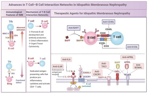
Idiopathic membranous nephropathy (IMN) is an organ-specific autoimmune podocytopathy mediated by autoantibodies. Current understanding integrates the traditional 'antibody-centric' theory into a broader working model, highlighting 'T-B cell network imbalance' as a potential upstream regulatory and amplifying framework. This review systematically discusses the proposed “dual-axis dysregulation” model involving enhanced Tfh-B-cell help and impaired Tfr/Breg-mediated restraint in IMN. It emphasizes that follicular helper T cells (especially the Tfh2/Tfh17 subsets) may support B-cell differentiation and plasma-cell generation via IL-21/ICOS/CD40L signaling. These immune alterations may support the persistent production of anti-PLA2R/THSD7A antibodies, which subsequently contribute to podocyte damage and proteinuria. Existing therapies, such as rituximab, do not directly eliminate long-lived plasma cells or fully restore immune tolerance, leaving a subset of high-risk or refractory patients vulnerable to incomplete response or relapse. Emerging precision-oriented strategies conceptually focus on three major directions: targeting co-stimulatory signaling molecules (e.g., anti-ICOS/L antibodies, CD40L inhibitors), modulating the cytokine network (e.g., BAFF/APRIL dual inhibitors like Telitacicept, IL-6/STAT3 antagonists), and targeting terminal effector cells (e.g., BCMA CAR-T, anti-CD38 monoclonal antibodies). Importantly, these approaches differ substantially in disease specificity and evidence maturity, ranging from direct but preliminary MN clinical evidence to cross-disease, preclinical, or conceptual support, and should not be interpreted as therapeutically equivalent. This article integrates the available basic and clinical evidence, providing a theoretical basis and translational perspective for the shift from generalized immune suppression to network-based precision regulation in IMN treatment.
Keywords: T cell-B cell interaction networks, immune dysregulation, precision therapeutics, idiopathic membranous nephropathy
1. Introduction
Idiopathic membranous nephropathy (IMN) accounts for approximately 75-80% of membranous nephropathy cases and represents one of the leading causes of nephrotic syndrome in non-diabetic adults globally [1]. It can progress to chronic kidney disease or ESRD in patients with persistent active disease [1-3]. Epidemiological studies have shown a sustained increase in its incidence, with a global annual incidence rate of 10-12 cases per million people [1]. Notably, in central and northeastern China, IMN has surpassed IgA nephropathy, accounting for 24.96% of cases, making it the leading cause of primary glomerular disease [4]. Although approximately 30-40% of patients experience spontaneous remission [5, 6], another 30-40% of patients, particularly those in high-risk cohorts, progress to ESRD within 5-10 years due to persistent proteinuria [7, 8], imposing a significant public health burden.
The clinical treatment of IMN currently faces significant challenges. Given the mechanism of podocyte injury mediated by anti-PLA2R/THSD7A antibodies [3], mainstream therapies still rely on nonspecific immunosuppressive approaches. While B-cell-targeted therapies, such as rituximab, are recommended by KDIGO as risk-adapted options for patients with moderate-to-high-risk IMN [9], their long-term efficacy is limited by B-cell regeneration and the inability to clear long-lived plasma cells that do not express CD20 (CD20⁻ LLPCs) [10, 11]. Traditional therapies, such as steroid and alkylating agent or calcineurin inhibitor combinations, can achieve a 60-70% remission rate, but are associated with bone marrow suppression, nephrotoxicity, and a recurrence risk of up to 50% [12-14]. Complement inhibitors only intervene in downstream damage, and the effectiveness of renin-angiotensin system (RAS) blockers is limited in patients with heavy proteinuria [15, 16]. The core issue lies in the fact that pathogenic autoantibodies are produced by autoreactive B cells, which account for only 0.1-0.5% of circulating B cells [17, 18], and current therapies fail to precisely eliminate them.
Recent studies suggest that the imbalance in T-B-cell interactions may serve as an important upstream regulatory and amplifying framework in the immunopathogenesis of IMN. This imbalance can be conceptualized as a “dual-axis dysregulation”: the pathogenic Tfh-B-cell axis appears to be enhanced, with follicular helper T cells, particularly the Tfh2/Tfh17 subsets, showing increased activity through IL-21/ICOS/CD40L signaling and potentially promoting B-cell differentiation into autoantibody-secreting plasma cells [19-21]. At the same time, the tolerogenic Tfr-Breg axis appears to be functionally impaired, as follicular regulatory T cells and regulatory B cells may exhibit reduced immunosuppressive capacity, potentially contributing to a feed-forward cycle of “T-cell overactivation → abnormal B-cell response → antibody deposition → podocyte injury” [22]. This dynamic dysregulation may help explain why B-cell depletion alone does not always achieve sustained remission.
Novel targeted therapeutic strategies aimed at this mechanism are being explored: targeting T-B cell co-signaling molecules (such as anti-ICOS antibodies) may modulate the pathogenic axis signaling [23]; dual inhibitors of BAFF/APRIL (such as Telitacicept) combined with B cell depletion may help reduce the recurrence rate [24]; BCMA-directed cellular therapies remain biologically plausible but exploratory approaches for refractory MN, and their IMN-specific efficacy requires further clinical validation [25]; and rebuilding immune tolerance mediated by Tfr/Breg cells holds promise for future immune-regulatory treatment. This review aims to summarize the molecular regulatory mechanisms of the T-B cell interaction network in IMN and systematically discuss the progress of targeted therapeutic strategies from basic research to clinical translation, providing new perspectives to overcome the current treatment bottleneck.
Throughout this review, primary human evidence in MN—including patient cohorts, prospective and controlled clinical studies, renal tissue and single-cell analyses, and antigen-specific B-cell investigations—is prioritized where available. Direct human evidence in MN is distinguished from cross-disease, preclinical, and conceptual evidence, which is used to provide mechanistic context and is not interpreted as establishing equivalent pathogenic or therapeutic roles in IMN. Within the human evidence base, associative observations are distinguished from functional, longitudinal, and interventional findings, and causal interpretations are calibrated to the design and strength of the available evidence.
2. Immunological Features of IMN
2.1 Autoantigen Presentation and the Initiation of T-B Cell Responses
IMN is an organ-specific autoimmune disease driven by autoantibodies targeting podocyte-associated antigens [5]. To date, at least 14 target antigens have been identified—comprehensively summarized in Supplementary Table S1—with phospholipase A2 receptor (PLA2R) and thrombospondin type-1 domain-containing 7A (THSD7A) being the most prominent [26, 27]. The expanding spectrum of autoantigens, including EXT1/EXT2 associated with systemic autoimmunity and SEMA3B enriched in pediatric cohorts, highlights the heterogeneous immune contexts in which T-B-cell responses may arise [28, 29]. Among these antigens, PLA2R remains the best-characterized clinical biomarker in IMN; serum anti-PLA2R antibody titers are associated with disease activity [30], treatment response [31], and long-term renal prognosis [32, 33]. Beyond the established MN-associated antigen spectrum, the recent identification of anti-vinculin autoantibodies in idiopathic nephrotic syndrome further supports the broader concept of podocyte-targeted autoimmunity; however, this evidence should be regarded as contextual rather than IMN-defining [34].
Rather than serving only as passive targets of antibody deposition, podocyte-associated antigens may also participate in shaping the initiation and maintenance of autoreactive T-B-cell responses. In principle, major podocyte antigens such as PLA2R and THSD7A may be captured and processed by antigen-presenting cells, including B cells with antigen-presenting capacity [35]. While dendritic cells may contribute to early antigen presentation, cognate interactions between antigen-presenting B cells and CD4⁺ T cells through MHC-II-TCR recognition could further support the selection, expansion, and maintenance of autoreactive Tfh-cell responses. In addition, podocyte injury may increase the release or urinary detectability of podocyte-derived antigenic material, including extracellular vesicle/exosome-associated PLA2R. Although these findings mainly support the biomarker value of urinary exosomal PLA2R rather than establishing a direct immunological mechanism, persistent antigen availability may provide a source of ongoing antigenic stimulation, thereby sustaining Tfh-B-cell interactions and contributing to impaired Tfh/Tfr balance within secondary lymphoid organs [36, 37].
The autoantibodies generated in IMN predominantly belong to the IgG4 subclass, a feature consistent with type-2/IL-4-associated humoral skewing [16, 36, 37]. After binding to podocyte antigens in situ, immune complexes are associated with complement activation and podocyte structural injury [16, 37]. Importantly, podocyte injury may further increase antigen release or exposure, creating a potential feedback loop between target-organ damage and autoreactive T-B-cell responses. This continuous interplay supports the need to consider upstream T-B-cell interactions together with the established downstream antibody-complement-podocyte injury cascade.
2.2 Characteristics of the Systemic and Renal Immune Microenvironment in IMN
Before discussing the subset-specific roles of T and B cells in the following sections, this section provides an overview of the immune microenvironment in which autoreactive T-B cell responses are generated and maintained. IMN is characterized not only by antibody-mediated podocyte injury but also by systemic immune dysregulation that creates an autoantibody-supportive milieu [16]. At the systemic level, enhanced Tfh/Th2-associated signals, including increased ICOS- and IL-21-related Tfh activity and Th2 cytokine responses, may favor B-cell help and IgG4-prone humoral immunity [36, 38]. In parallel, alterations in the Th17/Treg balance and regulatory cytokine pathways suggest a shift from immune tolerance toward a more inflammatory and less restrained immune state [39, 40]. B-cell survival and differentiation factors further shape this systemic humoral environment. Increased BAFF and APRIL expression or serum levels have been reported in membranous nephropathy and anti-PLA2R-positive IMN, and these factors have been associated with disease activity, treatment response, or renal outcomes in some cohorts [41, 42]. Thus, the circulating immune microenvironment in IMN can be broadly viewed as a convergence of helper signals, B-cell survival factors, and impaired regulatory restraint that supports autoreactive humoral immunity. These findings should be interpreted as a permissive immunological background rather than evidence that any single cytokine pathway independently drives disease progression.
At the renal level, single-cell transcriptomic studies of kidney tissue from patients with IMN have identified infiltrating myeloid, T-cell, and B-lineage populations and suggested chemokine and inflammatory programs involved in leukocyte recruitment and tissue remodeling [43, 44]. In addition, renal tissue studies in IMN have identified B-cell aggregates and tertiary lymphoid structures (TLS), whose presence has been associated with more severe clinicopathological features or unfavorable renal outcomes [45]. Together, these systemic and renal features provide the microenvironmental context in which autoreactive T-B-cell responses may arise and persist. The cell-specific roles of Tfh, Tfr, Treg, B-cell subsets, and plasma cells are discussed in Sections 3 and 4 and are not further developed here.
3. The Central Role of T Cells in IMN
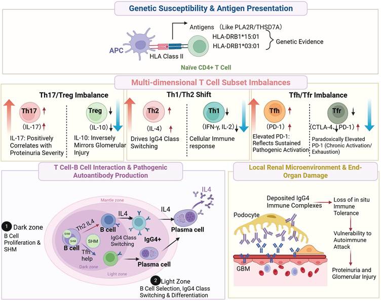
T cells play an important role in the autoimmune cascade of IMN, not only by contributing to local inflammatory responses but also by regulating pathogenic autoantibody production. Genetic susceptibility studies, including associations with HLA class II molecules such as HLA-DRB1*15:01 and HLA-DRB1*03:01 alleles, as well as evidence from animal models, support an important role for T cells in the pathogenesis of IMN [46-49]. HLA class II molecules present antigen-derived peptides to CD4⁺ T cells; in IMN, this mechanism provides a basis for the presentation of podocyte antigen-derived peptides, including those from PLA2R and potentially THSD7A, thereby promoting autoreactive T-cell activation and subsequent B-cell responses [35].
3.1 Imbalance of T Cell Subsets
During the pathogenesis of IMN, imbalance among CD4⁺ T-cell subsets creates an immune environment that favors autoreactive B-cell activation and autoantibody production. Among these abnormalities, Th17/Treg imbalance represents an important inflammation-tolerance disturbance. At the systemic immune level, antigenic stimulation may promote the skewing of naïve CD4⁺ T cells toward a pro-inflammatory Th17 phenotype, while the regulatory T-cell compartment becomes numerically reduced and functionally impaired. Rather than being merely a secondary inflammatory response, this shift may act as an important amplifier of disease activity [50]. Clinically, circulating IL-17 levels positively correlate with proteinuria severity, whereas IL-10 is inversely associated with proteinuria or disease activity-related parameters [51]. In anti-PLA2R-positive patients, reduced renal Foxp3⁺ Treg infiltration and the inverse correlation between circulating IL-10 and proteinuria further support impaired regulatory immune control in IMN [51-54]. This regulatory defect may be further amplified by reduced expression of immune checkpoint molecules such as PD-1 and CTLA-4 on peripheral Tregs, thereby weakening Treg-mediated restraint on B-cell activation and antibody production [55, 56]. At the local renal level, decreased Foxp3 expression, enhanced IL-17-related signals, and increased inflammatory GZMK⁺CD8⁺ T cells have been described, suggesting that systemic Th17/Treg imbalance may be accompanied by local renal immune activation [43]. Consistent with this effector-skewed state, an increased peripheral CD4⁺/CD8⁺ T-cell ratio has been reported in IMN, indicating broader CD4⁺ T-cell-dominated immune activation rather than an isolated Th17/Treg abnormality [36, 57].
Within this broader CD4⁺ T-cell abnormality, Th1/Th2 skewing constitutes another important layer of immune dysregulation in IMN. Th1 cells mainly participate in cellular immune responses, whereas Th2 cells preferentially support humoral immunity [38]. In IMN, the immune response may shift toward a type-2 profile, characterized by an increased proportion of IL-4-producing Th2 cells and reduced Th1-associated cytokines such as IFN-γ and IL-2, resulting in a decreased Th1/Th2 ratio that correlates with proteinuria severity [36, 58, 59]. This type-2-skewed immune milieu, particularly IL-4-associated signaling, may favor IgG4 class switching and thereby contribute to the IgG4-predominant autoantibody profile of IMN [60].
At the germinal-center level, Tfh/Tfr imbalance is particularly relevant to the balance between B-cell help and restraint. Tfh cells promote germinal-center reactions, B-cell differentiation, and antibody secretion [19, 61], whereas Tfr cells limit excessive germinal-center responses and plasma-cell differentiation [62, 63]. In patients with IMN, the proportion of peripheral Tfh cells is increased, whereas Tfr cells are reduced in number and/or functionally impaired. Unlike peripheral Tregs, which may lose suppressive function in association with PD-1/CTLA-4 downregulation, Tfr cells in a chronic autoimmune microenvironment may exhibit reduced CTLA-4 expression together with paradoxically increased PD-1 levels [64]. This seemingly divergent checkpoint pattern may require spatial interpretation. Within the germinal center, increased PD-1 expression does not necessarily translate into enhanced suppression; instead, abnormal PD-1/PD-L1 signaling may disturb chemokine-dependent positioning and displace Tfr cells from their regulatory niches [65]. Concurrently, diminished CTLA-4 may weaken competitive engagement with CD80/CD86 on B cells, allowing Tfh-driven CD28 co-stimulation to persist [66]. Together, this dual checkpoint alteration—elevated PD-1 and reduced CTLA-4—may impair Tfr-mediated immune restraint.
Taken together, T-cell imbalance in IMN can be summarized into three interconnected layers: Th17/Treg imbalance reflects disruption of inflammation-tolerance homeostasis; Th1/Th2 skewing contributes to type-2/IgG4-prone humoral responses; and Tfh/Tfr imbalance may influence the balance between B-cell help and restraint within germinal centers. This layered framework provides a cellular context for considering how T-cell abnormalities may support sustained B-cell activation, affinity maturation, and pathogenic autoantibody production (Figure 1).
Imbalance of T-cell subsets in IMN. Arrows indicate subset expansion (↑) or reduction (↓). Key homeostatic disturbances include: (1) Th17/Treg imbalance: Elevated Th17 cells (producing IL-17, TNF-α, IL-6) alongside diminished Treg cells (with reduced IL-10 and TGF-β1 secretion) impairs immunosuppression. (2) Th1/Th2 skewing: A Th2-dominant shift promotes autoantibody production. (3) Tfh/Tfr imbalance: Tfh expansion coupled with Tfr reduction (and decreased CTLA-4 expression) may favor plasma cell activation. Created with BioRender.com. Abbreviations: IMN, idiopathic membranous nephropathy; Th, T helper cell; Treg, regulatory T cell; Tfh, follicular helper T cell; Tfr, follicular regulatory T cell; IL, interleukin; TNF-α, tumor necrosis factor-alpha; TGF-β1, transforming growth factor-beta 1; CTLA-4, cytotoxic T-lymphocyte associated protein 4; GZMK, granzyme K.
3.2 T Cell-Mediated B Cell Activation
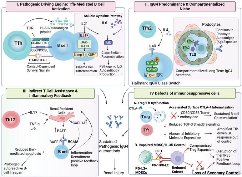
In the pathogenesis of IMN, aberrant B-cell activation and subsequent pathogenic autoantibody production are largely shaped by imbalanced T-cell help. This process depends on the integration of direct cell-to-cell co-stimulatory signals and regulation by soluble mediators, including cytokine-, survival factor-, and chemokine-dependent pathways [67-69]. Tfh cells serve as a key cellular source of B-cell help in this process. Several components of this pathway are supported by primary human studies in IMN. In IMN, circulating cTfh2 cells, particularly the CXCR3⁻CCR6⁻ subset, appear to be preferentially expanded and exhibit a B-cell-helper phenotype characterized by increased ICOS expression and reduced BTLA expression [38, 70, 71]. The proportion of ICOS⁺ cTfh2 cells has been correlated with 24-hour proteinuria, serum IgG4 levels, and the IgG4/IgG ratio, supporting the clinical relevance of this subset in IgG4-prone humoral responses [16, 71]. Mechanistically, this interaction begins with TCR-dependent recognition of B-cell-presented autoantigen-derived peptide-HLA class II complexes, which supports the selection and activation of antigen-specific autoreactive clones [72]. Subsequently, Tfh cells engage B cells through co-stimulatory pairs, including CD40L-CD40, ICOS-ICOSL, and OX40-OX40L, forming a structured immunological synapse that delivers key co-stimulatory survival and activation signals. The importance of this contact-dependent pathway is supported by preclinical evidence demonstrating that therapeutic blockade of the CD40L-CD40 interaction can suppress antibody production and mitigate renal injury [72].
Regarding soluble factor regulation, three soluble mediator-related mechanisms link T-cell abnormalities to B-cell activation in IMN. First, Tfh-derived IL-21 is a key cytokine regulating B-cell differentiation, maturation, and class-switch recombination [73-75]. By engaging IL-21R and activating the downstream JAK-STAT3 pathway, IL-21 induces transcription factors such as Blimp-1 and XBP-1, thereby promoting terminal B-cell differentiation into plasma cells. It may also cooperate with IL-6 to enhance activation-induced cytidine deaminase (AID) activity, supporting class-switch recombination and antibody diversification [76, 77]. The clinical relevance of this pathway is supported by observations that circulating IL-21⁺ Tfh cells are associated with anti-PLA2R antibody titers and can promote IgG4 production in autologous B-cell co-culture systems, providing clinical and functional support for a role of the Tfh-B-cell axis in autoreactive humoral responses in IMN. In parallel, PLA2R-specific IgG secretion by circulating antibody-secreting cells has been directly quantified in patients with IMN [57], providing antigen-specific B-cell evidence. However, a direct longitudinal link between these antibody-secreting cells and the Tfh abnormalities described above remains to be established.
Second, type-2/IL-4-associated signaling may help explain the IgG4-predominant humoral phenotype of IMN. IL-4 can activate the STAT6-GATA3 pathway in B cells and induce transcription of the immunoglobulin heavy-chain constant region Cγ4, thereby favoring IgG4 class switching [36, 78]. Thus, whereas IL-21 mainly supports general B-cell differentiation and plasma-cell generation, IL-4-associated signaling may contribute to the subclass bias of the autoantibody response. Finally, Th17 cells may influence B-cell responses indirectly by shaping the inflammatory microenvironment. IL-17, in cooperation with other pro-inflammatory cytokines, can activate renal resident cells, including podocytes and potentially mesangial cells, to upregulate B-cell survival and recruitment signals such as BAFF and CXCL13 [79]. BAFF supports the survival of autoreactive B cells, whereas CXCL13 promotes B-cell migration and accumulation at inflammatory sites. Together, these cytokine-, survival factor-, and chemokine-mediated pathways may create a feed-forward environment that sustains autoreactive humoral immunity.
In addition, defects in immune regulatory cells further exacerbate the uncontrolled B cell response, constituting a “dual hit” between immune activation and suppression. Specifically, Treg dysfunction reduces restraint on autoreactive B cells. The accelerated internalization of Treg surface CTLA-4 weakens the ability to clear B cell surface CD80/CD86 co-stimulatory molecules via trans-endocytosis, leading to sustained co-stimulatory signaling. Additionally, the reduced secretion of TGF-β by Treg cells diminishes their suppressive effect on B cells through the Smad3 pathway [80]. Similarly, Tfr cells also exhibit functional defects, with abnormal expression or accelerated internalization of surface inhibitory molecules (such as CTLA-4) [81, 82], preventing them from effectively regulating germinal center reactions, and thereby amplifying B cell activation driven by Tfh cells. Moreover, the immunosuppressive function of myeloid-derived suppressor cells (MDSCs) is impaired, with a reduced proportion of the PD-L2⁺ subset, compromising their ability to promote Treg expansion and secrete the inhibitory cytokine IL-35 via the PD-1/PD-L2 axis [83]. IL-35 is not only a key mediator of Treg function but also induces the formation of an iTR35 positive feedback inhibitory loop [80, 84, 85]. Emerging evidence suggests that the disruption of this loop may result in the loss of secondary regulatory control over the B cell response in IMN. In summary, T cell-mediated B cell activation in IMN is a multilayered process involving T-cell help linked to antigen presentation, cytokine polarization, co-stimulatory signaling, and impaired immune regulation, which may collectively support persistent IgG4-predominant autoantibody responses, thereby contributing to renal injury (Figure 2).
T cell-mediated B-cell activation pathways in IMN. Imbalanced T-cell help may support autoreactive B-cell activation and IgG4-predominant autoantibody responses through four interconnected modules. (I) Contact-dependent Tfh-B-cell interaction: antigen-presenting B cells display autoantigen-derived peptide-HLA class II complexes to Tfh cells, while co-stimulatory pairs, including CD40L-CD40, ICOS-ICOSL, and OX40-OX40L, provide survival and activation signals to B cells. Tfh-derived IL-21 further promotes plasma-cell differentiation through the IL-21R-JAK/STAT3-Blimp-1/XBP-1 pathway, and may cooperate with IL-6/AID signaling to support class-switch recombination. (II) IgG4-predominant humoral skewing: type-2/IL-4-associated signaling activates the IL-4R-STAT6-GATA3 pathway and favors Cγ4 transcription, thereby contributing to IgG4 class switching. Local antigen exposure and TLS-like niches may further support the persistence of IgG4-prone humoral responses. (III) Indirect inflammatory support: Th17-derived IL-17, together with TNF-α and IL-6, can activate renal resident cells to upregulate BAFF and CXCL13, promoting autoreactive B-cell survival, recruitment, and inflammatory feedback. (IV) Defective immune restraint: dysfunction of Treg, Tfr, and MDSC/IL-35-mediated regulatory pathways weakens suppression of Tfh-driven germinal-center responses and B-cell co-stimulation, thereby facilitating sustained pathogenic humoral immunity. Created with BioRender.com. Abbreviations: IMN, idiopathic membranous nephropathy; IgG4, immunoglobulin G4; CD40L, CD40 ligand; IL, interleukin; Tfh, follicular helper T cell; Tfh2, type 2 follicular helper T cell; Th17, T helper 17 cell; TNF-α, tumor necrosis factor-alpha; NF-κB, nuclear factor kappa B; BAFF, B cell activating factor; CXCL13, C-X-C motif chemokine ligand 13; Treg, regulatory T cell; Tfr, follicular regulatory T cell; MDSCs, myeloid-derived suppressor cells.
4. Abnormal B Cell Activation and Autoantibody Production: The Core of Humoral Immunity in IMN
The essence of IMN is an organ-specific autoimmune disease, with its core pathological feature being the B cell-mediated production of pathogenic antibodies and the subsequent deposition of immune complexes in the glomeruli. B cells play a triple pathogenic role in IMN: on one hand, they serve as the cells responsible for producing autoantibodies such as anti-PLA2R and anti-THSD7A antibodies [86, 87]; on the other hand, they act as professional antigen-presenting cells to activate CD4⁺ T cells [88, 89], forming T-B cell interactions; additionally, B cells contribute to the formation of ectopic lymphoid tissues, which promote the onset of immune-mediated disease [90-92].
4.1 Dynamic Changes in B Cell Subsets: Abnormal Distribution from Peripheral Blood to the Kidneys
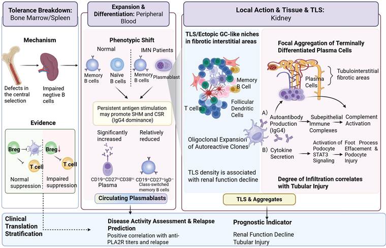
In the pathogenesis of IMN, the dynamic changes and abnormal distribution of B cell subsets are central to the production of pathogenic autoantibodies. This process may be closely related to the disruption of both central and peripheral B-cell tolerance against podocyte autoantigens. Defects in central selection mechanisms, such as clonal deletion and receptor editing of immature B cells in the bone marrow, may allow autoreactive B-cell clones to escape into the peripheral compartment; subsequently, impaired negative selection of transitional B cells in the spleen may further promote their persistence and clonal expansion [93]. A more direct peripheral manifestation of this process is the remodeling of circulating B-cell subsets. In IMN, an increase in transitional B cells (CD19+CD24hiCD38hi) has been observed, and these cells may display an altered cytokine profile, with increased IL-6/IL-12 and reduced IL-10 production, potentially favoring a pro-inflammatory B-cell milieu [94]. Flow-cytometric analysis has further shown that the proportion of plasmablasts (CD19⁺CD27ʰⁱCD38ʰⁱ) in peripheral blood is significantly increased, whereas germinal center-derived class-switched memory B cells (CD19⁺CD27⁺IgD⁻) are relatively reduced in patients with IMN [95]. Taken together, these circulating B-cell alterations may be consistent with sustained antigen-driven B-cell activation, although this relationship has not been directly established. Independent B-cell receptor repertoire analysis in MN demonstrated increased somatic hypermutation rates across multiple immunoglobulin heavy-chain isotypes [96]. In parallel, PLA2R-associated MN was characterized by IgG4-biased, higher-affinity anti-PLA2R antibodies, with antibody affinity and the IgG4 proportion associated with proteinuria and histologic severity [97]. Together, these observations are consistent with altered affinity maturation and immunoglobulin class switching in MN and suggest that these processes may contribute to persistent autoreactive antibody production and greater disease activity.
As the terminal target organ of immune injury, the kidney exhibits highly specific B cell infiltration and activation characteristics in its local microenvironment. Studies have shown that the proportion of memory B cells (CD19⁺CD27⁺) in renal tissue is higher compared to controls, suggesting local activation driven by continuous antigen exposure. Meanwhile, terminally differentiated plasma cells (CD20⁻CD38⁺CD138⁺) are focally aggregated around the renal interstitium and glomeruli. These plasma cells may serve as an important local source of autoantibodies and may also activate podocyte STAT3 signaling through inflammatory cytokines such as IL-6 and TNF-α, contributing to foot process effacement and podocyte injury [98, 99]. Importantly, within germinal center-like structures or tertiary lymphoid structures (TLS) formed in chronic inflammatory regions, there is oligoclonal expansion of B cells. This “ectopic” germinal center response is hypothesized to provide a specialized niche that facilitates the selection and expansion of autoreactive B cell clones, with the density of their infiltration showing a significant negative correlation with renal function decline in patients [100-104].
Immunohistochemical studies further confirm that B cell aggregates are predominantly distributed in fibrotic interstitial areas and around sclerotic glomeruli, with the degree of infiltration correlating positively with tubular injury [105]. This underscores the close relationship between B cell inflammation and the tissue repair/fibrosis process. In addition to the active presence of effector B cells, dysfunction of Breg further exacerbates immune imbalance. Under physiological conditions, Bregs exert key negative immunoregulatory effects mainly through IL-10 secretion, thereby inhibiting effector T-cell activation and reducing the antigen-presenting capacity of dendritic cells [106-112]. However, in IMN patients, the proportion of IL-10⁺ Bregs in peripheral blood is significantly reduced, leading to impaired immune suppression and an inability to effectively curb autoimmune inflammation. These findings provide a potential clinical rationale for B-cell subset analysis in IMN. For example, circulating plasma-cell levels may have value as biomarkers of disease activity. Its strong positive correlation with anti-PLA2R antibody titers and further elevation in patients with relapses suggests that it could serve as a prospective biomarker for predicting relapse risk (Figure 3).
Dynamic spatial distribution and clinical relevance of B-cell subsets in IMN. Disruption of central and peripheral B-cell tolerance may permit autoreactive B-cell clones to persist and expand. In peripheral blood, IMN is characterized by increased transitional B cells and circulating plasmablasts, relative reduction of class-switched memory B cells, and impaired Breg-mediated suppression, collectively favoring IgG4-predominant autoantibody production. In the kidney, B-cell aggregates, germinal center-like/TLS niches, and focally accumulated terminally differentiated plasma cells may support local immune persistence, oligoclonal expansion, and podocyte injury. Circulating plasmablasts may reflect disease activity and relapse risk, whereas renal TLS/B-cell aggregates are associated with tubular injury and renal function decline. Created with BioRender.com. Abbreviations: IMN, idiopathic membranous nephropathy; Bregs, regulatory B cells; TLS, tertiary lymphoid structures.
4.2 Key Mechanism of Autoantibody Production: Plasma Cell Heterogeneity and Treatment Resistance
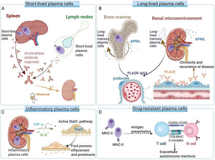
Plasma cells, as the terminal effector cells of B cell differentiation, are the direct source of pathogenic autoantibodies, and their heterogeneity is closely related to treatment resistance. Based on developmental origin, survival characteristics, and localization, plasma cells can be classified into different functional subsets: short-lived plasma cells can originate from extrafollicular activated B cells or early germinal center reactions, are typically localized in the spleen and lymph nodes, and are responsible for acute-phase antibody responses. Notably, terminally differentiated plasma cells are typically CD20-negative and therefore are not directly targeted by CD20-directed therapies such as rituximab. Monoclonal antibodies targeting CD20, such as rituximab, reduce autoantibody production mainly by depleting upstream CD20⁺ B-cell compartments, including activated and memory B cells, thereby limiting the generation of new antibody-secreting cells. This is one of the key mechanisms through which approximately 60-70% of high-risk IMN patients achieve long-term remission [7, 113, 114]. In contrast, long-lived plasma cells (LLPCs), characterized by a CD19⁻CD20⁻CD38⁺CD138⁺ phenotype, primarily reside in specialized survival niches such as the bone marrow and inflamed renal microenvironment, where they may be maintained long-term through APRIL/BAFF-enriched survival-niche signaling. These cells may continuously secrete anti-PLA2R IgG4 antibodies within APRIL/BAFF-enriched survival niches, thereby contributing to disease chronicity and relapse [115, 116]. Additionally, functionally specialized plasma-cell subsets have been identified in renal tissue in autoimmune nephritis, and inflammatory cytokines such as IL-6 and TNF-α may contribute to local renal injury; notably, IL-6/STAT3 signaling is activated in podocytes in experimental membranous nephropathy and may contribute to podocyte injury and proteinuria [57, 117, 118]. Some studies have also found that certain plasma cells highly express MHC-II molecules (CD138⁺CD38⁺MHC-IIhi), suggesting that they may retain some antigen-presenting capability and potentially sustain the local autoimmune response through interactions with T cells. These unique phenotypes may help explain their resistance to conventional treatments, though further research is needed to validate these emerging subsets and their functions.
Persistent autoantibody production in IMN may reflect both the continued generation of antibody-secreting cells and the persistence of established plasma-cell populations. Upstream abnormalities in B-cell tolerance, regulatory restraint, affinity maturation, and renal TLS-like niches are discussed in Section 4.1; here, their relevance is considered specifically in relation to the generation and persistence of short-lived and long-lived antibody-secreting plasma cells.
The formation of treatment resistance is closely associated with the characteristics of long-lived plasma cells (LLPCs) and their high adaptability to the microenvironment. Rituximab effectively depletes CD20⁺ B cells but has limited efficacy against CD20⁻ LLPCs; approximately 30% of patients continue to secrete autoantibodies at least in part because of the persistence of these cells [119]. LLPCs rely on survival signals such as APRIL to resist conventional immunosuppressive drugs, providing a rationale for dual inhibition of BAFF and APRIL, such as atacicept, to more comprehensively target their survival pathways [119-121]. Additionally, complement activation products in the kidneys (such as C5a and C3a) can promote the formation of fibrotic barriers by indirectly activating the BMP2/pSMAD1 signaling pathway. It is hypothesized that the resulting fibrotic barriers might provide a physical sanctuary, further protecting local plasma cells from drug-induced damage, similar to mechanisms observed in other plasma cell dyscrasias [122].
Currently, new therapeutic strategies targeting drug-resistant plasma cells are being explored. Anti-CD38 monoclonal antibodies (e.g., daratumumab) are being explored as plasma-cell-directed strategies in refractory MN [123-125]. Bispecific antibodies targeting BCMA/CD3 (e.g., Elranatamab) have shown high remission rates in clinical trials for relapsed/refractory multiple myeloma [126], but their efficacy in IMN is still under investigation. Additionally, exploratory strategies such as combining BAFF/APRIL pathway inhibitors (e.g., Atacicept) with anti-CD20 monoclonal antibodies aim to achieve synergistic effects through “dual-pathway blockade,” though the clinical value of this approach requires further research. These mechanistic observations provide the rationale for plasma-cell- and survival-niche-directed interventions; the disease specificity, evidence maturity, and translational limitations of anti-CD38, BCMA-directed, and BAFF/APRIL-targeted approaches are discussed further in Section 5 (Figure 4).
Heterogeneity of plasma cells in IMN. (A) Short-lived plasma cells (SLPCs) derived from activated B cells in secondary lymphoid organs; their continuous generation is limited by rituximab-mediated depletion of upstream CD20⁺ B-cell compartments. (B) CD20⁻ long-lived plasma cells (LLPCs) residing in the bone marrow and inflamed renal microenvironment, supporting chronic anti-PLA2R IgG4 production through APRIL/BAFF-enriched survival-niche signaling. (C) Inflammatory plasma cells may contribute to podocyte injury through IL-6 and TNF-α secretion. (D) Drug-resistant plasma cells expressing high levels of MHC-II, potentially sustaining local immune activation through antigen-presenting activity. Created with BioRender.com. Abbreviations: IMN, idiopathic membranous nephropathy; SLPCs, short-lived plasma cells; LLPCs, long-lived plasma cells; PLA2R, M-type phospholipase A2 receptor; IgG4, immunoglobulin G4; APRIL, a proliferation-inducing ligand; IL-6, interleukin-6; TNF-α, tumor necrosis factor-alpha; MHC-II, major histocompatibility complex class II.
4.3 The Dual-Axis Dysregulation Model in IMN: A Hypothesis-Generating Framework
In IMN, autoimmunity is organized around podocyte-associated antigens, such as PLA2R and THSD7A, whose cognate circulating autoantibodies bind in situ and contribute to subepithelial immune-complex deposition and podocyte-centered immune injury [27, 37, 127]. Against this antigen-specific background, the “dual-axis dysregulation” model serves as an IMN-oriented explanatory framework that integrates T-B-cell abnormalities with the disease's IgG4-biased humoral response and podocyte-centered injury [36, 128]. It includes a pathogenic axis of enhanced Tfh-cell-mediated B-cell help within a type-2-skewed immune milieu [71], and a regulatory axis of impaired Tfr- and Breg-mediated restraint [129]. Its disease specificity lies not in IMN-exclusive Tfh, Tfr, or Breg abnormalities, but in the coupling of these immune-regulatory disturbances with podocyte-associated autoantigens, IgG4-biased humoral responses, and in situ subepithelial immune-complex deposition. Accordingly, the dual-axis dysregulation model is presented as an IMN-oriented, hypothesis-generating framework rather than an established causal pathway. IMN-specific studies support several individual components of the model, including altered helper and regulatory lymphocyte phenotypes and their associations with autoreactive humoral activity. However, much of the available human evidence remains based on peripheral immune phenotyping, cross-sectional associations, and relatively small cohorts, while several detailed Tfr-, Breg-, checkpoint-, and cytokine-dependent mechanisms are informed by evidence from adjacent autoimmune diseases, animal models, or in vitro systems. Its relevance to IMN derives from its application to the disease context defined by podocyte-associated antigens, IgG4-predominant autoantibodies, and in situ subepithelial immune deposits, rather than from the assumption that the underlying immune-cell abnormalities are unique to IMN. Direct antigen-specific, tissue-resolved, and longitudinal evidence linking these immune states to PLA2R- or THSD7A-reactive B-cell or plasma-cell clones, LLPC persistence, treatment resistance, or relapse remains limited. Taken together, the dual-axis model links altered T-cell help and regulatory restraint to persistent autoreactive humoral activity within the antigen-specific context of IMN. It complements, rather than replaces, the established antibody-complement-podocyte injury paradigm and is used here as an integrative framework for the cell-specific evidence summarized in Sections 3 and 4, rather than as an additional independent layer of mechanism.
The framework may also connect peripheral immune abnormalities with the renal tubulointerstitial immune microenvironment. TLO/TLS-like aggregates, in which B cells may organize with T cells and dendritic cells, may provide local niches for antigen presentation and immune-cell persistence [45, 101]. Residual CD20-negative long-lived plasma cells are not directly depleted by anti-CD20 therapy [130, 131], while autoreactive B-cell reconstitution, persistent activity of residual plasma cells, and incomplete recovery of regulatory mechanisms may contribute to persistent or recurrent humoral activity after B-cell depletion [58, 86, 132]. Although the relative importance of these processes remains uncertain in IMN, they may inform future stratification among B-cell depletion, plasma-cell-directed therapy, and regulatory-axis restoration.
5. From Mechanism to Clinic: Precision Strategies Targeting the T-B Cell Interaction Network in IMN
The T-B cell interaction network in IMN operates through two principal modes of communication: direct cell-to-cell interactions mediated by co-signaling molecules and soluble mediator-dependent signaling involving cytokines and B-cell survival factors. Key Tfh-associated co-signaling pathways, including ICOS-ICOSL, PD-1-PD-L1, and CD40L-CD40, are proposed to regulate Tfh-B-cell interactions through direct cellular contact and immune-synapse formation [21]. In parallel, soluble mediator networks involving BAFF, IL-6, and IL-21 contribute to the maintenance and amplification of immune responses by regulating B-cell survival, T-cell polarization, and inflammatory-cell recruitment. Together, these interconnected mechanisms provide a theoretical rationale for exploring precision-oriented interventions. However, to avoid excessive extrapolation, the therapeutic candidates discussed below are evaluated along two complementary dimensions: disease specificity and evidence maturity. Disease specificity distinguishes direct clinical evidence in MN from cross-disease extrapolation and mechanistic concept transfer, whereas evidence maturity spans mechanistic and preclinical evidence, case reports and small case series, prospective early-phase studies, and controlled clinical studies. Accordingly, Section 5.1 summarizes therapeutics with direct clinical evidence in MN, Section 5.2 discusses interventions supported primarily by cross-disease or concept-transfer evidence, and Section 5.3 presents preclinical, mechanistic, and early exploratory candidates.
5.1 Therapeutics with Direct Clinical Evidence in MN
Because established B-cell-directed therapy provides the clinical benchmark for emerging precision strategies, randomized MN trial evidence is presented first. In the MENTOR trial, rituximab was noninferior to cyclosporine for inducing complete or partial remission at 12 months and was superior in maintaining remission at 24 months [114]. Emerging MN-specific therapies discussed below are positioned relative to this benchmark according to their disease specificity and evidence maturity.
5.1.1 Therapeutics Targeting the BAFF/APRIL Axis
In the immunopathology of IMN, cytokines form a complex soluble-mediator regulatory network, with the BAFF (B cell activating factor, also known as BLyS) and APRIL (a proliferation-inducing ligand) signaling pathways playing important roles in B cell homeostasis and autoantibody production. Both are members of the tumor necrosis factor (TNF) superfamily, primarily produced by monocytes, macrophages, and bone marrow stromal cells. They regulate B cell development [133, 134], survival [135], class switching, and antibody secretion [135, 136] by binding to three receptors—BAFF-R, TACI (transmembrane activator and calcium-modulator ligand interactor), and BCMA (B cell maturation antigen). At the molecular level, BAFF acts as a crucial survival factor for B cells, promoting the maturation of transitional B cells into memory B cells primarily through BAFF-R signaling [137]. Its abnormal overexpression is a key event that disrupts B cell immune tolerance. While APRIL is not essential for B cell development, it mediates the differentiation of marginal zone B cells into short-lived plasma cells through TACI and sustains the long-term survival of plasma cells through BCMA signaling, particularly in the inflammatory microenvironment [138, 139]. Both BAFF and APRIL can activate the NF-κB signaling pathway, thereby enhancing the production of antibodies, especially IgG subclasses, and contributing to autoimmune glomerular diseases.
The pathogenic relevance of this pathway in IMN is clinically supported: serum levels of BAFF and APRIL are significantly elevated in patients compared to healthy controls [41, 42, 140], and BAFF levels correlate positively with anti-PLA2R antibody titers and the severity of proteinuria, serving as an independent risk factor for disease prognosis [41, 42]. In contrast, APRIL levels do not show a significant correlation with clinical indicators [141, 142], suggesting a difference in their pathogenic mechanisms—BAFF primarily governs B cell survival, autoantibody production, and T cell co-stimulation [143, 144], while APRIL is more involved in plasma cell maintenance, antigen presentation, and immunoglobulin class switching [145, 146]. Transgenic animal models further support the central role of BAFF, as its overexpression can induce a lupus-like phenotype and renal immune complex deposition [147, 148].
Based on the mechanisms outlined above, targeting the BAFF/APRIL pathway represents a rational therapeutic strategy under clinical investigation in IMN. Belimumab, the first approved anti-BAFF monoclonal antibody for SLE [149], was evaluated in a prospective open-label experimental medicine study of PLA2R-Ab-positive primary MN patients with persistent nephrotic-range proteinuria (BEL116472; ClinicalTrials.gov identifier: NCT01610492). Treatment with belimumab for up to 2 years was associated with reductions in proteinuria and circulating PLA2R-Ab levels by 86% and 97%, respectively. Among participants who received more than 16 weeks of treatment, 9 of 11 achieved partial or complete remission during the study, including 1 complete remission. The decline in PLA2R-Ab preceded the reduction in proteinuria, supporting a potential therapeutic effect of belimumab [150]. These findings provide direct but preliminary clinical evidence for BAFF inhibition in IMN. Given the small sample size and the slower decline in anti-PLA2R antibodies at Week 28 compared with rituximab-treated patients in the GEMRITUX study, belimumab may currently be better positioned as an adjunctive or selected-patient strategy rather than a broadly established first-line replacement [150]. In addition to single-target blockade, dual BAFF/APRIL inhibitors have advanced into IMN-specific clinical evaluation, supporting their inclusion in the category of IMN-specific clinical evaluation. Notably, telitacicept, a novel recombinant TACI-Fc fusion protein, has been evaluated in SLE and IgA nephropathy [151, 152] and has been registered for evaluation in a multicenter, prospective, randomized clinical study specifically in patients with primary membranous nephropathy (NCT06614985). By simultaneously neutralizing both BAFF and APRIL, telitacicept may provide broader inhibition of B cell survival and long-lived plasma cell maintenance compared to anti-BAFF monotherapy [153]. Providing preliminary clinical support for this rationale, a recent case series of three refractory IMN patients reported that telitacicept (240 mg) induced complete remission (proteinuria <0.3 g/day) in one patient and partial remission in another, suggesting potential clinical benefit in heavily pretreated individuals [154]. In the future, biomarker-based strategies, such as using BAFF/APRIL levels and anti-PLA2R antibodies, may help accurately select patients who would benefit from these therapies and optimizing personalized treatment strategies (Figure 5, Table 1).
Overview of targeted therapeutic agents stratified by disease specificity and evidence maturity
| Disease specificity | Evidence maturity | Target | Therapeutic Agent | Mechanism of Action | Clinical / Preclinical Status for IMN | References |
|---|---|---|---|---|---|---|
| Direct clinical evidence in MN | Case series plus registered disease-specific clinical trial | BAFF/APRIL | Telitacicept | Recombinant TACI-Fc fusion protein that neutralizes both BAFF and APRIL, thereby targeting B-cell survival, plasma-cell maintenance, and autoreactive humoral responses | IMN-specific evidence remains preliminary: a small refractory IMN case series has reported partial or complete remission, and a disease-specific randomized clinical trial has been registered; larger controlled studies are needed | NCT06614985; [153] mechanism; [154] case series |
| Prospective open-label study | BAFF | Belimumab | Anti-BAFF monoclonal antibody that limits BAFF-mediated autoreactive B-cell survival and autoantibody production | Direct but preliminary clinical evidence in PLA2R-Ab-positive IMN; evaluated in a small prospective open-label study, with larger controlled trials still needed to establish its clinical positioning in IMN | [149, 150] | |
| Published Phase 1/2 open-label study plus ongoing Phase 2b/3 randomized trial | BAFF/APRIL | Povetacicept | Enhanced dual BAFF/APRIL antagonist designed to block BAFF- and APRIL-dependent B-cell and plasma-cell survival pathways | Direct early clinical evidence in pMN from a Phase 1/2 study; currently under evaluation in an ongoing Phase 2b/3 randomized active-controlled trial | [242], NCT07204275 | |
| Case-report level | CD38 | Daratumumab | Depletes CD38-expressing antibody-secreting plasma cells, including CD20- long-lived plasma cells | Case reports in refractory PLA2R- and THSD7A-associated MN; durability remains uncertain | [124, 158] | |
| Prospective phase 1b/2a study | CD38 | Felzartamab | Fully human anti-CD38 monoclonal antibody targeting plasmablasts and plasma cells | Phase 1b/2a evidence in high-risk anti-PLA2R-positive IMN; longer follow-up and controlled validation are needed | [159] | |
| Cross-Disease Extrapolation | Phase III randomized clinical evidence in IgAN | BAFF/APRIL | Atacicept | A recombinant TACI-Fc fusion protein providing dual inhibition of BAFF and APRIL; pharmacologically analogous to Telitacicept | Extrapolated from Phase III clinical evidence in IgAN; awaiting direct IMN validation | [243] |
| Phase III clinical evidence in SLE | CD40L | Dapirolizumab pegol | CD40L inhibitor that blocks CD40-CD40L co-stimulatory signaling and T-cell-dependent B-cell activation | Phase III evidence in moderate-to-severe SLE; no direct IMN-specific clinical evidence | [182] | |
| Phase Ib clinical evidence in SLE | ICOSL | AMG 557 | A fully human IgG2 monoclonal antibody neutralizing ICOSL to suppress downstream Tfh cell activation and functionality. | Evaluated in Phase Ib trials for SLE; theoretical concept transfer for IMN modulation | [191] | |
| Completed Phase II randomized clinical study in SLE | ICOS / CD28 | ALPN-101 | A dual-antagonist Fc fusion protein (human ICOSL variant) simultaneously blocking CD28 and ICOS pathways to restrict pathogenic T-B cell costimulation | Completed Phase II clinical evaluation in SLE; no direct IMN-specific clinical evidence | NCT04835441; [244] mechanism | |
| Preclinical, Mechanistic, and Early Exploratory Candidates | Registered early-phase clinical study including MN; no published MN-specific efficacy data | BCMA | BCMA CAR-T | Engineered T cells executing targeted cellular ablation of BCMA-expressing mature plasma cells | Early exploratory clinical evaluation includes MN in a registered dual CD19/BCMA CAR-T study; published MN-specific efficacy data are not yet available | NCT07241468; [235, 236] |
| Evidence from CKD studies and rare secondary MN contexts | IL-6 / IL-6R | Tocilizumab / Ziltivekimab | IL-6 ligand or IL-6R blockade, potentially modulating IL-6/STAT3-related inflammatory signaling, T-cell polarization, and podocyte-associated responses | Evidence mainly from advanced CKD studies and rare Castleman disease-associated MN cases; no direct IMN-specific clinical validation | [217-220] | |
| Preclinical evidence in diabetic kidney disease models | JAK1 /JAK2 | Ruxolitinib | A JAK1/JAK2 inhibitor that suppresses JAK/STAT signaling and downstream inflammatory responses | Preclinical evidence in diabetic kidney disease models; pending specific IMN validation | [221] |
Abbreviations: IMN, idiopathic membranous nephropathy; SLE, systemic lupus erythematosus; IgAN, IgA nephropathy; LN, lupus nephritis; mAb, monoclonal antibody; BAFF, B cell activating factor; APRIL, a proliferation-inducing ligand; BLyS, B lymphocyte stimulator; TACI, transmembrane activator and CAML interactor; BCMA, B cell maturation antigen; CKD, chronic kidney disease; N/A, not available/not applicable.
The BAFF/APRIL signaling network and targeted therapeutic candidates in IMN. BAFF signaling via BAFF-R promotes peripheral B-cell maturation and survival, supports memory B-cell development, and, when dysregulated, contributes to the breakdown of B-cell immune tolerance. Concurrently, APRIL and BAFF engage TACI to mediate short-lived plasma cell (PC) differentiation, while the APRIL/BCMA axis specifically sustains the long-term survival of PCs. These pathways converge on NF-κB activation to amplify IgG autoantibody production and autoreactive humoral responses. Key targeted interventions include anti-BAFF (belimumab, blisibimod), anti-APRIL (sibeprenlimab, zigakibart), and dual antagonists (atacicept, povetacicept, telitacicept); however, these agents differ substantially in their level of IMN-specific evidence, ranging from direct but preliminary clinical evidence in IMN to cross-disease extrapolation or early-phase pharmacological evidence. Created with BioRender.com. Abbreviations: IMN, idiopathic membranous nephropathy; BAFF, B cell activating factor; APRIL, a proliferation-inducing ligand; BAFF-R, BAFF receptor; TACI, transmembrane activator and CAML interactor; BCMA, B cell maturation antigen; NF-κB, nuclear factor kappa B; IgG, immunoglobulin G; PC, plasma cell.
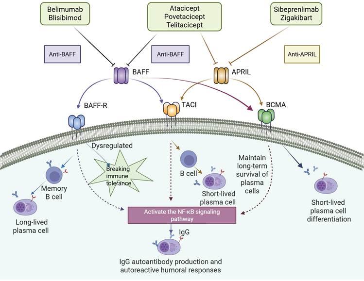
Other BAFF/APRIL-axis agents currently remain at different evidence levels and should not be regarded as established IMN therapies. APRIL-selective inhibitors, such as sibeprenlimab and zigakibart, have mainly been evaluated in IgA nephropathy, where they have shown proteinuria-lowering effects and advanced to Phase 3 clinical trials, thereby providing cross-disease support rather than direct IMN evidence. Similarly, other dual BAFF/APRIL inhibitors, such as atacicept [155, 156], and BAFF-targeting agents under development, such as blisibimod, are pharmacologically relevant to the same pathway but still await IMN-specific validation. These agents are therefore better regarded as cross-disease extrapolations or candidates for evidence tracking in Table 1 rather than therapies supported by direct MN clinical evidence.
5.1.2 Anti-CD38 Monoclonal Antibodies
Although anti-CD38 therapy does not directly block T-B cell co-stimulation, it targets CD38-expressing antibody-secreting plasma cells, the downstream effector cells responsible for persistent autoantibody production. In rituximab-resistant IMN, CD20- long-lived plasma cells may continue to produce pathogenic autoantibodies despite effective B-cell depletion and may contribute to sustained PLA2R antibody production in some patients, providing the rationale for plasma-cell-directed therapy [157]. In a reported case of multidrug-resistant PLA2R-associated membranous nephropathy, weekly intravenous daratumumab at 16 mg/kg induced a rapid decline in anti-PLA2R antibody levels and clinical improvement; however, antibody titers increased again after the dosing interval was extended. Immune phenotyping showed undetectable plasma cells with concomitant expansion of naïve B cells, supporting the authors' rationale for subsequent rituximab re-treatment [124]. Daratumumab has also been reported in refractory THSD7A-positive membranous nephropathy, with disappearance of anti-THSD7A antibodies by month 4, normalization of serum albumin, and reduction of proteinuria, although relapse occurred after treatment withdrawal [158]. In addition, the phase 1b/2a M-PLACE study showed that felzartamab, another anti-CD38 monoclonal antibody, induced rapid immunologic responses in high-risk anti-PLA2R-positive primary membranous nephropathy, with anti-PLA2R titer reductions ≥50% in 20 of 26 efficacy-evaluable patients and partial proteinuria remission in 9 of 26 patients; therapy was generally tolerated, but longer follow-up and controlled studies are still required to define its durability and safety [159, 160]. Thus, anti-CD38 therapy represents a direct but still early-stage MN-specific plasma-cell-directed strategy and may be particularly relevant to refractory or rituximab-resistant disease. Daratumumab is currently supported by case reports in PLA2R- and THSD7A-associated MN, whereas felzartamab has prospective phase 1b/2a evidence in high-risk anti-PLA2R-positive IMN. Longer follow-up and confirmatory controlled studies are required to establish the durability, optimal treatment sequencing, and role of anti-CD38 therapy in routine clinical practice.
5.2 Therapeutics Supported Primarily by Cross-Disease or Concept-Transfer Evidence
Clinical development in adjacent autoimmune or glomerular diseases does not establish therapeutic efficacy in IMN. Accordingly, the agents discussed in this section are presented as cross-disease or concept-transfer candidates. Their reported clinical phase refers to the source disease rather than to IMN, and their relevance to IMN remains to be tested in disease-specific studies.
5.2.1 CD40-CD40L Axis
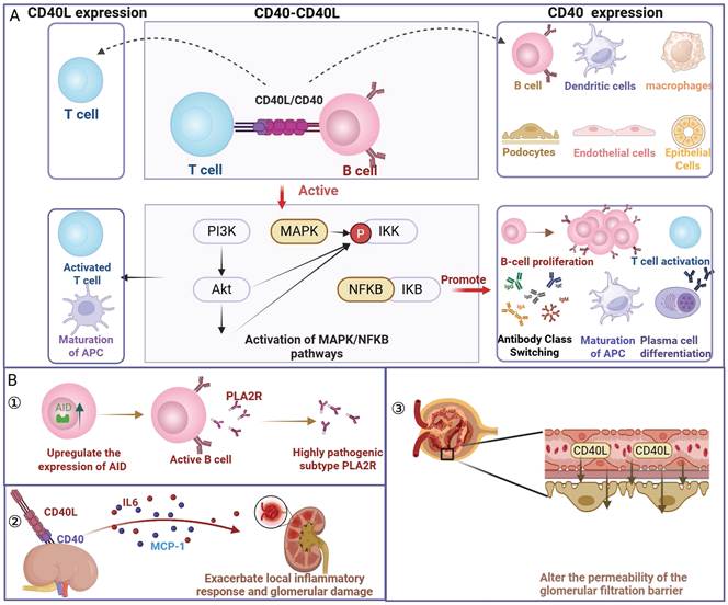
The CD40-CD40L signaling axis is a critical co-stimulatory pathway for the formation of immune synapses between T cells and B cells, playing an important role in the initiation and regulation of adaptive immune responses. CD40 belongs to the TNF receptor superfamily, whereas CD40L, also known as CD154, is a TNF superfamily ligand [161]. CD40L is primarily expressed on the surface of activated CD4⁺ T cells, while its receptor, CD40, is widely expressed on B cells, antigen-presenting cells (such as dendritic cells and macrophages), and renal resident cells (including podocytes, renal tubular epithelial cells, and endothelial cells) [162-165]. Under physiological conditions, the binding of CD40L to CD40 provides a key co-stimulatory signal for B-cell activation and T-cell-B-cell/APC crosstalk. Through the activation of downstream NF-κB and MAPK signaling pathways [166-168], this interaction promotes B cell proliferation, antibody class switching, and plasma cell differentiation [169], as well as enhances T cell activation and the maturation of antigen-presenting cells [170-173]. Notably, the expression of CD40L on T cells is transient, a characteristic that helps maintain the precision and controllability of immune responses [174]. In IMN, abnormal activation of the CD40-CD40L pathway may contribute to disease pathogenesis through several mechanisms. On one hand, this signaling supports the activation of autoreactive B cells and the production of pathogenic antibodies: by upregulating the expression of activation-induced cytidine deaminase within B cells, it promotes somatic hypermutation and antibody affinity maturation, thereby potentially favoring the maturation and class switching of autoreactive antibody responses [76, 77]. On the other hand, when CD40 on renal resident cells is activated by CD40L, it induces the release of pro-inflammatory cytokines such as IL-6 and MCP-1, further amplifying the local inflammatory response and glomerular injury [175-177]. Additionally, soluble CD40L (sCD40L) in circulation, as a proteolytic product of CD40L, has been proposed to act as a “circulating permeability factor,” potentially altering the permeability of the glomerular filtration barrier, providing new insights into understanding podocyte damage in IMN. Several intervention studies support the therapeutic potential of targeting this pathway. In MN-like animal models and lupus nephritis, the use of anti-CD40L monoclonal antibodies or soluble CD40 fusion proteins to block this axis inhibits the production of autoantibodies, reduce proteinuria, and alleviate renal tissue pathological damage [178-180]. Preclinical evidence also indicates that inhibiting the CD40-CD40L interaction can reduce B cell immunoglobulin secretion [181]. Based on these mechanisms, targeting this co-stimulatory axis represents a biologically plausible strategy extrapolated from systemic autoimmunity. Currently, clinical evidence for CD40L inhibitors such as dapirolizumab pegol is derived primarily from studies in systemic lupus erythematosus (SLE), including recent Phase III evidence [182]; thus, their application to IMN remains to be established in disease-specific studies. Furthermore, future clinical translation should carefully address the historical safety concerns regarding CD40L-mediated platelet activation and thromboembolism, and may require safer molecular designs or targeted delivery strategies to optimize the therapeutic index (Figure 6, Table 1).
The CD40-CD40L co-stimulatory axis in IMN pathogenesis. (A) Physiological signaling: Engagement of CD40L (on activated T cells) with CD40 activates the PI3K/Akt, MAPK, and NF-κB pathways. This cascade drives B-cell proliferation, class switching, plasma cell differentiation, and APC maturation. (B) Pathological dysregulation: Aberrant CD40-CD40L activation contributes to IMN-related immune dysregulation through three potential mechanisms: (1) Upregulation of B-cell AID promotes somatic hypermutation, antibody affinity maturation, and class-switch recombination, thereby supporting autoreactive antibody responses. (2) CD40 activation on renal resident cells triggers the release of pro-inflammatory mediators (IL-6, MCP-1), amplifying local glomerular injury. (3) Circulating soluble CD40L (sCD40L) has been proposed to act as a permeability factor that can disturb the glomerular filtration barrier. Created with BioRender.com. Abbreviations: IMN, idiopathic membranous nephropathy; CD40L, CD40 ligand; sCD40L, soluble CD40 ligand; PI3K, phosphoinositide 3-kinase; Akt, protein kinase B; APCs, antigen-presenting cells; NF-κB, nuclear factor kappa B; MAPK, mitogen-activated protein kinase; AID, activation-induced cytidine deaminase; PLA2R, M-type phospholipase A2 receptor; IL-6, interleukin-6; MCP-1, monocyte chemoattractant protein-1.
5.2.2 ICOS-ICOSL and PD-1/PD-L1 Axis: Bidirectional Regulation and Targeted Intervention
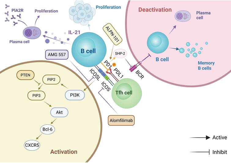
Precision intervention could be optimized by considering PD-1's context-dependent roles: its downregulation on peripheral Tregs versus pathological elevation on follicular Tfh cells contributes to the immune imbalance in IMN. Restoring subset-specific signaling equilibrium, rather than generalized modulation, provides the core pharmacological rationale for this strategy. ICOS and PD-1 are two critical, yet functionally distinct, immune receptors on Tfh cells. Rather than a simple binary opposition, high PD-1 expression is a fundamental hallmark of germinal center (GC) Tfh cell differentiation and activation, and may participate in restraining excessive Tfh-B-cell activation through PD-1/PD-L1 interactions. Simultaneously, ICOS provides a potent positive co-stimulatory signal driving Tfh functionality. Together, these axes are proposed to bidirectionally regulate the immunopathological cascade of IMN [183]. At the molecular mechanism level, the binding of ICOS on Tfh cells with ICOSL on B cells activates the PI3K-AKT signaling pathway [169, 184], which induces the expression of the transcription factor Bcl-6 [184], promotes CXCR5 upregulation, and drives Tfh cell differentiation. Additionally, it enhances IL-21 secretion, thereby supporting B cell proliferation and plasma cell differentiation, leading to the production of pathogenic antibodies (such as anti-PLA2R antibodies). In contrast, the PD-1/PD-L1 interaction can inhibit BCR signaling through SHP-2 phosphorylation, thereby limiting excessive B cell activation and regulating the localization of Tfh cells in the germinal center, helping to maintain immune homeostasis [70, 185, 186]. Importantly, the PD-1/PD-L1 axis may also exert regulatory effects on the B-cell side. B cells, particularly GC-associated or bystander B-cell populations, can express or upregulate PD-L1 to engage PD-1 on Tfh cells, while specific regulatory B-cell subsets may use PD-L1-dependent mechanisms to suppress pathogenic T-cell responses. Furthermore, the engagement of PD-1 expressed on B cells themselves recruits SHP-2 to the B cell receptor (BCR) signalosome, which acts as a physiological brake to terminate early activation and induce apoptosis of autoreactive B cell clones [187, 188]. In IMN, the expansion of Tfh cells leads to elevated absolute numbers of PD-1⁺ Tfh cells (reflecting an overactive GC response); however, the concurrent increased expression of ICOS results in a significantly increased ICOS⁺/PD-1⁺ Tfh ratio. This elevated ratio suggests that potent ICOS-driven co-stimulation may outweigh the physiological PD-1 inhibitory restraint, forming a pro-inflammatory Tfh-biased profile that may accelerate the autoimmune response [189]. Clinical observations further show that in newly diagnosed IMN patients, the frequencies of ICOS⁺ Tfh, PD-1⁺ Tfh, and IL-21⁺ Tfh cells in peripheral blood are significantly higher than in healthy controls. Moreover, the ICOS⁺/PD-1⁺ Tfh-cell ratio was positively correlated with 24-hour urinary protein levels and increased with proteinuria-stratified disease severity in IMN, suggesting that this ratio may serve as a sensitive indicator of IMN disease activity [183]. Experimental models provide further supportive evidence. In rats with passive Heymann nephritis (PHN), the proportion of splenic ICOS⁺ Tfh cells increases and correlates with proteinuria. The upregulation of Bcl-6 and IL-21 expression preceding overt proteinuria suggests their involvement in driving the autologous phase of antibody production [190]. In terms of treatment response, after receiving ACEI combined with immunosuppressive therapy, this ratio decreases as urine protein levels decrease, further supporting its close association with pathological activity [38, 183].
Based on the mechanisms described above, targeting the ICOS/ICOSL and PD-1/PD-L1 signaling axes offers an exploratory theoretical framework. It must be emphasized that this approach relies heavily on immunological concept transfer. While agents like the anti-ICOS-L antibody AMG 557 are being tested in SLE, and PD-1 pathway modulators are explored in oncology, their translational viability for IMN remains conceptual at this stage. Currently, several related targeted therapies have entered clinical development, but many are focused on other autoimmune diseases or oncology and therefore provide mechanistic reference rather than direct IMN evidence. For example, the anti-ICOS-L monoclonal antibody AMG 557 (Amgen) has shown acceptable safety and preliminary efficacy signals in Phase Ib studies for systemic lupus erythematosus, providing proof of concept for targeting the ICOS pathway in autoimmune diseases [191]. At the same time, novel agonists aimed at enhancing PD-1 signaling (such as GenSci120) have entered early clinical exploration in autoimmune diseases like rheumatoid arthritis, providing a conceptual reference for strategies aimed at restoring immune homeostasis in IMN through enhancement of PD-1 and other inhibitory receptor signaling [192]. The core challenge for future research lies in deeply analyzing the functional heterogeneity of PD-1⁺ Tfh cells (such as their resting and exhausted states) and exploring the combination of ICOS signaling blockade and PD-1 signaling enhancement as a regulatory strategy, with the aim of synergistically restoring immune homeostasis and improving disease control [183] (Figure 7, Table 1).
Bidirectional regulation and exploratory targeted intervention of the ICOS-ICOSL and PD-1/PD-L1 axes in IMN. (1) ICOS-ICOSL-mediated co-stimulation: Engagement of ICOS on Tfh cells with ICOSL on B cells activates the PI3K-AKT pathway, promotes Bcl-6 and CXCR5 expression, and supports Tfh differentiation, IL-21 secretion, B-cell proliferation, plasma-cell differentiation, and anti-PLA2R autoantibody production. (2) PD-1/PD-L1-mediated restraint: PD-1/PD-L1 signaling can recruit SHP-2 to dampen BCR signaling, thereby limiting excessive B-cell activation and helping maintain germinal-center immune homeostasis. (3) Concept-transfer interventions: ICOS-ICOSL blockade, represented by the anti-ICOSL monoclonal antibody AMG 557, and strategies aimed at enhancing PD-1 inhibitory signaling are shown as exploratory approaches extrapolated from other autoimmune diseases or oncology; their application in IMN remains conceptual and requires disease-specific validation. Created with BioRender.com. Abbreviations: IMN, idiopathic membranous nephropathy; ICOS, inducible T-cell costimulator; ICOSL, inducible T-cell costimulator ligand; PD-1, programmed cell death protein 1; PD-L1, programmed death-ligand 1; Tfh, follicular helper T cell; PI3K, phosphoinositide 3-kinase; AKT, protein kinase B; Bcl-6, B-cell lymphoma 6; CXCR5, C-X-C chemokine receptor type 5; IL-21, interleukin-21; PLA2R, M-type phospholipase A2 receptor; BCR, B-cell receptor; SHP-2, Src homology region 2 domain-containing phosphatase-2.
5.3 Preclinical, Mechanistic, and Early Exploratory Candidates
The pathways discussed below are supported predominantly by experimental, pharmacological, or indirect clinical evidence and are included to define mechanistic research questions rather than to imply established clinical applicability in IMN. To preserve the focus of this review, they are considered only where they may intersect with T-cell polarization, B-cell activation or differentiation, regulatory restraint, or plasma-cell persistence. Broader effects involving leukocyte recruitment, macrophage responses, fibrosis, or podocyte signaling are treated as contextual rather than core components of the T-B-cell interaction network. Accordingly, IL-6/STAT3 is discussed because of its potential relevance to T-cell polarization, B-cell differentiation, and plasma-cell survival, whereas CX3CL1/CX3CR1 is retained as a contextual pathway of renal immune-cell recruitment; direct IMN-specific evidence for these pathways remains limited.
5.3.1 IL-6/STAT3: Inflammation Cascade Amplifier and T Cell Polarization
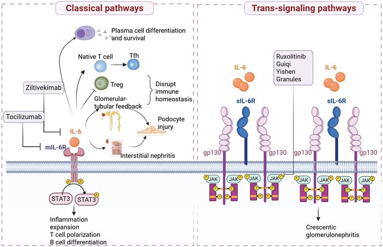
The IL-6/STAT3 signaling pathway is conceptualized as a key molecular bridge connecting immune abnormalities and podocyte damage in IMN. It may contribute to local inflammatory responses and plays an important role in modulating T-B cell immune interactions. IL-6 is a pleiotropic cytokine released after inflammatory stimulation and is an important mediator of inflammatory responses, T-cell polarization, B-cell differentiation, and T-B cell communication [142]. IL-6 is involved in the pathogenesis of various inflammatory glomerular diseases [193-196]. In addition to leukocyte-derived IL-6, glomerular endothelial cells and mesangial cells can also secrete IL-6 under stimulation by angiotensin II, and podocytes can produce IL-6 after endotoxin administration [197-199], indicating its potential paracrine and autocrine regulatory functions within the renal local microenvironment. Signal transducer and activator of transcription 3 (STAT3) is a canonical downstream signaling mediator of IL-6. Phosphorylated STAT3 translocates to the nucleus and regulates the transcription of target genes, leading to immune responses [200]. In IMN, both circulating and urinary IL-6 levels are significantly elevated [201-203], but urinary IL-6 levels do not correlate with plasma levels [202], highlighting its importance in local renal activation. Although there is a lack of obvious cellular infiltration in IMN pathology, IL-6 may still indirectly participate in podocyte injury by mediating glomerular-tubular feedback and interstitial inflammation. Furthermore, genetic studies suggest a potential pathogenic relevance of the IL-6/STAT3 pathway in IMN. Chen et al. reported that the IL-6 C-572G polymorphism might be a potential risk factor for IMN in the Han Chinese population [204], providing genetic evidence supporting further investigation of this pathway.
IL-6 signaling includes both the classical and trans-signaling pathways. The classical pathway is activated through membrane-bound IL-6 receptor (mIL-6R), while the trans-signaling pathway is mediated by soluble IL-6 receptor (sIL-6R) [205-208]. The classical signaling pathway relies on the dimerization of gp130 and activation of the JAK/STAT3 cascade, which in turn is proposed to drive inflammation amplification, T cell polarization, and B cell differentiation [209]. In T-B cell interactions, IL-6 promotes the production of Th2-type cytokines [210], induces the differentiation of naïve T cells into follicular helper T cells (Tfh) [199, 211, 212], and suppresses Treg activity, thereby disrupting immune homeostasis. Furthermore, IL-6 plays a critical role in the differentiation of antibody-secreting cells and the long-term survival of plasma cells [213]. Notably, podocytes may participate in IL-6-related signaling: cultured glomerular epithelial cells can express IL-6 and IL-6R, whereas in vivo podocytes express gp130 and can activate downstream STAT3 signaling, potentially mainly through IL-6 trans-signaling [205, 208]. Within this working model, persistent activation of STAT3 is hypothesized to mediate podocyte apoptosis, cytoskeletal remodeling, and slit diaphragm damage, a mechanism that has been validated in multiple glomerular models [200, 214]. The trans-signaling pathway amplifies inflammatory effects through the sIL-6R/gp130 axis and is negatively regulated by soluble gp130 (sgp130). This mechanism has been confirmed in crescentic glomerulonephritis [215, 216], suggesting its potential role in the interstitial inflammation of IMN.
Currently, therapeutic strategies targeting the IL-6/STAT3 axis have made progress in fields such as CKD, IgAN, and polycystic kidney disease, but direct studies on IMN remain limited. Anti-IL-6 inhibitors such as ziltivekimab [217, 218] have demonstrated anti-inflammatory effects in patients with advanced CKD. The IL-6R monoclonal antibody tocilizumab has also shown clinical efficacy in some cases of Castleman disease-associated membranous nephropathy [219, 220]. Additionally, JAK inhibitors (e.g., ruxolitinib) [221], and multi-target intervention strategies, such as traditional Chinese medicine compounds (e.g., Guiqi Yishen Granules) [222], provide new directions for the precision treatment of IMN (Figure 8, Table 1).
Mechanistic model and candidate interventions targeting the IL-6/STAT3 axis in IMN. (1) Classical IL-6 signaling: IL-6 binds to membrane-bound IL-6R (mIL-6R) and gp130, activating the JAK/STAT3 pathway and thereby contributing to inflammatory amplification, T-cell polarization, B-cell differentiation, Tfh skewing, Treg suppression, and plasma-cell differentiation and survival. This pathway may also participate in podocyte injury through glomerular-tubular feedback and interstitial inflammation. IL-6- or IL-6R-targeting agents, including ziltivekimab and tocilizumab, are shown as candidate interventions extrapolated from other renal or inflammatory diseases. (2) IL-6 trans-signaling: IL-6 complexed with soluble IL-6R (sIL-6R) activates gp130-dependent signaling and may amplify inflammatory responses; this mechanism has been implicated in crescentic glomerulonephritis and provides mechanistic reference for IMN rather than direct disease-specific evidence. (3) Downstream pathway modulation: JAK inhibitors, such as ruxolitinib, and multi-target candidate approaches, such as Guiqi Yishen Granules, may suppress downstream JAK/STAT3 activation, but their application in IMN remains exploratory and requires disease-specific validation. Created with BioRender.com. Abbreviations: IMN, idiopathic membranous nephropathy; IL-6, interleukin-6; mIL-6R, membrane-bound IL-6 receptor; sIL-6R, soluble IL-6 receptor; gp130, glycoprotein 130; STAT3, signal transducer and activator of transcription 3; JAK, Janus kinase; Tfh, follicular helper T cell; Treg, regulatory T cell.
5.3.2 CX3CL1/CX3CR1: Recruiters of Local Renal Inflammation
CX3CL1/CX3CR1 is discussed here as a contextual pathway of renal immune-cell recruitment rather than as a core component of the T-B-cell interaction network. Its direct contribution to autoreactive T-B-cell cooperation and autoantibody production in IMN remains insufficiently established. The CX3C chemokine ligand 1 (CX3CL1, also known as fractalkine) and its specific receptor, CX3C chemokine receptor 1 (CX3CR1), form a signaling axis involved in immune-cell chemotaxis and adhesion, as well as renal inflammatory and fibrotic remodeling [223]. CX3CL1 is the only member of the CX3C subfamily and is expressed in stromal cells, glomerular cells, and endothelial cells, and is distributed on the surface of glomerular, renal tubular epithelial, and mesangial cells [224]. This molecule anchors to endothelial cell membranes through its unique tripeptide sequence and mucin-like stalk structure, exhibiting dual functions as both a chemokine and an adhesion molecule [225]. CX3CL1 mediates the directed migration of immune cells, such as T cells, monocytes/macrophages, and natural killer cells, by binding to CX3CR1, directly capturing these cells in circulation, and promoting their extravasation into tissues. CX3CR1 is primarily expressed on the surface of leukocytes, including monocytes, lymphocytes, and dendritic cells, where it plays a specific chemotactic role [226, 227]. In normal kidney tissue, CX3CL1 expression is minimal or undetectable, whereas inflammatory stimulation induces its expression on renal endothelial and tubular epithelial cells.
Within the proposed working model for IMN, glomerular endothelial cells may upregulate the expression of CX3CL1 in response to immune complex deposition, forming an early signal to recruit inflammatory cells. Infiltrating CX3CR1⁺ cells are diffusely distributed, widely present in both the glomeruli and renal interstitial regions, and their localization highly correlates with that of T cells and monocytes/macrophages [228]. This widespread cross-regional distribution contrasts sharply with CCR5 (which is primarily enriched in the interstitium, absent in the glomeruli) and CCR2 (preferentially distributed in the glomeruli), highlighting the broad cell recruitment function of CX3CR1 in the early inflammation of IMN [229]. Experimental frameworks suggest that CX3CL1 further recruits macrophages to the glomeruli by binding to CX3CR1 on the surface of infiltrating monocytes. These macrophages are hypothesized to polarize into the M2 phenotype and secrete TGF-β1, which activates the STAT3 signaling pathway in podocytes, inducing high expression of type IV collagen (COL4A3/COL4A4) and promoting the thickening of the glomerular basement membrane [24].
In addition to its role in early inflammatory recruitment, the CX3CL1/CX3CR1 axis may also contribute to renal interstitial fibrosis. This possibility is supported by the upregulation of CX3CR1 in fibrotic human kidney tissues and by preclinical evidence suggesting that CX3CR1 blockade can attenuate renal fibrosis progression [230-232].
The therapeutic potential of targeting the CX3CL1/CX3CR1 axis has been supported in experimental models of severe glomerular inflammation. For instance, in crescentic glomerulonephritis Wistar Kyoto rats, blocking CX3CR1 reduced proteinuria and glomerular damage, with effects superior to CCR5 blockade [233, 234]. The core mechanism lies in CX3CR1's role in leukocyte adhesion and extravasation (the initial step of inflammation), whereas receptors like CCR2/CCR5 regulate subsequent cell microenvironmental positioning. This stage-specific action makes CX3CR1 a plausible target for intervening in early inflammation [229, 233]. In terms of translational strategies, combining a CX3CR1 antagonist such as AZD8797 with anti-CD20 therapy may provide a conceptual framework for targeting complementary components of the renal immune response: CX3CR1 blockade may reduce CD8⁺ T-cell and macrophage recruitment and thereby potentially limit local inflammatory and antigen-presenting activity (Figure 9), whereas anti-CD20 therapy depletes upstream CD20⁺ B-cell compartments and may reduce the generation of new antibody-secreting cells. However, this proposed combination remains conceptual, and its relationship to autoreactive T-B-cell cooperation in IMN requires disease-specific validation.
Mechanistic model and candidate intervention targeting the CX3CL1/CX3CR1 axis in IMN. (1) Renal CX3CL1 expression and leukocyte recruitment: In the proposed IMN inflammatory microenvironment, immune-complex deposition and inflammatory stimulation may induce CX3CL1 expression in renal resident cells, particularly endothelial and tubular epithelial cells. The CX3CL1-CX3CR1 interaction mediates the adhesion, capture, and tissue extravasation of CX3CR1⁺ circulating leukocytes, including monocytes, lymphocytes, and dendritic cells. (2) Macrophage-podocyte crosstalk and GBM remodeling: Recruited monocytes/macrophages may polarize toward an M2-like phenotype and secrete TGF-β1, which can activate STAT3 signaling in podocytes. This pathway may promote COL4A3/COL4A4 upregulation and contribute to glomerular basement membrane (GBM) thickening. (3) Candidate therapeutic targeting: Pharmacological blockade of CX3CR1, represented by AZD8797, is shown as an exploratory strategy that may reduce leukocyte infiltration, macrophage recruitment, and downstream inflammatory-fibrotic remodeling; its therapeutic relevance in IMN remains mechanistic and requires disease-specific validation. Created with BioRender.com. Abbreviations: IMN, idiopathic membranous nephropathy; CX3CL1, C-X3-C motif chemokine ligand 1; CX3CR1, C-X3-C motif chemokine receptor 1; TGF-β1, transforming growth factor-beta 1; STAT3, signal transducer and activator of transcription 3; COL4A3/COL4A4, collagen type IV alpha 3/alpha 4 chain; GBM, glomerular basement membrane.
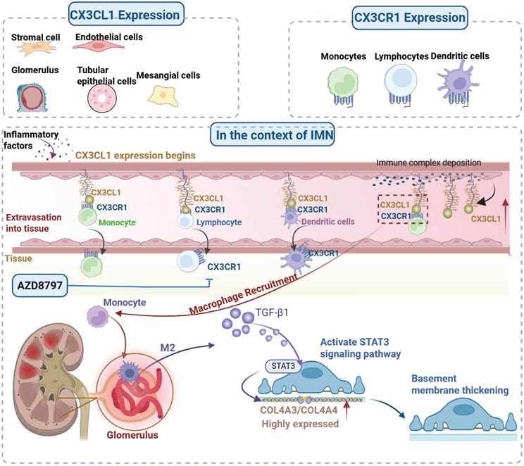
5.3.3 BCMA CAR-T Therapy: Exploratory Cellular Ablation
BCMA is mainly expressed on plasmablasts and plasma cells and is involved in the survival of long-lived antibody-producing cells. This makes BCMA a biologically plausible target in refractory IMN, where persistent anti-PLA2R or anti-THSD7A antibody production may be maintained by CD20- long-lived plasma cells that escape conventional anti-CD20 therapy [235]. The main conceptual advance of dual CD19/BCMA-directed CAR-T therapy is that it may simultaneously target CD19⁺ B-cell compartments and BCMA⁺ downstream antibody-secreting cells, thereby extending depletion beyond conventional CD20-directed therapy [160].
In MN, a BCMA-containing CAR-T strategy has entered early clinical exploration, as one registered AIKD trial includes MN and evaluates dual CD19/BCMA CAR-T therapy [235, 236]. However, published MN-specific efficacy data are not yet available. In parallel, antigen-specific CAAR-based strategies incorporating PLA2R1 or THSD7A domains have shown in vitro elimination of corresponding autoantibody-secreting cells, suggesting a more selective approach to pathogenic B-cell targeting [237]. However, because MN is typically not rapidly progressive, does not usually require lifelong or continuous immunosuppression, often affects an older population, LLPC-directed depletion may compromise protective humoral immunity, and only a subset of patients are truly refractory, CAR-T approaches are not currently considered to address a broad unmet clinical need in most patients with MN. BCMA-based CAR-T strategies should therefore remain early exploratory approaches, potentially limited to exceptional patients with severe nephrotic syndrome refractory to anti-B-cell therapy and cyclophosphamide/glucocorticoid-based regimens [160].
5.3.4 Treg-Targeted Immunomodulation: An Exploratory Strategy for Tolerance Restoration
IMN pathogenesis also involves impaired immune tolerance. As key mediators of peripheral tolerance, Treg cells are reduced in patients with IMN, accompanied by decreased FOXP3 expression and Th17/Treg imbalance [39, 238]. These findings provide a rationale for considering Treg expansion or functional restoration as a complementary strategy to pathogenic B-cell or plasma-cell depletion. However, no Treg-targeted therapy has been established for MN. Recent experimental studies suggest that engineered IL-2 muteins can selectively expand functional Tregs without broad effector T-cell activation; in a passive Heymann nephritis model resembling MN-like disease, once-weekly subcutaneous STS718 alleviated proteinuria, hypoalbuminemia, hypercholesterolemia, and mild renal dysfunction, accompanied by reduced nephrin loss, foot process effacement, GBM thickening, and C5b-9 deposition [239]. Low-dose IL-2 has also shown preliminary signals of clinical efficacy and tolerability in autoimmune diseases such as SLE, accompanied by Treg expansion, but this should be interpreted as cross-disease supportive evidence rather than direct evidence for IMN [240, 241]. Therefore, Treg-targeted immunomodulation remains a hypothesis-generating strategy requiring disease-specific validation.
6. Conclusion
The understanding of the immunopathogenesis of IMN has expanded to integrate the traditional “antibody-centric theory” with an explanatory framework in which imbalance of the T-B-cell interaction network may act as an upstream regulatory and amplifying process. Within the proposed “dual-axis dysregulation” model, enhanced Tfh-B-cell help, including Tfh2/Tfh17-associated IL-21/ICOS/CD40L signaling, and impaired Tfr/Breg-mediated restraint may together support persistent anti-PLA2R/THSD7A autoantibody production and subsequent podocyte injury. This framework should therefore be regarded as hypothesis-generating rather than as a confirmed causal pathway. It complements, rather than replaces, the established antibody-complement-podocyte injury paradigm and may help contextualize the current therapeutic bottleneck: B-cell-targeted therapies such as rituximab do not directly eliminate long-lived plasma cells (LLPCs) or fully restore immune tolerance, which may contribute to incomplete response, persistent autoantibody production, or relapse in a subset of high-risk or refractory patients. This mechanistic grouping does not imply comparable clinical readiness; the relevance of each intervention should be interpreted according to its disease specificity and evidence maturity. Precision therapeutic strategies can be conceptually grouped into three major directions: (1) targeting co-stimulatory signaling pathways (e.g., anti-ICOS antibodies, agents targeting the CD40-CD40L axis, or dual CD28/ICOS antagonists like ALPN-101) to theoretically disrupt the pathogenic Tfh-B cell axis; (2) remodeling the cytokine network (e.g., exploring BAFF/APRIL dual inhibitors like Telitacicept alongside B-cell depletion to potentially mitigate relapse risk, and IL-6/STAT3 antagonists to evaluate their role in inhibiting Tfh polarization and podocyte damage); and (3) targeting terminal effector cells (e.g., evaluating BCMA CAR-T or the anti-CD38 antibody Daratumumab as exploratory approaches to deplete LLPCs). Overall, the potential upstream role of the T-B cell interaction network provides an emerging mechanistic framework for IMN, but translating these mechanistic insights into clinical practice remains a promising direction that requires prospective validation. Future efforts should focus on multi-omics techniques, particularly single-cell sequencing analysis of local T-B cell clonal interactions in the kidneys, develop kidney-targeted delivery systems, and explore a sequential therapeutic model of “signal blockade - tolerance restoration - plasma cell eradication,” driving the transformation of IMN from generalized immune suppression to network precision regulation. Related advances in adjacent glomerular diseases, including genetic risk-score-based stratification in IgA nephropathy and single-cell/machine-learning analyses in lupus nephritis, further support the value of integrating molecular profiling into precision nephrology; however, IMN-specific validation remains essential [245-247].
7. Limitations
Despite current advances in understanding the T-B cell interaction network, several critical limitations must be acknowledged. First, there remains a significant translational gap between established animal models and human IMN pathology. Historically, foundational mechanistic insights have heavily relied on the passive Heymann nephritis model, which is induced by heterologous antibody injection and fails to fully recapitulate the spontaneous, T-cell-dependent immune complex formation characteristic of human in situ autoimmunity. Second, although IMN-specific studies support individual components of the dual-axis model, the integrated framework has not yet been validated through antigen-specific, tissue-resolved, and longitudinal analyses. Future studies combining PLA2R/THSD7A-specific lymphocyte assays, paired blood-kidney profiling, immune-receptor clonotyping, and serial immune and serological measurements are needed to determine whether these immune states are directly linked to autoantibody persistence, treatment resistance, or relapse. Third, translating these pathophysiological findings into clinical success is heavily hindered by the challenge of clearing LLPCs. These terminally differentiated cells often reside in specialized protective survival niches within the bone marrow and inflamed renal tissues, rendering them highly refractory to conventional B-cell depleting agents like rituximab. Overcoming these niche-mediated protections to effectively eradicate LLPCs remains a formidable therapeutic bottleneck.
Supplementary Material
Supplementary table s1.

Acknowledgements
Funding
This work was supported by the National Natural Science Foundation of China (grant numbers 82374396, 82503899, 82503492); Shanghai Shenkang Hospital Development Center-Joint Program on Emerging Frontiers (grant numbers SHDC12025121); Plucked Project of the Oriental Talent Program in 2023(grant numbers 2446); Longhua Hospital-Minhang” Traditional Chinese Medicine Specialty (and Specific Diseases) Alliance(grant numbers 2024LM02); Shanghai Shenkang Hospital Development Center Clinical Diagnosis and Treatment Technology Promotion and Optimization Management(grant numbers SHDC12025121); Xuhui District Famous Traditional Chinese Medicine Practitioner Studio Construction(No project number); Bethune·Saint Create Future — Foundation of Chronic Kidney Disease Research(grant numbers SCWLKY-007-002);the Guangdong Basic and Applied Basic Research Foundation (Grant No. 2023A1515111044, 2025A1515012665); the Shenzhen Science and Technology Program (Grant No. RCBS20231211090733052, JCYJ20240813150221028, JCYJ20240813150310014);Shenzhen Medical Research Fund (Grant No. A2603111).
Author contributions
Lin Wang: Writing - original draft, Visualization, Data curation; Qin Tian: Visualization, Supervision; Ao Xu: Visualization, Investigation, Writing - original draft; Lianli Shen: Formal analysis, writing - review & editing; Yue Xing: Formal analysis, Writing - review & editing; Hangxing Yu: Writing - review & editing; Tianxingjian Ke: Writing - review & editing; Yuling Liang: Writing - review & editing; Jinjie Gao: Writing - review & editing; Ming Pei: Writing - review & editing; Hongtao Yang: Conceptualization, Project administration; Jinxiang Wang: Conceptualization, Resources; Yifei Zhong: Conceptualization, Funding acquisition, Project administration.
Competing Interests
The authors have declared that no competing interest exists.
References
1. Couser WG. Primary membranous nephropathy. Clin J Am Soc Nephrol. 2017;12:983-97
2. Cattran DC, Brenchley PE. Membranous nephropathy: Integrating basic science into improved clinical management. Kidney Int. 2017;91:566-74
3. Alsharhan L, Beck LH Jr. Membranous nephropathy: Core curriculum 2021. Am J Kidney Dis. 2021;77:440-53
4. Hu R, Quan S, Wang Y, Zhou Y, Zhang Y, Liu L. et al. Spectrum of biopsy proven renal diseases in central china: A 10-year retrospective study based on 34,630 cases. Sci Rep. 2020;10:10994
5. Radhakrishnan Y, Zand L, Sethi S, Fervenza FC. Membranous nephropathy treatment standard. Nephrol Dial Transplant. 2024;39:403-13
6. Storrar J, Gill-Taylor T, Chinnadurai R, Chrysochou C, Poulikakos D, Rainone F. et al. A low rate of end-stage kidney disease in membranous nephropathy: A single centre study over 2 decades. PLoS One. 2022;17:e0276053
7. Troyanov S, Wall CA, Miller JA, Scholey JW, Cattran DC. Idiopathic membranous nephropathy: Definition and relevance of a partial remission. Kidney Int. 2004;66:1199-205
8. Floege J, Barbour SJ, Cattran DC, Hogan JJ, Nachman PH, Tang SCW. et al. Management and treatment of glomerular diseases (part 1): Conclusions from a kidney disease: Improving global outcomes (kdigo) controversies conference. Kidney Int. 2019;95:268-80
9. Kdigo 2021 clinical practice guideline for the management of glomerular diseases. Kidney Int. 2021; 100: S1-s276.
10. Dass S, Vital EM, Emery P. Rituximab: Novel b-cell depletion therapy for the treatment of rheumatoid arthritis. Expert Opin Pharmacother. 2006;7:2559-70
11. Pescovitz MD. Rituximab, an anti-cd20 monoclonal antibody: History and mechanism of action. Am J Transplant. 2006;6:859-66
12. von Groote TC, Williams G, Au EH, Chen Y, Mathew AT, Hodson EM. et al. Immunosuppressive treatment for primary membranous nephropathy in adults with nephrotic syndrome. Cochrane Database Syst Rev. 2021;11:Cd004293
13. Floege J, Rovin BH. The starmen trial: Rethinking calcineurin inhibitor therapy in membranous nephropathy. Kidney Int. 2021;99:811-3
14. Caravaca-Fontán F, Fernández-Juárez GM, Floege J, Goumenos D, Kronbichler A, Turkmen K. et al. The management of membranous nephropathy-an update. Nephrol Dial Transplant. 2022;37:1033-42
15. Koppe L, Fouque D. The role for protein restriction in addition to renin-angiotensin-aldosterone system inhibitors in the management of ckd. Am J Kidney Dis. 2019;73:248-57
16. Ronco P, Beck L, Debiec H, Fervenza FC, Hou FF, Jha V. et al. Membranous nephropathy. Nat Rev Dis Primers. 2021;7:69
17. Perico L, Casiraghi F, Sônego F, Todeschini M, Corna D, Cerullo D. et al. Bi-specific autoantigen-t cell engagers as targeted immunotherapy for autoreactive b cell depletion in autoimmune diseases. Front Immunol. 2024;15:1335998
18. Pollmann R, Walter E, Schmidt T, Waschke J, Hertl M, Möbs C. et al. Identification of autoreactive b cell subpopulations in peripheral blood of autoimmune patients with pemphigus vulgaris. Front Immunol. 2019;10:1375
19. Song W, Craft J. T follicular helper cell heterogeneity: Time, space, and function. Immunol Rev. 2019;288:85-96
20. Crotty S. T follicular helper cell biology: A decade of discovery and diseases. Immunity. 2019;50:1132-48
21. Mintz MA, Cyster JG. T follicular helper cells in germinal center b cell selection and lymphomagenesis. Immunol Rev. 2020;296:48-61
22. Wu Y, Jiang H, Hu Y, Dai H, Zhao Q, Zheng Y. et al. B cell dysregulation and depletion therapy in primary membranous nephropathy: Prospects and potential challenges. Int Immunopharmacol. 2024;140:112769
23. Zhang H, Ruan L, Fu X, Yu J, Ruan Q, Li Y. et al. Mechanisms and clinical translation of icos/icosl signaling pathway blockade in delaying vascular complications of type 2 diabetes. Diabetol Metab Syndr. 2025;17:328
24. Cai A, Meng Y, Zhou H, Cai H, Shao X, Wang Q. et al. Podocyte pathogenic bone morphogenetic protein-2 pathway and immune cell behaviors in primary membranous nephropathy. Adv Sci (Weinh). 2024;11:e2404151
25. Qin C, Dong MH, Zhou LQ, Wang W, Cai SB, You YF. et al. Single-cell analysis of refractory anti-srp necrotizing myopathy treated with anti-bcma car-t cell therapy. Proc Natl Acad Sci U S A. 2024;121:e2315990121
26. Sethi S, Beck LH Jr, Glassock RJ, Haas M, De Vriese AS, Caza TN. et al. Mayo clinic consensus report on membranous nephropathy: Proposal for a novel classification. Kidney Int. 2023;104:1092-102
27. Meyer-Schwesinger C, Lambeau G, Stahl RA. Thrombospondin type-1 domain-containing 7a in idiopathic membranous nephropathy. N Engl J Med. 2015;372:1074-5
28. Sethi S, Debiec H, Madden B, Vivarelli M, Charlesworth MC, Ravindran A. et al. Semaphorin 3b-associated membranous nephropathy is a distinct type of disease predominantly present in pediatric patients. Kidney Int. 2020;98:1253-64
29. Sethi S, Madden BJ, Debiec H, Charlesworth MC, Gross L, Ravindran A. et al. Exostosin 1/exostosin 2-associated membranous nephropathy. J Am Soc Nephrol. 2019;30:1123-36
30. Radice A, Trezzi B, Maggiore U, Pregnolato F, Stellato T, Napodano P. et al. Clinical usefulness of autoantibodies to m-type phospholipase a2 receptor (pla2r) for monitoring disease activity in idiopathic membranous nephropathy (imn). Autoimmun Rev. 2016;15:146-54
31. Ruggenenti P, Fervenza FC, Remuzzi G. Treatment of membranous nephropathy: Time for a paradigm shift. Nat Rev Nephrol. 2017;13:563-79
32. Hoxha E, Thiele I, Zahner G, Panzer U, Harendza S, Stahl RA. Phospholipase a2 receptor autoantibodies and clinical outcome in patients with primary membranous nephropathy. J Am Soc Nephrol. 2014;25:1357-66
33. Hofstra JM, Beck LH Jr, Beck DM, Wetzels JF, Salant DJ. Anti-phospholipase a₂ receptor antibodies correlate with clinical status in idiopathic membranous nephropathy. Clin J Am Soc Nephrol. 2011;6:1286-91
34. Meng H, Wang D, Zheng C, Zhou C, Mao X, Gu J. et al. Autoantibodies targeting vinculin reveal novel insight into the mechanisms of autoimmune podocytopathies. Research (Wash D C). 2025;8:0722
35. Dendrou CA, Petersen J, Rossjohn J, Fugger L. Hla variation and disease. Nat Rev Immunol. 2018;18:325-39
36. Kuroki A, Iyoda M, Shibata T, Sugisaki T. Th2 cytokines increase and stimulate b cells to produce igg4 in idiopathic membranous nephropathy. Kidney Int. 2005;68:302-10
37. Hoxha E, Reinhard L, Stahl RAK. Membranous nephropathy: New pathogenic mechanisms and their clinical implications. Nat Rev Nephrol. 2022;18:466-78
38. Zhang Z, Shi Y, Yang K, Crew R, Wang H, Jiang Y. Higher frequencies of circulating icos(+), il-21(+) t follicular helper cells and plasma cells in patients with new-onset membranous nephropathy. Autoimmunity. 2017;50:458-67
39. Motavalli R, Etemadi J, Soltani-Zangbar MS, Ardalan MR, Kahroba H, Roshangar L. et al. Altered th17/treg ratio as a possible mechanism in pathogenesis of idiopathic membranous nephropathy. Cytokine. 2021;141:155452
40. Cremoni M, Brglez V, Perez S, Decoupigny F, Zorzi K, Andreani M. et al. Th17-immune response in patients with membranous nephropathy is associated with thrombosis and relapses. Front Immunol. 2020;11:574997
41. Han SS, Yang SH, Jo HA, Oh YJ, Park M, Kim JY. et al. Baff and april expression as an autoimmune signature of membranous nephropathy. Oncotarget. 2018;9:3292-302
42. Netti GS, Infante B, Spadaccino F, Godeas G, Corallo MG, Prisciandaro C. et al. Serum levels of baff and april predict clinical response in anti-pla2r-positive primary membranous nephropathy. J Immunol Res. 2019;2019:8483650
43. Shi M, Wang Y, Zhang H, Ling Z, Chen X, Wang C. et al. Single-cell rna sequencing shows the immune cell landscape in the kidneys of patients with idiopathic membranous nephropathy. Front Immunol. 2023;14:1203062
44. Gu Q, Wen Y, Cheng X, Qi Y, Cao X, Gao X. et al. Integrative profiling of untreated primary membranous nephropathy at the single-cell transcriptome level. Clin Kidney J. 2024;17:sfae168
45. Wang ZF, Cheng YC, Li YQ, Liu L, Ge SW, Xu G. Characteristics and prognostic value of tertiary lymphoid organs in membranous nephropathy: A retrospective study. Front Med (Lausanne). 2021;8:803929
46. Sollid LM, Pos W, Wucherpfennig KW. Molecular mechanisms for contribution of mhc molecules to autoimmune diseases. Curr Opin Immunol. 2014;31:24-30
47. Cui Z, Xie LJ, Chen FJ, Pei ZY, Zhang LJ, Qu Z. et al. Mhc class ii risk alleles and amino acid residues in idiopathic membranous nephropathy. J Am Soc Nephrol. 2017;28:1651-64
48. Le WB, Shi JS, Zhang T, Liu L, Qin HZ, Liang S. et al. Hla-drb1*15:01 and hla-drb3*02:02 in pla2r-related membranous nephropathy. J Am Soc Nephrol. 2017;28:1642-50
49. Quiza CG, Leenaerts PL, Hall BM. The role of t cells in the mediation of glomerular injury in heymann's nephritis in the rat. Int Immunol. 1992;4:423-32
50. Li H, Wu H, Guo Q, Yu H, Xu Y, Yu J. et al. Myeloid-derived suppressor cells promote the progression of primary membranous nephropathy by enhancing th17 response. Front Immunol. 2020;11:1777
51. Ma DH, Yang XD, Hua QJ, Hou YL, Liu Y, Xu QY. et al. Changes and significance of treg and th17 in adult patients with primary membranous nephropathy. Clin Nephrol. 2021;96:155-64
52. Jurubiță R, Obrișcă B, Achim C, Micu G, Sorohan B, Bobeică R. et al. Anti-phospholipase a2 receptor antibody screening in nephrotic syndrome may identify a distinct subset of patients with primary membranous nephropathy. Int Urol Nephrol. 2022;54:1713-23
53. Bobart SA, Han H, Tehranian S, De Vriese AS, Roman JCL, Sethi S. et al. Noninvasive diagnosis of pla2r-associated membranous nephropathy: A validation study. Clin J Am Soc Nephrol. 2021;16:1833-9
54. Georgiev P, Charbonnier LM, Chatila TA. Regulatory t cells: The many faces of foxp3. J Clin Immunol. 2019;39:623-40
55. Jonuleit H, Schmitt E, Kakirman H, Stassen M, Knop J, Enk AH. Infectious tolerance: Human cd25(+) regulatory t cells convey suppressor activity to conventional cd4(+) t helper cells. J Exp Med. 2002;196:255-60
56. Shi X, Zhang M, Wang G, Chen G, Hu R, Lee X. et al. The diversity of peripheral blood t cells and myeloid-derived suppressor cells in patients with idiopathic membranous nephropathy. Clin Nephrol. 2024;101:287-97
57. Cantarelli C, Jarque M, Angeletti A, Manrique J, Hartzell S, O'Donnell T. et al. A comprehensive phenotypic and functional immune analysis unravels circulating anti-phospholipase a2 receptor antibody secreting cells in membranous nephropathy patients. Kidney Int Rep. 2020;5:1764-76
58. Rosenzwajg M, Languille E, Debiec H, Hygino J, Dahan K, Simon T. et al. B- and t-cell subpopulations in patients with severe idiopathic membranous nephropathy may predict an early response to rituximab. Kidney Int. 2017;92:227-37
59. Masutani K, Taniguchi M, Nakashima H, Yotsueda H, Kudoh Y, Tsuruya K. et al. Up-regulated interleukin-4 production by peripheral t-helper cells in idiopathic membranous nephropathy. Nephrol Dial Transplant. 2004;19:580-6
60. Gascan H, Gauchat JF, Roncarolo MG, Yssel H, Spits H, de Vries JE. Human B cell clones can be induced to proliferate and to switch to IgE and IgG4 synthesis by interleukin 4 and a signal provided by activated CD4+ T cell clones. J Exp Med. 1991;173:747-750 https://doi.org/10.1084/jem.173.3.747
61. Crotty S. T follicular helper cell differentiation, function, and roles in disease. Immunity. 2014;41:529-42
62. Clement RL, Daccache J, Mohammed MT, Diallo A, Blazar BR, Kuchroo VK. et al. Follicular regulatory t cells control humoral and allergic immunity by restraining early b cell responses. Nat Immunol. 2019;20:1360-71
63. Sage PT, Ron-Harel N, Juneja VR, Sen DR, Maleri S, Sungnak W. et al. Suppression by t(fr) cells leads to durable and selective inhibition of b cell effector function. Nat Immunol. 2016;17:1436-46
64. Xu L, Tayier D, Yang S, Zhang X, Lu C. Follicular helper t cells and follicular regulatory t cells involved in immune disorders of idiopathic membranous nephropathy. Indian J Pediatr. 2024;91:702-8
65. Shi J, Hou S, Fang Q, Liu X, Liu X, Qi H. Pd-1 controls follicular t helper cell positioning and function. Immunity. 2018;49:264-74.e4
66. Hosseini A, Gharibi T, Marofi F, Babaloo Z, Baradaran B. Ctla-4: From mechanism to autoimmune therapy. Int Immunopharmacol. 2020;80:106221
67. Swain SL, Dutton RW. Consequences of the direct interaction of helper t cells with b cells presenting antigen. Immunol Rev. 1987;99:263-80
68. Noelle RJ, Daum J, Bartlett WC, McCann J, Shepherd DM. Cognate interactions between helper t cells and b cells. V. Reconstitution of t helper cell function using purified plasma membranes from activated th1 and th2 t helper cells and lymphokines. J Immunol. 1991;146:1118-24
69. Kishimoto T, Hirano T. Molecular regulation of b lymphocyte response. Annu Rev Immunol. 1988;6:485-512
70. Mintz MA, Felce JH, Chou MY, Mayya V, Xu Y, Shui JW. et al. The hvem-btla axis restrains t cell help to germinal center b cells and functions as a cell-extrinsic suppressor in lymphomagenesis. Immunity. 2019;51:310-23.e7
71. Deng B, Huang H, Deng L, Zhao Z, Liu M, Lin H. et al. Imbalance of t follicular helper cell subsets trigger the differentiation of pathogenic b cells in idiopathic membranous nephropathy. Inflamm Res. 2024;73:485-98
72. Wang Y, Liu J, Burrows PD, Wang JY. B cell development and maturation. Adv Exp Med Biol. 2020;1254:1-22
73. Morita R, Schmitt N, Bentebibel SE, Ranganathan R, Bourdery L, Zurawski G. et al. Human blood cxcr5(+)cd4(+) t cells are counterparts of t follicular cells and contain specific subsets that differentially support antibody secretion. Immunity. 2011;34:108-21
74. Ren HM, Lukacher AE, Rahman ZSM, Olsen NJ. New developments implicating il-21 in autoimmune disease. J Autoimmun. 2021;122:102689
75. Quast I, Dvorscek AR, Pattaroni C, Steiner TM, McKenzie CI, Pitt C. et al. Interleukin-21, acting beyond the immunological synapse, independently controls t follicular helper and germinal center b cells. Immunity. 2022;55:1414-30.e5
76. Leong YA, Chen Y, Ong HS, Wu D, Man K, Deleage C. et al. Cxcr5(+) follicular cytotoxic t cells control viral infection in b cell follicles. Nat Immunol. 2016;17:1187-96
77. Caielli S, Veiga DT, Balasubramanian P, Athale S, Domic B, Murat E. et al. A cd4(+) t cell population expanded in lupus blood provides b cell help through interleukin-10 and succinate. Nat Med. 2019;25:75-81
78. Ma X, Nakayamada S. Multi-source pathways of t follicular helper cell differentiation. Front Immunol. 2021;12:621105
79. Li DD, Bechara R, Ramani K, Jawale CV, Li Y, Kolls JK. et al. Rtec-intrinsic il-17-driven inflammatory circuit amplifies antibody-induced glomerulonephritis and is constrained by regnase-1. JCI Insight. 2021 6
80. Ryba-Stanisławowska M, Sakowska J, Zieliński M, Ławrynowicz U, Trzonkowski P. Regulatory t cells: The future of autoimmune disease treatment. Expert Rev Clin Immunol. 2019;15:777-89
81. Wing JB, Ise W, Kurosaki T, Sakaguchi S. Regulatory t cells control antigen-specific expansion of tfh cell number and humoral immune responses via the coreceptor ctla-4. Immunity. 2014;41:1013-25
82. Eschweiler S, Clarke J, Ramírez-Suástegui C, Panwar B, Madrigal A, Chee SJ. et al. Intratumoral follicular regulatory t cells curtail anti-pd-1 treatment efficacy. Nat Immunol. 2021;22:1052-63
83. Youn JI, Nagaraj S, Collazo M, Gabrilovich DI. Subsets of myeloid-derived suppressor cells in tumor-bearing mice. J Immunol. 2008;181:5791-802
84. Collison LW, Workman CJ, Kuo TT, Boyd K, Wang Y, Vignali KM. et al. The inhibitory cytokine il-35 contributes to regulatory t-cell function. Nature. 2007;450:566-9
85. Ma Y, Chen L, Xie G, Zhou Y, Yue C, Yuan X. et al. Elevated level of interleukin-35 in colorectal cancer induces conversion of t cells into itr35 by activating stat1/stat3. Oncotarget. 2016;7:73003-15
86. So BYF, Yap DYH, Chan TM. B cells in primary membranous nephropathy: Escape from immune tolerance and implications for patient management. Int J Mol Sci. 2021 22
87. Ronco P, Debiec H. Pathophysiological advances in membranous nephropathy: Time for a shift in patient's care. Lancet. 2015;385:1983-92
88. Cohen CD, Calvaresi N, Armelloni S, Schmid H, Henger A, Ott U. et al. Cd20-positive infiltrates in human membranous glomerulonephritis. J Nephrol. 2005;18:328-33
89. Glassock RJ. Human idiopathic membranous nephropathy-a mystery solved? N Engl J Med. 2009;361:81-3
90. Jelcic I, Al Nimer F, Wang J, Lentsch V, Planas R, Jelcic I. et al. Memory b cells activate brain-homing, autoreactive cd4(+) t cells in multiple sclerosis. Cell. 2018;175:85-100.e23
91. Pieper K, Grimbacher B, Eibel H. B-cell biology and development. J Allergy Clin Immunol. 2013;131:959-71
92. Shen P, Fillatreau S. Antibody-independent functions of b cells: A focus on cytokines. Nat Rev Immunol. 2015;15:441-51
93. Vincent FB, Morand EF, Schneider P, Mackay F. The baff/april system in sle pathogenesis. Nat Rev Rheumatol. 2014;10:365-73
94. Deng B, Deng L, Liu M, Zhao Z, Huang H, Tu X. et al. Elevated circulating cd19(+)cd24(hi)cd38(hi) b cells display pro-inflammatory phenotype in idiopathic membranous nephropathy. Immunol Lett. 2023;261:58-65
95. Wang H, Lan L, Wang J, Chen J, Xiao L, Han F. Alterations of b-cell subsets in peripheral blood from adult patients with idiopathic membranous nephropathy. Immunol Lett. 2024;266:106838
96. Su Z, Jin Y, Zhang Y, Guan Z, Li H, Chen X. et al. The diagnostic and prognostic potential of the b-cell repertoire in membranous nephropathy. Front Immunol. 2021;12:635326
97. Cheng YJ, Wang M, Wang J, Cui Z, Zhao MH. The characters of antibodies against pla2r in healthy individuals and in the patient with pla2r associated membranous nephropathy. Eur J Med Res. 2023;28:128
98. Shim JS, Kim EJ, Lee LE, Kim JY, Cho Y, Kim H. et al. The oxidative phosphorylation inhibitor im156 suppresses b-cell activation by regulating mitochondrial membrane potential and contributes to the mitigation of systemic lupus erythematosus. Kidney Int. 2023;103:343-56
99. Yang X, Tang X, Li T, Man C, Yang X, Wang M. et al. Circulating follicular t helper cells are possibly associated with low levels of serum immunoglobulin g due to impaired immunoglobulin class-switch recombination of b cells in children with primary nephrotic syndrome. Mol Immunol. 2019;114:162-70
100. Kolovou K, Laskari K, Roumelioti M, Tektonidou MG, Panayiotidis P, Boletis JN. et al. B-cell oligoclonal expansions in renal tissue of patients with immune-mediated glomerular disease. Clin Immunol. 2020;217:108488
101. Segerer S, Schlöndorff D. B cells and tertiary lymphoid organs in renal inflammation. Kidney Int. 2008;73:533-7
102. Prunotto M, Farina A, Lane L, Pernin A, Schifferli J, Hochstrasser DF. et al. Proteomic analysis of podocyte exosome-enriched fraction from normal human urine. J Proteomics. 2013;82:193-229
103. Sethi S. New 'antigens' in membranous nephropathy. J Am Soc Nephrol. 2021;32:268-78
104. Ronco P, Debiec H. Molecular pathogenesis of membranous nephropathy. Annu Rev Pathol. 2020;15:287-313
105. Wang B, Zuo K, Wu Y, Huang Q, Qin WS, Zeng CH. et al. Correlation between b lymphocyte abnormality and disease activity in patients with idiopathic membranous nephropathy. J Int Med Res. 2011;39:86-95
106. Blair PA, Noreña LY, Flores-Borja F, Rawlings DJ, Isenberg DA, Ehrenstein MR. et al. Cd19(+)cd24(hi)cd38(hi) b cells exhibit regulatory capacity in healthy individuals but are functionally impaired in systemic lupus erythematosus patients. Immunity. 2010;32:129-40
107. Iwata Y, Matsushita T, Horikawa M, Dilillo DJ, Yanaba K, Venturi GM. et al. Characterization of a rare il-10-competent b-cell subset in humans that parallels mouse regulatory b10 cells. Blood. 2011;117:530-41
108. Lindner S, Dahlke K, Sontheimer K, Hagn M, Kaltenmeier C, Barth TF. et al. Interleukin 21-induced granzyme b-expressing b cells infiltrate tumors and regulate t cells. Cancer Res. 2013;73:2468-79
109. Aravena O, Ferrier A, Menon M, Mauri C, Aguillón JC, Soto L. et al. Tim-1 defines a human regulatory b cell population that is altered in frequency and function in systemic sclerosis patients. Arthritis Res Ther. 2017;19:8
110. Matsumoto M, Baba A, Yokota T, Nishikawa H, Ohkawa Y, Kayama H. et al. Interleukin-10-producing plasmablasts exert regulatory function in autoimmune inflammation. Immunity. 2014;41:1040-51
111. van de Veen W, Stanic B, Yaman G, Wawrzyniak M, Söllner S, Akdis DG. et al. Igg4 production is confined to human il-10-producing regulatory b cells that suppress antigen-specific immune responses. J Allergy Clin Immunol. 2013;131:1204-12
112. Griffin DO, Rothstein TL. Human “orchestrator” cd11b(+) b1 cells spontaneously secrete interleukin-10 and regulate t-cell activity. Mol Med. 2012;18:1003-8
113. Rojas-Rivera JE, Carriazo S, Ortiz A. Treatment of idiopathic membranous nephropathy in adults: Kdigo 2012, cyclophosphamide and cyclosporine a are out, rituximab is the new normal. Clin Kidney J. 2019;12:629-38
114. Fervenza FC, Appel GB, Barbour SJ, Rovin BH, Lafayette RA, Aslam N. et al. Rituximab or cyclosporine in the treatment of membranous nephropathy. N Engl J Med. 2019;381:36-46
115. Hiepe F, Radbruch A. Plasma cells as an innovative target in autoimmune disease with renal manifestations. Nat Rev Nephrol. 2016;12:232-40
116. Mei HE, Wirries I, Frölich D, Brisslert M, Giesecke C, Grün JR. et al. A unique population of igg-expressing plasma cells lacking cd19 is enriched in human bone marrow. Blood. 2015;125:1739-48
117. Ma K, Li J, Wang X, Lin X, Du W, Yang X. et al. Tlr4(+)cxcr4(+) plasma cells drive nephritis development in systemic lupus erythematosus. Ann Rheum Dis. 2018;77:1498-506
118. Zhao Q, Dai H, Jiang H, Zhang N, Hou F, Zheng Y. et al. Activation of the il-6/stat3 pathway contributes to the pathogenesis of membranous nephropathy and is a target for mahuang fuzi and shenzhuo decoction (mfsd) to repair podocyte damage. Biomed Pharmacother. 2024;174:116583
119. Zhang L, Chen M, Le W, Zhang M, Tu Y, Zuo K. et al. Efficacy of long-term repeated rituximab treatment in refractory phospholipase a2 receptor-antibody-related membranous nephropathy. Immunotherapy. 2022;14:1237-44
120. Isenberg D, Gordon C, Licu D, Copt S, Rossi CP, Wofsy D. Efficacy and safety of atacicept for prevention of flares in patients with moderate-to-severe systemic lupus erythematosus (sle): 52-week data (april-sle randomised trial). Ann Rheum Dis. 2015;74:2006-15
121. Nestorov I, Munafo A, Papasouliotis O, Visich J. Pharmacokinetics and biological activity of atacicept in patients with rheumatoid arthritis. J Clin Pharmacol. 2008;48:406-17
122. Yang Z, Cao G, Tan X, Orfanos S, Jude J, Barbet G. et al. Distinct mural cells and fibroblasts promote pathogenic plasma cell accumulation in idiopathic pulmonary fibrosis. Eur Respir J. 2025 65
123. Benoit SW, Khandelwal P, Grimley MS. A case of treatment-resistant membranous nephropathy associated with graft versus host disease successfully treated with daratumumab. Pediatr Transplant. 2022;26:e14263
124. Vink CH, van Cranenbroek B, van der Heijden JW, Koenen H, Wetzels JFM. Daratumumab for multidrug-resistant phospholipase-a2 receptor-related membranous nephropathy. Kidney Int. 2022;101:646-7
125. Stehlé T, Grimbert P, Remy P, Moktefi A, Audard V, El Karoui K. Anti-cd38 therapy for pla2r-positive membranous nephropathy resistant to conventional immunosuppression. Kidney Int. 2022;101:416-8
126. Lesokhin AM, Tomasson MH, Arnulf B, Bahlis NJ, Miles Prince H, Niesvizky R. et al. Elranatamab in relapsed or refractory multiple myeloma: Phase 2 magnetismm-3 trial results. Nat Med. 2023;29:2259-67
127. Beck LH Jr, Bonegio RG, Lambeau G, Beck DM, Powell DW, Cummins TD. et al. M-type phospholipase a2 receptor as target antigen in idiopathic membranous nephropathy. N Engl J Med. 2009;361:11-21
128. Zhao Q, Dai H, Liu X, Jiang H, Liu W, Feng Z. et al. Helper t cells in idiopathic membranous nephropathy. Front Immunol. 2021;12:665629
129. Ramachandran R, Kaundal U, Girimaji N, Rakha A, Rathi M, Gupta KL. et al. Regulatory b cells are reduced and correlate with disease activity in primary membranous nephropathy. Kidney Int Rep. 2020;5:872-8
130. Cragg MS, Walshe CA, Ivanov AO, Glennie MJ. The biology of cd20 and its potential as a target for mab therapy. Curr Dir Autoimmun. 2005;8:140-74
131. Oracki SA, Walker JA, Hibbs ML, Corcoran LM, Tarlinton DM. Plasma cell development and survival. Immunol Rev. 2010;237:140-59
132. Buse M, Dounousi E, Kramann R, Floege J, Stamellou E. Newer b-cell and plasma-cell targeted treatments for rituximab-resistant patients with membranous nephropathy. Clin Kidney J. 2025;18:sfaf088
133. Schneider P, MacKay F, Steiner V, Hofmann K, Bodmer JL, Holler N. et al. Baff, a novel ligand of the tumor necrosis factor family, stimulates b cell growth. J Exp Med. 1999;189:1747-56
134. Moore PA, Belvedere O, Orr A, Pieri K, LaFleur DW, Feng P. et al. Blys: Member of the tumor necrosis factor family and b lymphocyte stimulator. Science. 1999;285:260-3
135. Mackay F, Schneider P, Rennert P, Browning J. Baff and april: A tutorial on b cell survival. Annu Rev Immunol. 2003;21:231-64
136. Castigli E, Wilson SA, Scott S, Dedeoglu F, Xu S, Lam KP. et al. Taci and baff-r mediate isotype switching in b cells. J Exp Med. 2005;201:35-9
137. Rowland SL, Leahy KF, Halverson R, Torres RM, Pelanda R. Baff receptor signaling aids the differentiation of immature b cells into transitional b cells following tonic bcr signaling. J Immunol. 2010;185:4570-81
138. Medema JP, Planelles-Carazo L, Hardenberg G, Hahne M. The uncertain glory of april. Cell Death Differ. 2003;10:1121-5
139. Varfolomeev E, Kischkel F, Martin F, Seshasayee D, Wang H, Lawrence D. et al. April-deficient mice have normal immune system development. Mol Cell Biol. 2004;24:997-1006
140. Oniszczuk J, Beldi-Ferchiou A, Audureau E, Azzaoui I, Molinier-Frenkel V, Frontera V. et al. Circulating plasmablasts and high level of baff are hallmarks of minimal change nephrotic syndrome in adults. Nephrol Dial Transplant. 2021;36:609-17
141. Li Z, Chen P, Zhang Y, Chen J, Zheng S, Li W. et al. Serum baff levels are associated with the prognosis of idiopathic membranous nephropathy. Ren Fail. 2024;46:2391069
142. Zhao Q, Dai H, Hu Y, Jiang H, Feng Z, Liu W. et al. Cytokines network in primary membranous nephropathy. Int Immunopharmacol. 2022;113:109412
143. Batten M, Groom J, Cachero TG, Qian F, Schneider P, Tschopp J. et al. Baff mediates survival of peripheral immature b lymphocytes. J Exp Med. 2000;192:1453-66
144. Mackay F, Tangye SG. The role of the baff/april system in b cell homeostasis and lymphoid cancers. Curr Opin Pharmacol. 2004;4:347-54
145. Baert L, Manfroi B, Casez O, Sturm N, Huard B. The role of april - a proliferation inducing ligand - in autoimmune diseases and expectations from its targeting. J Autoimmun. 2018;95:179-90
146. Dillon SR, Gross JA, Ansell SM, Novak AJ. An april to remember: Novel tnf ligands as therapeutic targets. Nat Rev Drug Discov. 2006;5:235-46
147. Khare SD, Sarosi I, Xia XZ, McCabe S, Miner K, Solovyev I. et al. Severe b cell hyperplasia and autoimmune disease in tall-1 transgenic mice. Proc Natl Acad Sci U S A. 2000;97:3370-5
148. Mackay F, Woodcock SA, Lawton P, Ambrose C, Baetscher M, Schneider P. et al. Mice transgenic for baff develop lymphocytic disorders along with autoimmune manifestations. J Exp Med. 1999;190:1697-710
149. Navarra SV, Guzmán RM, Gallacher AE, Hall S, Levy RA, Jimenez RE. et al. Efficacy and safety of belimumab in patients with active systemic lupus erythematosus: A randomised, placebo-controlled, phase 3 trial. Lancet. 2011;377:721-31
150. Barrett C, Willcocks LC, Jones RB, Tarzi RM, Henderson RB, Cai G. et al. Effect of belimumab on proteinuria and anti-phospholipase a2 receptor autoantibody in primary membranous nephropathy. Nephrol Dial Transplant. 2020;35:599-606
151. Zan J, Liu L, Li G, Zheng H, Chen N, Wang C. et al. Effect of telitacicept on circulating gd-iga1 and iga-containing immune complexes in iga nephropathy. Kidney Int Rep. 2024;9:1067-71
152. Lv J, Liu L, Hao C, Li G, Fu P, Xing G. et al. Randomized phase 2 trial of telitacicept in patients with iga nephropathy with persistent proteinuria. Kidney Int Rep. 2023;8:499-506
153. Shi F, Xue R, Zhou X, Shen P, Wang S, Yang Y. Telitacicept as a blys/april dual inhibitor for autoimmune disease. Immunopharmacol Immunotoxicol. 2021;43:666-73
154. Sun L, Chen F, Zhang X. Single-dose telitacicept therapy for refractory idiopathic membranous nephropathy: A case series. Clin Case Rep. 2024;12:e9553
155. Barratt J, Tumlin J, Suzuki Y, Kao A, Aydemir A, Pudota K. et al. Randomized phase ii janus study of atacicept in patients with iga nephropathy and persistent proteinuria. Kidney Int Rep. 2022;7:1831-41
156. Lafayette R, Barbour S, Israni R, Wei X, Eren N, Floege J. et al. A phase 2b, randomized, double-blind, placebo-controlled, clinical trial of atacicept for treatment of iga nephropathy. Kidney Int. 2024;105:1306-15
157. Zand L, Fervenza FC, Sethi S. What's new in membranous nephropathy and how to incorporate new antigen discoveries into clinical practice: A review. Am J Kidney Dis. 2026;87:93-101
158. Ribes D, Faguer S. First report of daratumumab therapy in refractory thsd7a-positive membranous nephropathy. Kidney Int Rep. 2025;10:4315-6
159. Rovin BH, Ronco PM, Wetzels JFM, Adler SG, Ayoub I, Zaoui P. et al. Phase 1b/2a study assessing the safety and efficacy of felzartamab in anti-phospholipase a2 receptor autoantibody-positive primary membranous nephropathy. Kidney Int Rep. 2024;9:2635-47
160. Floege J, Ayoub I, Brix SR, Campbell KN, Furie R, Nachman PH. et al. Targeting b cells in immune-mediated kidney diseases: Conclusions from a kidney disease: Improving global outcomes (kdigo) controversies conference. Kidney Int. 2026
161. Anand SX, Viles-Gonzalez JF, Badimon JJ, Cavusoglu E, Marmur JD. Membrane-associated cd40l and scd40l in atherothrombotic disease. Thromb Haemost. 2003;90:377-84
162. Van Kooten C, Banchereau J. Cd40-cd40 ligand: A multifunctional receptor-ligand pair. Adv Immunol. 1996;61:1-77
163. Grewal IS, Flavell RA. Cd40 and cd154 in cell-mediated immunity. Annu Rev Immunol. 1998;16:111-35
164. van Kooten C, van der Linde X, Woltman AM, van Es LA, Daha MR. Synergistic effect of interleukin-1 and cd40l on the activation of human renal tubular epithelial cells. Kidney Int. 1999;56:41-51
165. Kairaitis L, Wang Y, Zheng L, Tay YC, Wang Y, Harris DC. Blockade of cd40-cd40 ligand protects against renal injury in chronic proteinuric renal disease. Kidney Int. 2003;64:1265-72
166. Deambrosis I, Scalabrino E, Deregibus MC, Camussi G, Bussolati B. Cd40-dependent activation of phosphatidylinositol 3-kinase/akt pathway inhibits apoptosis of human cultured mesangial cells induced by oxidized ldl. Int J Immunopathol Pharmacol. 2005;18:327-37
167. Berberich I, Shu GL, Clark EA. Cross-linking cd40 on b cells rapidly activates nuclear factor-kappa b. J Immunol. 1994;153:4357-66
168. Sutherland CL, Heath AW, Pelech SL, Young PR, Gold MR. Differential activation of the erk, jnk, and p38 mitogen-activated protein kinases by cd40 and the b cell antigen receptor. J Immunol. 1996;157:3381-90
169. Wan Z, Lin Y, Zhao Y, Qi H. T(fh) cells in bystander and cognate interactions with b cells. Immunol Rev. 2019;288:28-36
170. Schönbeck U, Libby P. Cd40 signaling and plaque instability. Circ Res. 2001;89:1092-103
171. Schönbeck U, Libby P. The cd40/cd154 receptor/ligand dyad. Cell Mol Life Sci. 2001;58:4-43
172. Klaus SJ, Berberich I, Shu G, Clark EA. Cd40 and its ligand in the regulation of humoral immunity. Semin Immunol. 1994;6:279-86
173. Durie FH, Foy TM, Masters SR, Laman JD, Noelle RJ. The role of cd40 in the regulation of humoral and cell-mediated immunity. Immunol Today. 1994;15:406-11
174. Lederman S, Yellin MJ, Krichevsky A, Belko J, Lee JJ, Chess L. Identification of a novel surface protein on activated cd4+ t cells that induces contact-dependent b cell differentiation (help). J Exp Med. 1992;175:1091-101
175. van Kooten C, Gerritsma JS, Paape ME, van Es LA, Banchereau J, Daha MR. Possible role for cd40-cd40l in the regulation of interstitial infiltration in the kidney. Kidney Int. 1997;51:711-21
176. Abbate M, Zoja C, Rottoli D, Corna D, Perico N, Bertani T. et al. Antiproteinuric therapy while preventing the abnormal protein traffic in proximal tubule abrogates protein- and complement-dependent interstitial inflammation in experimental renal disease. J Am Soc Nephrol. 1999;10:804-13
177. Woltman AM, S DEH, Boonstra JG, Gobin SJP, Daha MR, Kooten CV. Interleukin-17 and cd40-ligand synergistically enhance cytokine and chemokine production by renal epithelial cells. J Am Soc Nephrol. 2000;11:2044-55
178. Early GS, Zhao W, Burns CM. Anti-cd40 ligand antibody treatment prevents the development of lupus-like nephritis in a subset of new zealand black x new zealand white mice. Response correlates with the absence of an anti-antibody response. J Immunol. 1996;157:3159-64
179. Biancone L, Andres G, Ahn H, DeMartino C, Stamenkovic I. Inhibition of the cd40-cd40ligand pathway prevents murine membranous glomerulonephritis. Kidney Int. 1995;48:458-68
180. Kalled SL, Cutler AH, Datta SK, Thomas DW. Anti-cd40 ligand antibody treatment of snf1 mice with established nephritis: Preservation of kidney function. J Immunol. 1998;160:2158-65
181. Contin C, Pitard V, Delmas Y, Pelletier N, Defrance T, Moreau JF. et al. Potential role of soluble cd40 in the humoral immune response impairment of uraemic patients. Immunology. 2003;110:131-40
182. Clowse MEB, Isenberg DA, Merrill JT, Dörner T, Petri M, Vital EM. et al. Efficacy and safety of the cd40 ligand inhibitor dapirolizumab pegol in systemic lupus erythematosus (phoenycs go): A randomised, double-blind, placebo-controlled, phase 3 trial. Lancet. 2026;407:2291-304
183. Shi X, Qu Z, Zhang L, Zhang N, Liu Y, Li M. et al. Increased ratio of icos(+) /pd-1(+) follicular helper t cells positively correlates with the development of human idiopathic membranous nephropathy. Clin Exp Pharmacol Physiol. 2016;43:410-6
184. Weber JP, Fuhrmann F, Feist RK, Lahmann A, Al Baz MS, Gentz LJ. et al. Icos maintains the t follicular helper cell phenotype by down-regulating krüppel-like factor 2. J Exp Med. 2015;212:217-33
185. Yokosuka T, Takamatsu M, Kobayashi-Imanishi W, Hashimoto-Tane A, Azuma M, Saito T. Programmed cell death 1 forms negative costimulatory microclusters that directly inhibit t cell receptor signaling by recruiting phosphatase shp2. J Exp Med. 2012;209:1201-17
186. Hui E, Cheung J, Zhu J, Su X, Taylor MJ, Wallweber HA. et al. T cell costimulatory receptor cd28 is a primary target for pd-1-mediated inhibition. Science. 2017;355:1428-33
187. Ogishi M, Kitaoka K, Good-Jacobson KL, Rinchai D, Zhang B, Wang J. et al. Impaired development of memory b cells and antibody responses in humans and mice deficient in pd-1 signaling. Immunity. 2024;57:2790-807.e15
188. Xiang G, Cui Y, Wang P, Feng Y, Zhang C, Lou J. et al. Nanomedicine targeting the pd-1/pd-l1 axis in autoimmune diseases: Breaking conventional barriers to restore immune tolerance. J Nanobiotechnology. 2025;23:664
189. Wang B, Liu Z, Wu Y, Zuo K, Huang Q, Qin W. et al. Changes in regulatory t cells and b lymphocytes in patients with idiopathic membranous nephropathy and their clinical significance. Chin J Nephrol Dial Transplant. 2009;18:322-8
190. Deng L, Deng B, Zhao Z, Huang H, Wang X, Tian R. et al. Expansion of t follicular helper cells is associated with disease progression in rat experimental membranous nephropathy model. Adv Clin Exp Med. 2024;33:889-99
191. Cheng LE, Amoura Z, Cheah B, Hiepe F, Sullivan BA, Zhou L. et al. Brief report: A randomized, double-blind, parallel-group, placebo-controlled, multiple-dose study to evaluate amg 557 in patients with systemic lupus erythematosus and active lupus arthritis. Arthritis Rheumatol. 2018;70:1071-6
192. Jiang W, Chu X, Song L, Li X, Xue W, Li L. et al. Gensci120, a humanized pd-1 agonistic monoclonal antibody, enhances the binding of both pd-l1 and pd-l2 to pd-1, and mitigates the symptoms of gvhd in a mouse model. J Immunol. 2024;212:0718_6289-0718_6289
193. Eitner F, Westerhuis R, Burg M, Weinhold B, Gröne HJ, Ostendorf T. et al. Role of interleukin-6 in mediating mesangial cell proliferation and matrix production in vivo. Kidney Int. 1997;51:69-78
194. Fukatsu A, Matsuo S, Tamai H, Sakamoto N, Matsuda T, Hirano T. Distribution of interleukin-6 in normal and diseased human kidney. Lab Invest. 1991;65:61-6
195. Malide D, Russo P, Bendayan M. Presence of tumor necrosis factor alpha and interleukin-6 in renal mesangial cells of lupus nephritis patients. Hum Pathol. 1995;26:558-64
196. Takemura T, Yoshioka K, Murakami K, Akano N, Okada M, Aya N. et al. Cellular localization of inflammatory cytokines in human glomerulonephritis. Virchows Arch. 1994;424:459-64
197. Lee SJ, Borsting E, Declèves AE, Singh P, Cunard R. Podocytes express il-6 and lipocalin 2/ neutrophil gelatinase-associated lipocalin in lipopolysaccharide-induced acute glomerular injury. Nephron Exp Nephrol. 2012;121:e86-96
198. Moriyama T, Fujibayashi M, Fujiwara Y, Kaneko T, Xia C, Imai E. et al. Angiotensin ii stimulates interleukin-6 release from cultured mouse mesangial cells. J Am Soc Nephrol. 1995;6:95-101
199. Colombo PC, Onat D, Harxhi A, Demmer RT, Hayashi Y, Jelic S. et al. Peripheral venous congestion causes inflammation, neurohormonal, and endothelial cell activation. Eur Heart J. 2014;35:448-54
200. He FF, Bao D, Su H, Wang YM, Lei CT, Zhang CY. et al. Il-6 increases podocyte motility via mlc-mediated focal adhesion impairment and cytoskeleton disassembly. J Cell Physiol. 2018;233:7173-81
201. Roca N, Martinez C, Jatem E, Madrid A, Lopez M, Segarra A. Activation of the acute inflammatory phase response in idiopathic nephrotic syndrome: Association with clinicopathological phenotypes and with response to corticosteroids. Clin Kidney J. 2021;14:1207-15
202. Honkanen E, Teppo AM, Meri S, Lehto T, Grönhagen-Riska C. Urinary excretion of cytokines and complement sc5b-9 in idiopathic membranous glomerulonephritis. Nephrol Dial Transplant. 1994;9:1553-9
203. Kalavrizioti D, Gerolymos M, Rodi M, Kalliakmani P, Provatopoulou S, Eleftheriadis T. et al. T helper (th)-cytokines in the urine of patients with primary glomerulonephritis treated with immunosuppressive drugs: Can they predict outcome? Cytokine. 2015;76:260-9
204. Chen SY, Chen CH, Huang YC, Chuang HM, Lo MM, Tsai FJ. Effect of il-6 c-572g polymorphism on idiopathic membranous nephropathy risk in a han chinese population. Ren Fail. 2010;32:1172-6
205. Moutabarrik A, Nakanishi I, Ishibashi M. Interleukin-6 and interleukin-6 receptor are expressed by cultured glomerular epithelial cells. Scand J Immunol. 1994;40:181-6
206. Garbers C, Hermanns HM, Schaper F, Müller-Newen G, Grötzinger J, Rose-John S. et al. Plasticity and cross-talk of interleukin 6-type cytokines. Cytokine Growth Factor Rev. 2012;23:85-97
207. Scheller J, Rose-John S. Interleukin-6 and its receptor: From bench to bedside. Med Microbiol Immunol. 2006;195:173-83
208. Nagayama Y, Braun GS, Jakobs CM, Maruta Y, van Roeyen CR, Klinkhammer BM. et al. Gp130-dependent signaling in the podocyte. Am J Physiol Renal Physiol. 2014;307:F346-55
209. Oberg HH, Wesch D, Grüssel S, Rose-John S, Kabelitz D. Differential expression of cd126 and cd130 mediates different stat-3 phosphorylation in cd4+cd25- and cd25high regulatory t cells. Int Immunol. 2006;18:555-63
210. Rincón M, Anguita J, Nakamura T, Fikrig E, Flavell RA. Interleukin (il)-6 directs the differentiation of il-4-producing cd4+ t cells. J Exp Med. 1997;185:461-9
211. Kimura A, Kishimoto T. Th17 cells in inflammation. Int Immunopharmacol. 2011;11:319-22
212. Miossec P, Korn T, Kuchroo VK. Interleukin-17 and type 17 helper t cells. N Engl J Med. 2009;361:888-98
213. Nguyen DC, Joyner CJ, Sanz I, Lee FE. Factors affecting early antibody secreting cell maturation into long-lived plasma cells. Front Immunol. 2019;10:2138
214. Dai Y, Gu L, Yuan W, Yu Q, Ni Z, Ross MJ. et al. Podocyte-specific deletion of signal transducer and activator of transcription 3 attenuates nephrotoxic serum-induced glomerulonephritis. Kidney Int. 2013;84:950-61
215. Luig M, Kluger MA, Goerke B, Meyer M, Nosko A, Yan I. et al. Inflammation-induced il-6 functions as a natural brake on macrophages and limits gn. J Am Soc Nephrol. 2015;26:1597-607
216. Braun GS, Nagayama Y, Maruta Y, Heymann F, van Roeyen CR, Klinkhammer BM. et al. Il-6 trans-signaling drives murine crescentic gn. J Am Soc Nephrol. 2016;27:132-42
217. Ridker PM, Devalaraja M, Baeres FMM, Engelmann MDM, Hovingh GK, Ivkovic M. et al. Il-6 inhibition with ziltivekimab in patients at high atherosclerotic risk (rescue): A double-blind, randomised, placebo-controlled, phase 2 trial. Lancet. 2021;397:2060-9
218. Pergola PE, Devalaraja M, Fishbane S, Chonchol M, Mathur VS, Smith MT. et al. Ziltivekimab for treatment of anemia of inflammation in patients on hemodialysis: Results from a phase 1/2 multicenter, randomized, double-blind, placebo-controlled trial. J Am Soc Nephrol. 2021;32:211-22
219. Saiki R, Katayama K, Hirabayashi Y, Oda K, Fujimoto M, Murata T. et al. Membranous nephropathy associated with multicentric castleman's disease that was successfully treated with tocilizumab: A case report and review of the literature. BMC Nephrol. 2021;22:216
220. Tosaki T, Okabe M, Suzuki T, Shimizu A, Koike K, Tsuboi N. et al. Membranous nephropathy with thrombotic microangiopathy-like lesions successfully treated with tocilizumab in a patient with idiopathic multicentric castleman disease. CEN Case Rep. 2021;10:265-72
221. El-Kady MM, Naggar RA, Guimei M, Talaat IM, Shaker OG, Saber-Ayad M. Early renoprotective effect of ruxolitinib in a rat model of diabetic nephropathy. Pharmaceuticals (Basel). 2021 14
222. Cao JW, Tang YW, Jiang MY, Lu MD, Yang RC, Yu DR. et al. Integrative network pharmacology and transcriptomics reveal the mechanism of gui-qi-yi-shen granules in the treatment of iga nephropathy. Drug Des Devel Ther. 2025;19:6269-82
223. Zhuang Q, Cheng K, Ming Y. Cx3cl1/cx3cr1 axis, as the therapeutic potential in renal diseases: Friend or foe? Curr Gene Ther. 2017;17:442-52
224. Chung AC, Lan HY. Chemokines in renal injury. J Am Soc Nephrol. 2011;22:802-9
225. Imai T, Hieshima K, Haskell C, Baba M, Nagira M, Nishimura M. et al. Identification and molecular characterization of fractalkine receptor cx3cr1, which mediates both leukocyte migration and adhesion. Cell. 1997;91:521-30
226. Kim YG, Alvarez M, Suzuki H, Hirose S, Izui S, Tomino Y. et al. Pathogenic role of a proliferation-inducing ligand (april) in murine iga nephropathy. PLoS One. 2015;10:e0137044
227. Trinchieri G. Critical role for cx₃cr1⁺ mononuclear phagocytes in intestinal homeostasis. J Exp Med. 2014;211:1500-1
228. Segerer S, Hughes E, Hudkins KL, Mack M, Goodpaster T, Alpers CE. Expression of the fractalkine receptor (cx3cr1) in human kidney diseases. Kidney Int. 2002;62:488-95
229. Segerer S, Mac KM, Regele H, Kerjaschki D, Schlöndorff D. Expression of the c-c chemokine receptor 5 in human kidney diseases. Kidney Int. 1999;56:52-64
230. Oh DJ, Dursun B, He Z, Lu L, Hoke TS, Ljubanovic D. et al. Fractalkine receptor (cx3cr1) inhibition is protective against ischemic acute renal failure in mice. Am J Physiol Renal Physiol. 2008;294:F264-71
231. Engel DR, Krause TA, Snelgrove SL, Thiebes S, Hickey MJ, Boor P. et al. Cx3cr1 reduces kidney fibrosis by inhibiting local proliferation of profibrotic macrophages. J Immunol. 2015;194:1628-38
232. Koziolek MJ, Vasko R, Bramlage C, Müller GA, Strutz F. The cx(3)c-chemokine fractalkine in kidney diseases. Mini Rev Med Chem. 2009;9:1215-28
233. Feng L, Chen S, Garcia GE, Xia Y, Siani MA, Botti P. et al. Prevention of crescentic glomerulonephritis by immunoneutralization of the fractalkine receptor cx3cr1 rapid communication. Kidney Int. 1999;56:612-20
234. Chen S, Bacon KB, Li L, Garcia GE, Xia Y, Lo D. et al. In vivo inhibition of cc and cx3c chemokine-induced leukocyte infiltration and attenuation of glomerulonephritis in wistar-kyoto (wky) rats by vmip-ii. J Exp Med. 1998;188:193-8
235. Tondolo F, Gessaroli E, Maritati F, Colombini I, Shkjau M, La Manna G. et al. Car-t cell therapy in nephrology. Ren Fail. 2025;47:2594261
236. Gu R, Shen J, Zhang J, Mao J, Ye Q. Revolutionizing autoimmune kidney disease treatment with chimeric antigen receptor-t cell therapy. Research (Wash D C). 2025;8:0712
237. Seifert L, Riecken K, Zahner G, Hambach J, Hagenstein J, Dubberke G. et al. An antigen-specific chimeric autoantibody receptor (caar) nk cell strategy for the elimination of anti-pla2r1 and anti-thsd7a antibody-secreting cells. Kidney Int. 2024;105:886-9
238. Li Y, Liu H, Yan H, Xiong J. Research advances on targeted-treg therapies on immune-mediated kidney diseases. Autoimmun Rev. 2023;22:103257
239. Kuang H, Lin CX, Huang J, Li WX, Zhu YG, Jiang N. et al. In vivo expansion of regulatory t cells via an engineered il-2 mutein to suppress autoimmune glomerulonephritis. Theranostics. 2026;16:4787-803
240. Lykhopiy V, Malviya V, Humblet-Baron S, Schlenner SM. “Il-2 immunotherapy for targeting regulatory t cells in autoimmunity”. Genes Immun. 2023;24:248-62
241. He J, Zhang R, Shao M, Zhao X, Miao M, Chen J. et al. Efficacy and safety of low-dose il-2 in the treatment of systemic lupus erythematosus: A randomised, double-blind, placebo-controlled trial. Ann Rheum Dis. 2020;79:141-9
242. Madan A, Yalavarthy R, Kim DK, Moon JY, Park I, Mandayam S. et al. Povetacicept for iga nephropathy and primary membranous nephropathy. Kidney Int Rep. 2026;11:106-16
243. Lafayette R, Barbour SJ, Brenner RM, Campbell KN, Doan T, Eren N. et al. A phase 3 trial of atacicept in patients with iga nephropathy. N Engl J Med. 2026;394:647-57
244. Dillon SR, Evans LS, Lewis KE, Debrot S, Blair TC, Mudri S. et al. Non-redundant roles of t cell costimulation pathways in inflammatory arthritis revealed by dual blockade of icos and cd28 with acazicolcept (alpn-101). Arthritis Rheumatol. 2023;75:1344-56
245. Xu L, Gan T, Chen P, Liu Y, Qu S, Shi S. et al. Clinical application of polygenic risk score in iga nephropathy. Phenomics. 2024;4:146-57
246. Berthier CC, Ju W. Towards risk stratification in clinical care for iga nephropathy: Genetic risk scores: Comments on the study “clinical application of polygenic risk score in iga nephropathy”. Phenomics. 2024;4:527-30
247. Mou L, Chen Z, Tian X, Lai Y, Pu Z, Wang M. Phosphorylation-related genes in lupus nephritis: Single-cell and machine learning insights. Genes Dis. 2025;12:101385
248. Sethi S, Debiec H, Madden B, Charlesworth MC, Morelle J, Gross L. et al. Neural epidermal growth factor-like 1 protein (nell-1) associated membranous nephropathy. Kidney Int. 2020;97:163-74
249. Caza TN, Hassen SI, Kuperman M, Sharma SG, Dvanajscak Z, Arthur J. et al. Neural cell adhesion molecule 1 is a novel autoantigen in membranous lupus nephritis. Kidney Int. 2021;100:171-81
250. Caza TN, Hassen SI, Kenan DJ, Storey A, Arthur JM, Herzog C. et al. Transforming growth factor beta receptor 3 (tgfbr3)-associated membranous nephropathy. Kidney360. 2021;2:1275-86
251. Zhang L, Zhang M, Xie Q, Wang Y, Ni Z, Liu S. et al. Target antigens of membranous nephropathy with syphilis infection. Kidney Int Rep. 2024;9:401-9
252. Gu Y, Xu H, Tang D. Mechanisms of primary membranous nephropathy. Biomolecules. 2021 11
253. Prunotto M, Carnevali ML, Candiano G, Murtas C, Bruschi M, Corradini E. et al. Autoimmunity in membranous nephropathy targets aldose reductase and sod2. J Am Soc Nephrol. 2010;21:507-19
254. Sethi S, Madden B, Debiec H, Morelle J, Charlesworth MC, Gross L. et al. Protocadherin 7-associated membranous nephropathy. J Am Soc Nephrol. 2021;32:1249-61
255. Al-Rabadi LF, Caza T, Trivin-Avillach C, Rodan AR, Andeen N, Hayashi N. et al. Serine protease htra1 as a novel target antigen in primary membranous nephropathy. J Am Soc Nephrol. 2021;32:1666-81
256. Reinhard L, Machalitza M, Wiech T, Gröne HJ, Lassé M, Rinschen MM. et al. Netrin g1 is a novel target antigen in primary membranous nephropathy. J Am Soc Nephrol. 2022;33:1823-31
257. Shida R, Iwakura T, Ohashi N, Ema C, Aoki T, Tashiro T. et al. Anti-contactin 1 antibody-associated membranous nephropathy in chronic inflammatory demyelinating polyneuropathy with several autoantibodies. Intern Med. 2024;63:699-705
258. Sethi S, Madden B, Casal Moura M, Nasr SH, Klomjit N, Gross L. et al. Hematopoietic stem cell transplant-membranous nephropathy is associated with protocadherin fat1. J Am Soc Nephrol. 2022;33:1033-44
259. Sethi S, Casal Moura M, Madden B, Debiec H, Nasr SH, Larsen CP. et al. Proprotein convertase subtilisin/kexin type 6 (pcsk6) is a likely antigenic target in membranous nephropathy and nonsteroidal anti-inflammatory drug use. Kidney Int. 2023;104:343-52
Author contact
Corresponding authors: Yifei Zhong (yifeililycom); Jinxiang Wang (wangjx258sysu.edu.cn); Hongtao Yang (tjtcmhtcom).