Impact Factor ISSN: 1449-2288




Int J Biol Sci 2027; 23(2):733-752. doi:10.7150/ijbs.134566 This issue Cite
Review
Research progress on engineered Extracellular Vesicles-based drug delivery systems
Lian Wang1,2*, Xiangyu Li4*, Qionghui Chen3,5, Liwen Zhang2, Xianchuan Xu2, Peng Zhang2, Jing Jiang2✉, Qizhou Lian2,3 
1. Department of Biomedical Engineering, Southern University of Science and Technology, Shenzhen, 518055, Guangdong, PR China.
2. Faculty of Synthetic Biology, Shenzhen University of Advanced Technology, Shenzhen, 518107, China.
3. State Key Laboratory of Quantitative Synthetic Biology, Shenzhen Institute of Synthetic Biology, Shenzhen Institutes of Advanced Technology, Chinese Academy of Sciences, Shenzhen, 518055, China.
4. State Key Laboratory of Food Nutrition and Safety, Key Laboratory of Industrial Fermentation Microbiology of the Ministry of Education, Tianjin Key Laboratory of Industrial Microbiology, College of Biotechnology, Tianjin University of Science and Technology, Tianjin 300457, PR China.
5. State Key Laboratory of Ecological Pest Control for Fujian and Taiwan Crops, Fujian Key Laboratory of Pathogenic Fungi and Mycotoxins, School of Life Sciences, Fujian Agriculture and Forestry University, Fuzhou, 350002, China.
*Lian Wang and Xiangyu Li contributed equally to this work.
Received 2026-3-17; Accepted 2026-6-3; Published 2027-1-1
Abstract
Extracellular vesicles (EVs) have emerged as promising carriers for small-molecule drugs, nucleic acids, proteins, and immunomodulators because of their biocompatibility, low immunogenicity, and ability to cross biological barriers. However, natural EVs face several limitations, including low yield, limited targeting, and reduced loading efficiency. To overcome these challenges, multiple engineering strategies have been developed, including source-cell engineering, therapeutic cargo loading, surface functionalization, membrane fusion, and hybrid vesicle construction. This review summarizes recent progress in engineered EV-based drug delivery systems, with emphasis on EV isolation and purification, source-cell engineering, surface modification, cargo-loading strategies, and applications in tumors, neurodegenerative diseases, musculoskeletal disorders, inflammatory diseases, and other pathological conditions. We further discuss the translational challenges of engineered EV therapeutics, including scalable and GMP-compliant production, product standardization, potency assays, regulatory considerations, and long-term safety, aiming to provide a systematic reference for the development of engineered EVs from basic research to clinical application.
Keywords: extracellular vesicles, exosomes, drug delivery, engineering oriented, targeted modification, disease treatment, synthetic biology
Introduction
EVs, including exosomes, are cell-derived nanoscale lipid-bilayer vesicles that mediate intercellular communication by transferring proteins, nucleic acids, lipids, and metabolites to recipient cells[1-3]. Owing to their endogenous origin, biocompatibility, low immunogenicity, favorable biodistribution, and ability to cross biological barriers, EVs have attracted growing interest as drug-delivery platforms for small-molecule drugs, nucleic acids, proteins, and immune modulators[4-7].
Clinical translation of native EVs is hampered by scarce yields, restricted endogenous loading, poor target selectivity, and rapid systemic clearance[8, 9]. To maximize their therapeutic utility, molecular engineering including synthetic biology approach is being exploited to reprogram vesicle biogenesis, cargo composition, and surface architecture[10, 11]. The resulting "EV therapeutics" display markedly elevated production titers, superior drug-loading capacities, and ligand-directed homing that collectively boost therapeutic index. These engineered platforms now serve as versatile shuttles for chemotherapeutics, nucleic-acid drugs, and immune modulators, positioning EVs as next-generation pharmacological carriers[12, 13].
In recent years, engineered EV-based therapeutic strategies have shown significant therapeutic potential in various disease models such as tumors[14], neurodegenerative diseases[15], musculoskeletal disorders[16], and others. Multiple studies have entered preclinical and early clinical trial stages[17]. In this review, we update recent progress in EV research, covering engineered exosome-based drug delivery systems and how they are used to treat certain diseases. We then go over the translational challenges for engineered EV therapeutics such as scalable GMP-compliant production, product standardization, potency assays, regulatory issues, and long-term safety, aiming to offer a systematic reference for moving engineered EVs from basic research into clinical use.
Exosomes
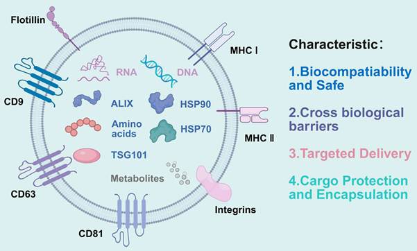
Exosomes are nanoscale EVs actively released by virtually all cell types, with diameters spanning 30-150 nm[18]. Their existence was first recognized in the 1980s, when vesicles shed during in vitro culture of sheep reticulocytes were isolated and christened “exosomes”[19]. Morphologically, they display a cup-shaped silhouette under electron microscopy, enclosed by a classic lipid-bilayer membrane[20]. This membrane is enriched in tetraspanins-CD9, CD63, and CD81-together with ESCRT-associated adaptor proteins (TSG101, Alix) and cytosolic chaperones (HSP70, HSP90) that collectively serve as canonical exosome markers[21-23]. Internally, exosomes ferry a complex molecular payload: regulatory RNAs (miRNA, mRNA, lncRNA, circRNA) and trace mitochondrial/nuclear DNAs that can reprogram gene expression in recipient cells[24, 25], sphingolipids, cholesterol, and phosphatidylserine that stabilize the vesicle and facilitate target-cell recognition; and a proteome that mirrors the physiological or pathological state of the parent cell. Thus, exosome composition is both a fingerprint and a messenger of cellular identity and condition[26, 27].
EVs, as an endogenous nanocarrier, exhibit unique advantages in drug delivery that synthetic nanocarriers cannot match[28]. Their most notable characteristics are excellent biocompatibility and low immunogenicity[29, 30]. Due to the fact that EVs originate from the body's own cells, the protein and lipid components carried on their membrane surface can effectively mimic parental cells, thereby avoiding or significantly reducing the body's immune rejection response and reducing side effects[6, 31].
EVs possess an innate capacity to negotiate biological barriers that are impermeable to most drugs, with the blood-brain barrier (BBB) being the most formidable[15, 32]. By hijacking receptor-mediated endocytosis and transcytotic pathways enriched in brain microvascular endothelium, EVs ferry their therapeutic cargo directly into the cerebral parenchyma, achieving concentrations that free drugs cannot reach[33, 34]. This tropism has created new therapeutic corridors for glioblastoma, Parkinson's, and Alzheimer's disease. Beyond the BBB, EVs similarly traverse mucosal linings of the gut, airways, and ocular surface, enabling non-invasive, site-specific therapy for digestive, respiratory, and ophthalmic disorders[35, 36].
Exosomes are endowed with built-in homing and high-efficiency cytosolic delivery[37, 38]. Membrane-anchored integrins, tetraspanins, and glycoproteins engage complementary receptors on target cells, directing vesicle docking to selected tissues[39]. Many exosomes also display a “boomerang” preference for their parental cell type, a property already exploited to concentrate anticancer agents within autologous tumors[40]. Once internalized, exosomes either fuse directly with the plasma membrane or escape late endosomes by back-fusion with the endosomal limiting membrane, emptying their payload straight into the cytoplasm. This shortcut bypasses destructive lysosomal routing and can raise the bioavailability of encapsulated drugs by orders of magnitude[41, 42].
The protective ability of exosomes towards their internal cargo is also one of their key advantages. Their lipid bilayer structure forms a stable internal environment, effectively protecting encapsulated unstable biological macromolecules, such as RNA and proteins, from degradation by enzymes in the circulatory system, thereby extending their half-life and ensuring their biological activity[1, 43]. This natural protective mechanism makes exosomes an ideal carrier for delivering siRNA, mRNA-based nucleic acid drugs, and protein drugs[44, 45].
Exosome formation process
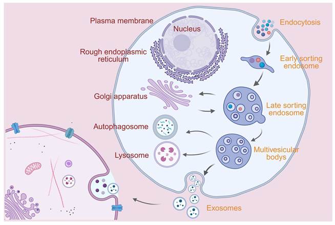
The exosome secretion process begins with the formation of early sorting endosomes (ESE) through the invagination of the plasma membrane. ESE can mature into late sorting endosomes (LSE) and ultimately produce multivesicular bodies (MVB), also known as multivesicular endosomes. MVB is formed through the inward invagination of the endosomal limiting membrane (i.e., the double invagination of the plasma membrane)[46]. The MVB membrane undergoes ubiquitin-mediated protein sorting and membrane inward budding mediated by endosomal sorting complex required for transport (ESCRT) proteins (ESCRT-0/I/II/III) to generate intraluminal vesicles (ILV)[47]. MVB trafficking is regulated by Rab GTPases (such as Rab7) and SNARE (soluble N-ethylmaleimide-sensitive factor attachment protein receptor) proteins[48]. Some MVB fuse with lysosomes, leading to ILV degradation, while others release ILV into the extracellular space by fusing with the plasma membrane to form exosomes[46, 49]. In addition to the ESCRT-dependent mechanism, EV biogenesis can also proceed through ESCRT-independent pathways. Ceramide generated by neutral sphingomyelinase 2 (nSMase2) can promote negative membrane curvature and inward budding of endosomal membranes, thereby facilitating ILV formation independently of canonical ESCRT machinery[50, 51]. Tetraspanin-enriched microdomains, particularly those organized by CD9, CD63, CD81, and related tetraspanins, may also function as membrane platforms for selective cargo sorting and vesicle formation[52].
Exosome structure diagram. Exosomes are nanoscale vesicles with a bilayer lipid membrane structure. Their membrane surface expresses characteristic proteins including CD9, CD63, CD81, Flotillin, MHC I/II, and Integrins, while the inner lumen encapsulates bioactive molecules such as RNA, DNA, amino acids, metabolites, and functional proteins (e.g., HSP70, HSP90, ALIX, TSG101). These inherent features endow exosomes with core advantages including excellent biocompatibility and safety, the ability to cross biological barriers, targeted delivery, and protection of encapsulated cargoes. Created in BioRender. lian, W. (2026) https://BioRender.com/a5hzzat.
Schematic diagram of the exosome biogenesis pathway. The biogenesis of exosomes originates from the endocytic process of cells: extracellular materials are internalized via endocytosis to form early sorting endosomes, which further mature into late sorting endosomes. The membrane of late endosomes invaginates and buds to form multivesicular bodies (MVBs), which encapsulate cytoplasmic contents such as nucleic acids and proteins. MVBs can either fuse with lysosomes for degradation or interact with autophagosomes, and ultimately fuse with the plasma membrane to release the internal vesicles into the extracellular space, thereby generating exosomes. Created in BioRender. lian, W. (2026) https://BioRender.com/a5hzzat.
EV isolation and purification technologies
Separation method
The effective isolation, precise characterization, and rigorous quality control of exosomes are crucial prerequisites for their transition from basic research to clinical application. Currently, exosome isolation can be achieved through various methods (see Table 1). Due to the small size of exosomes and their widespread presence in complex biological fluids, efficiently and purifyingly obtaining exosomes and conducting standardized assessments represent the core technical challenges in this field[53, 54].
EV isolation and purification methods
| Isolation Method | Brief Description | Advantages | Disadvantages | Recovery | Purity | Clinical suitability |
|---|---|---|---|---|---|---|
| Ultracentrifugation[55-58] | Isolate EVs through differential centrifugation forces | Widely used, no chemical reagents required, compatible with many sample types | Time-consuming, small separation volume, requirement for large scale equipment | Recovery varies with rotor type, speed, and protocol | Moderate; protein aggregates and similarly sized vesicles may co-precipitate | Suitable for laboratory-scale research but less suitable as a stand-alone GMP process |
| Ultrafiltration[18, 59, 60] | Isolate EVs by centrifugation with ultrafiltration membranes of appropriate pore sizes selected based on exosome particle size and molecular weight | Simple operation, short time consumption | Mechanical shear force causes damage to EVs | Moderate to high when optimized | Moderate; soluble proteins and small contaminants may remain | Useful for concentration and scalable workflows, but usually requires further purification |
| Immunoaffinity Capture[18, 61, 62] | Capture EVs based on the principle of antigen-antibody binding | High specificity, fast separation speed | High cost, additional steps for antibody removal, difficulty in large-scale application | Depends on marker abundance and binding efficiency | High for target-positive subpopulations | Suitable for diagnostics and specific EVs subpopulation studies, but difficult for bulk manufacturing |
| Polymer Precipitation[56, 62, 63] | Precipitate EVs by adding polymers to alter their solubility, surface charge or surface properties | Scalable, simple operation | High cost, potential impurity inclusion, low purity | High apparent yield | Low to moderate. | Limited suitability for therapeutic-grade EVs preparation |
| Size Exclusion Chromatography[26, 60] | Separate EVs based on molecular weight and particle size | High purity, preserved structural integrity | High cost | Moderate | High compared with precipitation and standard differential ultracentrifugation | Suitable for high-purity preparations and can be combined with concentration methods. |
| Microfluidic Isolation[62, 64, 65] | Isolate EVs through size-based filtration or microfluidic capture principles | High efficiency, rapid processing, automated operation | Low sample processing capacity | High for small clinical samples but limited bulk output | Variable to high depending on chip design and capture principle | High for diagnostics and point-of-care analysis; currently limited for large-scale therapeutic production |
EV cargo-loading methods
| Drug Loading Method | Drug | Advantages | Disadvantages | Typical loading performance | Effect on EV integrity | Translational suitability |
|---|---|---|---|---|---|---|
| Incubation[131-133] | Paclitaxel, Curcumin | Simple operation | Low loading efficiency | Low to moderate; highly dependent on cargo hydrophobicity, incubation time, and EV source | Generally well preserved | Suitable for preliminary loading of hydrophobic small-molecule drugs; limited for hydrophilic macromolecules |
| Transfection[133, 134] | miRNA | High loading efficiency | Cytotoxicity of reagents | Moderate to high for selected RNA cargos when producer cells are efficiently engineered | EV membrane is usually preserved because loading occurs during biogenesis | Suitable for endogenous RNA/protein cargo enrichment but requires strict control of producer cells |
| Electroporation[133, 135, 136] | Doxorubicin, Nucleic acid | Simple operation | Membrane damage, exosome aggregation | Moderate, affected by voltage, pulse number, buffer, and cargo type. | May be compromised under harsh parameters | Useful for nucleic acids in small-scale studies; reproducibility and aggregation control are major concerns |
| Ultrasonication[121, 133] | Paclitaxel | High loading capacity | Membrane damage | Moderate to high | Partially affected; membrane recovery depends on sonication conditions | Suitable when higher loading is required, but process optimization is essential |
| Extrusion[133, 137] | Enzyme | High loading capacity | Membrane damage, alteration of membrane potential | Moderate to high | Often affected because membrane reorganization occurs during extrusion | Suitable for engineered/hybrid vesicle preparation; requires rigorous characterization for translational use |
| Freeze-thaw[133, 138] | Curcumin | Simple operation | Exosome aggregation, protein inactivation | Low to moderate | Frequently affected by aggregation and membrane stress | Suitable for exploratory studies; limited for clinical-grade production unless carefully optimized |
| Drug Loading Method | Drug | Advantages | Disadvantages | Typical loading performance | Effect on EV integrity | Translational suitability |
| Surface Treatment[128, 133] | Porphyrin | Rapid and efficient | Potential membrane disruption | Variable; may improve loading for selected compounds | May be affected depending on surfactant type and concentration. | Useful for specific membrane-associated or permeabilization-assisted loading; safety and residual reagent control are needed |
| Microfluidics[130, 133] | mRNA | Small volume, low energy consumption, fast reaction rate | High production cost | Moderate to high depending on chip design and cargo type | Generally controllable under optimized conditions | Promising for standardized and scalable EV engineering, particularly when combined with surface modification or mRNA loading |
Ultracentrifugation (UC). Ultracentrifugation is the "gold standard" method for isolating exosomes. Its principle relies on the size and density differences of particles, precipitating particles of different sizes by gradually increasing the centrifugal force[66]. Differential ultracentrifugation is the most widely used UC technique, but its operation is cumbersome and time-consuming, and high centrifugal force may lead to aggregation or damage of exosomes, affecting their biological activity[67]. In addition, this method is prone to co-precipitating protein aggregates and other vesicles of similar size, resulting in low purity of the product[17]. Density gradient centrifugation, as an improved method of UC, can obtain exosomes with higher purity, but the operation is more complex and the yield is lower[66].
Ultrafiltration (UF). Ultrafiltration utilizes a filter membrane with a specific pore size to concentrate and purify exosomes by centrifugation or pressurization. It allows molecules smaller than the membrane pore size to pass through while retaining the exosomes, thus achieving the desired concentration and purification[66, 68]. UF is easy and fast to operate, but the filter membrane is prone to clogging, and exosomes may undergo adsorption on the membrane surface or deformation under shear forces, affecting recovery rate and integrity[54, 66].
Immunoaffinity Capture. Immunoaffinity capture is a method that utilizes antibodies to specifically bind to exosome surface markers (such as CD9, CD63, CD81) to achieve highly specific separation[54, 68]. This method can isolate subpopulations derived from specific cells or tissues with extremely high purity, and is particularly suitable for separating exosomes of specific origin from complex body fluids. For example, capturing L1CAM-positive exosomes derived from neurons for the diagnosis of Parkinson's disease[69, 70]. Its main limitations lie in high cost, low throughput, and the potential for biased capture of the entire exosome population due to targeting a single marker[53].
Polymer-based Precipitation. The polymer precipitation method, typically available in the form of commercialized reagent kits, operates on the principle of utilizing hydrophilic polymers like polyethylene glycol (PEG) to occupy water molecules, thereby reducing the solubility of other macromolecules (including exosomes) and causing them to precipitate[66, 71]. This method is straightforward and rapid, requiring no specialized equipment, but it suffers from poor selectivity, co-precipitating a significant amount of impurities such as non-vesicular proteins and lipoproteins, leading to extremely low product purity[72]. Despite this, it is still employed in some preliminary screening studies due to its convenience.
Size Exclusion Chromatography. Size exclusion chromatography (SEC) is a size-based separation technique that utilizes porous gel media to allow large molecules (such as exosomes) to elute preferentially, while small molecules (such as soluble proteins) enter the gel pores, thus achieving separation[66, 68]. SEC is considered a mild separation method that can well maintain the integrity and biological activity of exosomes, and has high purity[53]. Its main disadvantages are limited sample processing capacity and potential sample dilution issues, which result in poor separation performance for low-concentration samples[54].
Microfluidics-based Separation. Microfluidic technology achieves efficient, rapid, and automated separation of exosomes by precisely manipulating fluids within micron-scale channels[64, 65]. This type of technology can be divided into active and passive methods. Passive methods primarily utilize microstructures and fluid dynamics principles, such as size-based filtration (micro-nano filtration membranes), deterministic lateral displacement (DLD), and inertial focusing[73]. Active methods, on the other hand, rely on external physical fields, such as acoustic, electric, or magnetic field[74]. Microfluidic chips can integrate sample processing, separation, concentration, and even downstream analysis into one, greatly simplifying the operation process and reducing the amount of sample required, making them particularly suitable for rapid diagnosis of clinical samples with small amounts[65].
EV engineering strategies
Engineering of source cells
Engineering the source cells is a strategy to overcome the low yield and limited functionality of natural exosomes[75, 76]. Selecting the appropriate source cells is the first step towards optimization. Mesenchymal stem cells (MSCs), dendritic cells (DCs), HEK293 cells, and specific tumor cells are widely used due to their distinct biological characteristics[77-79]. Exosomes derived from MSCs possess natural tissue repair and immune regulatory capabilities[80], while those derived from DCs are rich in immune stimulatory molecules, making them suitable for tumor immunotherapy[81]. HEK293F cells, with their rapid growth rate, ease of genetic modification, and suspension culture, have become an ideal source for producing high-yield, high-purity engineered exosomes[77].
Research has shown that overexpressing key proteins involved in multivesicular body (MVB) formation and transport, such as ESCRT pathway-related proteins Alix, Rab GTPases family members Rab27a, Rab27b, or sphingomyelinase (nSMase2), can significantly promote the production and release of exosomes[82, 83].
Optimizing cell culture conditions is also an effective way to increase exosome production. For example, exposing cells to specific physical or chemical stress environments, such as hypoxia, low pH, heat shock, or specific cytokine stimulation, can induce cells to secrete more exosomes[82]. Three-dimensional (3D) cell culture technology not only increases cell density but also enhances the secretion efficiency and biological activity of exosomes[84]. In a semi-continuous culture mode with high-density fixed phase, the exosome production of HEK293F cells has been maximized[77]. Exosome mimetics, also known as nanovesicles, are produced on a large scale by subjecting cells to serial extrusion, a method that achieves yields significantly higher than those of naturally secreted exosomes[85]. These mimetics retain the surface protein profile and topology of the parent cell membrane. Consequently, they exhibit comparable targeting capabilities and immune evasion characteristics, which has led to their successful application in the targeted delivery of anticancer drugs and the modulation of the immune microenvironment[86, 87]. The combination of these cell engineering and culture optimization strategies provides a feasible technical route for the large-scale and high-quality production of functional exosomes.
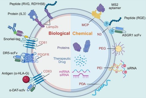
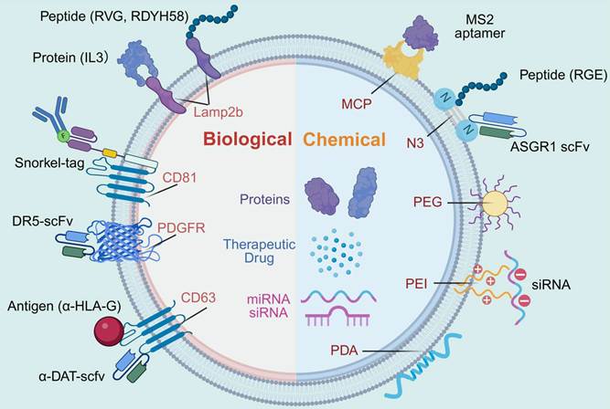
Schematic diagram of EV surface targeting engineering. Biological modification achieves specific recognition of target cells by fusing targeting peptides (e.g., RVG, RDYH58, RGE), functional proteins (e.g., IL3), single-chain antibody fragments (e.g., DR5-scFv, ASGR1 scFv), receptors (e.g., PDGFR), and tag proteins (e.g., Snorkel-tag, Lamp2b) onto the membrane surface. Chemical modification, by means of chemical molecules such as PEG, PEI, and PDA, as well as click chemistry (N3) and nucleic acid aptamers (MS2 aptamer), enhances the stability of exosomes while endowing them with targeted delivery capability. The interior of exosomes can encapsulate therapeutic drugs, proteins, miRNAs, siRNAs and other active substances, thereby providing a platform for precision targeted therapy. Created in BioRender. lian, W. (2026) https://BioRender.com/a5hzzat.
Surface targeting engineering
To enhance the targeted delivery efficiency of exosomes in complex biological environments, researchers have developed a variety of surface engineering strategies, which are primarily categorized into genetic engineering, chemical modification, and membrane fusion approaches. These technologies enable the display of functional molecules on the vesicle surface, guiding the precise recognition and interaction of exosomes with diseased tissues and thereby improving therapeutic outcomes.
Surface modification also serves to prolong the circulatory half-life of exosomes in vivo. Conjugation of hydrophilic polymers such as polyethylene glycol (PEG) to the exosome surface effectively reduces their recognition and clearance by the mononuclear phagocyte system (MPS)[31, 88]. Studies have demonstrated that the formation of polymer chains on exosome surfaces via reversible activation of atom transfer radical polymerization (ATRP) not only strengthens their storage stability and resistance to enzymatic degradation but also extends their circulatory half-life by fourfold[88].
Genetic Engineering. This method achieves high modification efficiency, excellent ligand targeting, and preserves the natural integrity of exosomes. Recent studies have successfully utilized PTGFRN and BASP1 as novel scaffold proteins to display antibody fragments and cytokines on the membrane surface, significantly enhancing the targeted recognition capability of exosomes[89].
Genetic engineering including synthetic biology approach represents the most commonly employed and well-established surface modification strategy, involving the fusion of gene sequences encoding targeting ligands with those encoding exosomal membrane proteins such as Lamp2b, CD63, or CD81[90, 91]. When parental cells express these fusion proteins, the targeting ligands are subsequently displayed on the surface of secreted exosomes. Lamp2b is the most widely utilized anchor protein, having been successfully applied to display a variety of targeting moieties, including the RDYH58 peptide for targeting myofibroblasts[92], IL3 for targeting leukemia cells[93], Her2 or E7 for tumor targeting[94], and the rabies virus glycoprotein (RVG) peptide for blood-brain barrier penetration[95]. Tetraspanin proteins, such as CD63 and CD81, are also frequently used as anchors. Notably, the CD81-anchored Snorkel-tag system provides a versatile platform for rapid conjugation of fluorophores or arbitrary targeting molecules, enabling specific binding to HER2-positive or CCR2-positive cells[96]. In addition to classical anchors such as Lamp2b, CD63, and CD81, large-scale scaffold screening has identified additional EVs-enriched scaffold proteins that may improve cargo sorting and surface display. Zheng et al. compared 244 candidate proteins and identified 24 proteins with conserved EVs-sorting capacity across five producer cell types; among them, TSPAN2 and TSPAN3 outperformed the commonly used CD63 scaffold and supported both luminal cargo transfer and surface display of functional entities[97]. More recently, scaffold-protein screening based on functional delivery further highlighted TSPAN2, TSPAN4, and TSPAN9 as candidates that can improve intracellular delivery of luminal proteins compared with CD63, supporting the idea that scaffold selection should be regarded as an active engineering variable rather than a passive anchoring choice[91]. Additionally, the transmembrane domain of PDGFR has been harnessed to display a single-chain variable fragment (scFv) against DR5 on the surface of NK cell-derived vesicles, effectively targeting and inducing apoptosis in diverse DR5+ cancer cells, myeloid-derived suppressor cells (MDSCs), and cancer-associated fibroblasts (CAFs)[98]. A study genetically engineered HEK 293T cells to overexpress α-HLA-G Nb-chimeric CD63 fusion protein, resulting in the production of EVs enriched with the fusion protein. Modified EVs (α-HLA-G EV) were loaded with chemotherapeutic drugs to treat different types of cancer. Drugs encapsulated in α-HLA-G EV (drug@α-HLA-G EV) target HLA-G-positive cancer cells by recognizing HLA-G on the cancer cell membrane. The drug@α-HLA-G EV are then internalized by cancer cells, delivering chemotherapeutic drugs to kill cancer cells[99]. In one study, an extracellular vesicle (EV)-based dopamine transporter (DAT)-targeted drug delivery system, derived from genetically engineered HEK293T cells, expressing DAT-targeting single-chain variable fragments (scFv) on the EVs surface, is developed[100]. The successful functionalization of exosomes via genetic engineering strategies has further expanded the application potential of genetically engineered modifications.
Chemical Modification. Chemical modification is suitable for engineering plant-derived or milk-derived exosomes, as well as exosomes that are difficult to genetically modify. Bioorthogonal chemistry has also advanced from simple ligand conjugation to more complex surface editing. Click chemistry is a highly efficient and specific chemical conjugation approach. In one study, superparamagnetic iron oxide nanoparticles (SPIONs) and curcumin (Cur) were loaded into exosomes. The exosome membrane was then conjugated with the neuropilin-1-targeting peptide (RGERPPR, RGE) via click chemistry, resulting in glioma-targeted exosomes with imaging and therapeutic capabilities[101]. Through metabolic glycan engineering, azide groups are introduced onto the cell membrane, enabling the subsequent covalent conjugation of targeting molecules such as ASGR1 scFv onto the surface of secreted exosomes via click chemistry. This approach achieves precise targeting to hepatocytes[102]. An alternative strategy involves non-covalent conjugation based on specific molecular affinity interactions. Researchers have developed an MS2 system in which the MS2 coat protein (MCP) fused to an exosomal membrane protein can capture cargo proteins containing the MS2 aptamer with high affinity. By modulating this affinity, efficient loading and controlled release of CRISPR-Cas9 complexes can be achieved[103, 104]. Polyethylenimine (PEI) -based modification is an electrostatic adsorption method that alters the surface charge of vesicles and is employed to load negatively charged siRNA[105]. Additionally, modifying biological scaffolds with distinct affinity molecules such as PDA and calcium phosphate (CaP) enables the regulation of exosome release kinetics[106]. Another representative strategy is the adaptive design of mRNA-loaded EVs, in which different targeting ligands are docked onto CD64-displaying EVs to guide cell-specific delivery. This approach enabled mRNA-loaded EVs to preferentially target glioblastoma cells and generate antitumor activity in vivo, thereby linking surface functionalization with therapeutic nucleic acid delivery[107]. Collectively, these recent advances indicate that future EVs surface engineering may increasingly rely on modular scaffold selection, bioorthogonal conjugation, and adaptive ligand docking to improve targeting specificity, cargo compatibility, and translational flexibility.
Membrane Fusion. Biomimetic membrane technology involves fusing exosomes with the cell membranes of specific cell types, thereby endowing the resulting vesicles with innate biological functions such as immune evasion and inflammatory targeting. Neutrophil membranes possess inherent inflammatory chemotactic properties, and coating exosomes with these membranes significantly enhances vesicle accumulation at inflammatory sites[108, 109]. Platelet membranes, owing to their affinity for damaged vascular endothelium and circulating tumor cells, have been utilized to construct delivery systems capable of targeting ischemic myocardium[110]. Similarly, macrophage membranes have been employed to develop drug delivery systems that can cross the blood-brain barrier and target gliomas[111]. Hybrid membrane systems have also been engineered. Examples include fusing mesenchymal stem cell-derived exosomes with astrocyte-derived exosomal membranes[112]. These approaches integrate the advantages of vesicles from different cellular origins to achieve synergistic multifunctional therapies. Another research direction involves hybrid vesicles composed of lipid nanoparticles and exosomes (LNP-EVs). By inducing fusion between LNPs and exosomes under low pH conditions, hybrid carriers can be constructed that retain the biocompatibility and targeting capabilities of exosomes while incorporating the efficient mRNA encapsulation properties of LNPs[113].
EV cargo loading
To transform exosomes into effective drug delivery carriers, it is necessary to efficiently load therapeutic cargo into them. Currently, drug loading strategies mainly fall into two categories: post-isolation loading and endogenous loading[8, 114]. Post-isolation loading refers to the introduction of drugs into the vesicles through physical or chemical methods after the exosomes have been isolated and purified. Common techniques include co-incubation, electroporation, ultrasonic treatment, saponin permeabilization, freeze-thaw cycles, and extrusion methods[114]. Endogenous loading involves genetically engineering the source cells to actively package specific therapeutic proteins or nucleic acids into exosomes during their biosynthetic process[76]. This method utilizes the natural cargo sorting mechanism within the cell, enabling efficient enrichment and stable loading of target cargo[115]. A technology platform called "GIFTed-Exos" successfully achieves efficient loading and active delivery of multiple functional proteins in engineered exosomes by fusing the target protein with CD9[116]. The advantage of endogenous loading lies in its stable and uniform cargo loading, as well as maintaining the natural structure and function of exosomes, but the technology is relatively complex[117].
Incubation. The incubation method is applicable to a diverse range of types and can successfully load therapeutic nucleic acids, small molecule drugs, proteins, peptides, and the like into exosomes. Studies have shown that the efficiency of drug loading varies depending on incubation time, drug characteristics, and other procedural details[118]. Incubation is a straightforward method in which purified EVs are incubated with drugs to allow them to enter the vesicle membrane[119].
Transfection. Transfection is a method that utilizes a designed vector to introduce active molecules such as proteins, peptides, and nucleic acids into cells. The specific process involves the use of transfection reagents to transduce specific plasmids into cells, prompting the cells to express the required substances, which are then packaged into exosomes[120].
Schematic diagram of exosome drug loading methods. The left panel illustrates that exosomes can encapsulate various types of therapeutic substances, including hydrophobic drugs, hydrophilic drugs, and RNAs. The right panel summarizes mainstream exosome drug loading technologies, such as drug co-incubation, ultrasonic incubation, transfection, freeze-thaw cycles, saponin treatment, extrusion, microfluidics, and electroporation. These techniques enable efficient loading of drugs with diverse physicochemical properties into exosomes, providing versatile technical options for exosomes as drug delivery vehicles. Created in BioRender. lian, W. (2026) https://BioRender.com/a5hzzat.
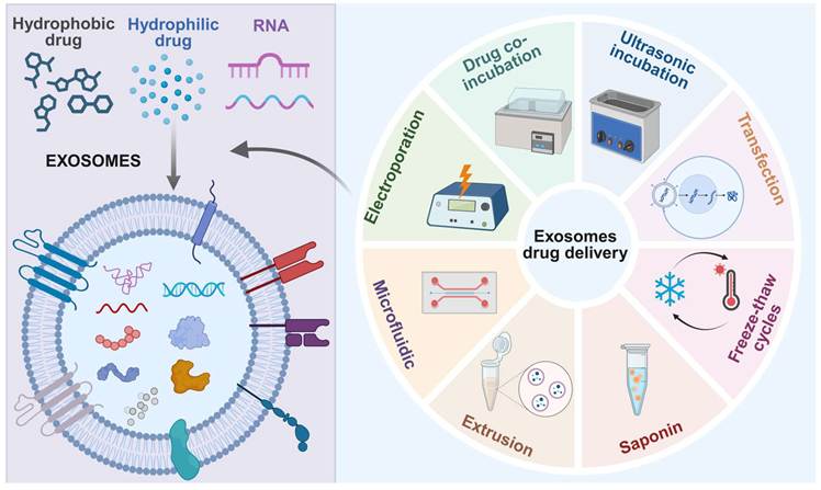
Electroporation. Electroporation involves the application of brief electrical pulses to induce micropores on the surface of isolated exosomes, thereby enabling drug migration into the exosomes. This method has been applied to drugs such as curcumin and paclitaxel, and drugs, nucleic acids, and nanomaterials can all be loaded using this approach[101, 121]. However, when applying this method, parameters such as condenser capacity, voltage, pulse number, pulse duration, and interval time need to be optimized. Moreover, electroporation can affect the stability and integrity of the exosome membrane, which imposes certain limitations on aggregate formation and loading efficiency[122].
Ultrasonic. Ultrasonic treatment involves the use of a homogenizing probe to subject the load and exosomes to ultrasonic manipulation. The mechanical shear force generated by the ultrasonic probe weakens the exosome membrane, allowing the load to penetrate into the exosome. Drugs, proteins, and nanomaterials can all be loaded using this method. However, the additional mechanical shear stress generated by ultrasonication can disrupt the integrity of the exosome membrane[123]. A study compared the application of ultrasonic treatment, incubation, and electroporation in paclitaxel loading, and found that ultrasonic loading (with a loading capacity of 28%) was significantly more effective than incubation (with a loading capacity of 1%) and electroporation (with a loading capacity of 5%)[121].
Extrusion. The extrusion method involves extruding the mixture of exosomes and payload through a 100-400 nanometer pore. During this process, the exosome membrane is damaged, and after repeated extrusion under specific parameters, the drug can be uniformly loaded into the exosomes. Although this method has high loading efficiency, the reorganization of the exosome surface structure may alter its immune status, making it more easily recognized by immune cells[124, 125].
Freeze-Thaw. The freeze-thaw method utilizes rapid temperature changes to directly load drugs, peptides, and proteins onto exosomes. During the operation, the load is first incubated with the exosomes at room temperature, then quickly frozen in a -80 ℃ environment or liquid nitrogen, and subsequently thawed at room temperature. This cycle is repeated at least three times. This method is simple to operate, but the drug loading efficiency is not high. Furthermore, repeated freeze-thaw cycles tend to cause exosome aggregation, resulting in protein inactivation[126]. In addition, this method can also be used to prepare exosome mimetic particles by fusing the membranes of exosomes and liposomes[127].
Surface treatment. Surface treatment involves the use of surfactants such as saponin and Triton to redistribute exosome membrane molecules, forming pores on the exosome surface to enhance membrane permeability. This method can significantly increase the loading capacity of exosomes for various compounds. However, the use of surfactants may lead to drug degradation or inactivation, thereby reducing the therapeutic effect of the drug[128].
Microfluidics. Microfluidics technology, which enables precise fluid manipulation at the microscale, provides an advanced platform for engineering exosomes. Microfluidic chips incorporating nanostructured channels and micromixing elements (e.g., ExoSE) can efficiently introduce functionalized lipids into exosomal membranes via mechanical perforation, while simultaneously facilitating rapid covalent conjugation of targeting ligands. This approach significantly outperforms conventional incubation methods in both modification efficiency and processing speed[129]. Furthermore, high-throughput microfluidic electroporation systems have been developed to enable the large-scale production of engineered exosomes loaded with mRNA[130].
Based on the above discussion of source-cell engineering, surface functionalization, membrane fusion, and cargo-loading strategies, the major EV engineering approaches can be further compared in terms of their effects on EV yield, cargo-loading performance, in vivo targeting fidelity, scalability, and translational limitations (Table 3).
Comparative overview of major EV engineering strategies for drug delivery
| Engineering strategy | Main purpose | Effect on EV yield | Cargo-loading performance | In vivo targeting fidelity | Major limitations |
|---|---|---|---|---|---|
| Source-cell engineering[97, 139] | Enhance EV production and intrinsic biological function | Moderate to high; depends on producer-cell type and engineered pathway | Indirectly improved through parental-cell programming or scaffold-assisted endogenous sorting | Variable; influenced by parental-cell tropism and surface composition | Genetic manipulation may alter EV composition and increase quality-control complexity |
| Culture optimization and bioreactor production[140, 141] | Increase production yield and batch consistency | High; hollow-fiber can markedly increase harvestable EVs output | No direct improvement | Limited direct effect | Requires GMP-compatible media, process control, scalable purification, defined release criteria |
| Engineering strategy | Main purpose | Effect on EV yield | Cargo-loading performance | In vivo targeting fidelity | Major limitations |
| Endogenous cargo programming[97, 139] | Package therapeutic RNAs or proteins during EV biogenesis | Usually unchanged or moderately affected | Moderate to high for selected protein cargos when efficient scaffold proteins are used | Depends on additional targeting modification | Cargo amount, scaffold selection, and batch consistency require strict validation |
| Genetic surface display[91, 97] | Improve ligand-directed recognition and tissue targeting | Usually unchanged | Usually unchanged unless coupled with luminal or RNA-loading systems | Moderate to high; depends on ligand density, orientation, and receptor expression | Potential immunogenicity, altered biodistribution, and the need to verify ligand density and function |
| Chemical or bioorthogonal modification[128, 139] | Add targeting ligands or functional molecules after EV isolation | No direct effect | Variable; mainly supports surface conjugation or cargo association rather than intrinsic vesicle production | Moderate to high; depends on conjugation efficiency and ligand specificity | May affect membrane integrity, surface charge, storage stability, and batch reproducibility |
| Membrane fusion or hybrid EV systems[113, 139] | Combine EVs biocompatibility with enhanced targeting, immune evasion, or synthetic-nanoparticle loading capacity | Variable | Moderate to high; hybrid EV-LNP systems may improve mRNA loading and functional delivery | Potentially high but formulation-dependent | Product heterogeneity, complex characterization, and regulatory uncertainty remain major barriers |
Progress in the application of engineered EVs in disease treatment
Cardiovascular diseases
The application of EVs in the field of cardiomyopathy therapy is currently one of the most promising approaches. Previously mesenchymal stromal cells (MSCs) transplantation improved heart function was considered attributable by MSC-secreted paracrine factors[142, 143]. Later, many studies indicated the MSC-secreted EVs play a key role[144, 145]. Cardiosphere-derived cell therapy has shown therapeutic potential in Duchenne muscular dystrophy-associated cardiomyopathy, and its beneficial effects may be partly related to paracrine mechanisms, including EV-mediated signaling. Capricor Therapeutics is developing deramiocel (CAP-1002), an allogeneic cell therapy that primarily works by secreting extracellular vesicles (exosomes) with anti-fibrotic and immunomodulatory properties. The therapy is designed to treat cardiomyopathy in Duchenne muscular dystrophy (DMD), a leading cause of heart failure in these patients. After completing the pivotal Phase III HOPE-3 trial, which showed a significant slowing of both upper limb decline and cardiac disease progression, Capricor resubmitted its Biologics License Application (BLA) to the FDA. The agency accepted the resubmission in March 2026, reinstated its review, and set a PDUFA target action date of August 22, 2026 for a potential approval decision[145, 146]. Exosomes also show promising prospects in the treatment of pulmonary hypertension (PH). Targeted delivery of drugs to pulmonary blood vessels through biomimetic nanodelivery systems (BNDS) is expected to overcome the limitations of low bioavailability in traditional therapies and provide new therapeutic strategies for PH[147].
Tumor diseases
The application of EVs in the field of tumor therapy is currently a hot research topic, and its strategies mainly cover three levels: as drug delivery carriers, as therapeutic vaccine platforms, and directly as therapeutic agents or targets[1, 4]. Due to its natural targeting, biocompatibility, and ability to cross physiological barriers, engineered exosomes have become an ideal tool for delivering various anti-cancer drugs[148]. Researchers use exosomes derived from tumor cells to deliver chemotherapy drugs such as doxorubicin or paclitaxel, enhancing drug enrichment at the tumor site and reducing systemic toxicity through their homing effect[149]. In refractory tumors with dense matrix barrier, such as pancreatic cancer, the small size and surface characteristics of exosomes enable them to effectively penetrate the matrix and deliver drugs to the tumor core area[150, 151]. Regarding the issue of drug resistance, EVs have also been used to deliver drugs or nucleic acids that can reverse drug resistance. Delivery of siRNA targeting PDK4 can reverse rituximab resistance in diffuse large B-cell lymphoma[152].
EVs play multiple roles in tumor immunotherapy. Dendritic cells exosomes (DEX) or exosomes derived from tumor cells themselves are rich in tumor associated antigens and immune stimulatory molecules, which can serve as cell-free tumor vaccines to activate the body's anti-tumor immune response[81, 153]. Engineered EVs can be used to deliver immune checkpoint inhibitors (ICIs) or their antagonists, such as PD-L1 antibodies or siRNA, directly targeting the tumor microenvironment (TME) and blocking the immunosuppressive signaling pathway[154, 155]. A study constructed an EVs nanoplatform responsive to the release of anti-PD-L1 antibodies in acidic TME, and combined with photodynamic therapy (PDT), successfully achieved synergistic therapy for non-small cell lung cancer[154]. EVs can also be used to reshape immunosuppressive TMEs, such as delivering drugs to regulate the polarization direction of tumor associated macrophages (TAMs) (from pro tumorigenic M2 type to anti-tumor M1 type), or targeting and inhibiting the function of cancer associated fibroblasts (CAFs)[155, 156]. A more innovative strategy is to use exosomes derived from CAR-M cells to construct CAR-EDC (CAR exosome drug conjugate), which not only achieves precise targeting of tumors through CAR molecules, but also utilizes CXCL10 carried by exosomes and covalently linked chemotherapy drug SN-38 to achieve strong synergy between chemotherapy and immunotherapy[157].
For brain tumors such as glioblastoma (GBM), EVs demonstrate unique therapeutic advantages due to their ability to cross the blood-brain barrier[158]. Researchers loaded chemotherapy drugs into engineered exosomes and successfully delivered the drugs to the intracranial tumor site through intravenous, effectively inhibiting tumor growth and prolonging survival[159]. Exosomes with surface modified anti EGFR scFv can efficiently deliver LPCAT1 siRNA and inhibit lung cancer brain metastasis[160]. Despite numerous preclinical research findings, the double-edged sword effect of tumor derived EVs in promoting tumor progression, metastasis, and immune escape also needs to be cautious[62]. At present, multiple studies on tumor diagnosis and treatment based on EVs have entered the clinical trial stage, indicating their enormous potential for clinical translation[17, 161].
Neurodegenerative diseases
The biggest challenge in the treatment of neurodegenerative diseases (NDDs) such as Alzheimer's disease (AD), Parkinson's disease (PD), and amyotrophic lateral sclerosis (ALS) is the difficulty of drugs effectively crossing the blood-brain barrier (BBB) into the central nervous system (CNS)[15, 32]. EVs are considered revolutionary carriers for delivering neuroprotective agents and gene therapy drugs into the brain due to their endogenous origin, low immunogenicity, and natural BBB crossing ability. Research has shown that EVs can carry drug molecules and efficiently penetrate the BBB through mechanisms such as receptor-mediated endocytosis, delivering therapeutic drugs precisely to neurons or glial cells[162-164].
In the treatment of Parkinson's disease, EVs are used to address the two core pathological problems of dopaminergic neuron loss and neuroinflammation. An innovative study has developed a biomimetic nanomodulator that utilizes neuronal derived exosomes as carriers to co deliver tyrosine hydroxylase (TH) mRNA and STING antagonist H-151. TH mRNA is translated into functional proteins in target cells, supplementing dopamine synthesis; H-151 inhibits the pro-inflammatory activation of microglia and promotes their transformation towards the anti-inflammatory M2 phenotype. The system significantly improved motor dysfunction in a PD mouse model, demonstrating the enormous potential for synergistic effects of gene therapy and anti-inflammatory therapy[165]. Encapsulating traditional Chinese medicine monomers and other neuroprotective agents in EVs can effectively improve their water solubility and BBB permeability, thereby exerting anti-inflammatory, anti-apoptotic, and antioxidant effects in various neurological disease models[166].
For Alzheimer's disease, EVs therapy mainly focuses on clearing β - amyloid (A β) and tau protein aggregates, as well as regulating neuroimmunity[36, 167]. Through engineering modification, the surface of EVs can display ligands that can bind to A β, thereby acting as nano scavengers to promote the clearance of A β[168]. EVs derived from stem cells contain various neurotrophic factors and anti-inflammatory molecules, which have been proven to alleviate neuroinflammation, promote nerve regeneration, and improve cognitive function[35].
In addition to being used directly as therapeutic agents or drug carriers, EVs also play an important role in the diagnosis and pathological mechanism research of NDDs. Brain derived EVs that can be detected in peripheral blood, carrying specific proteins or nucleic acids, can serve as non-invasive biomarkers for early diagnosis and monitoring of disease progression[169]. At the same time, research has found that EVs derived from the peripheral system can also cross the BBB, affecting central nervous system pathology and revealing the complex interaction network between the peripheral and central nervous systems, providing new targets and perspectives for the treatment of NDDs[170]. Although EVs have shown great potential in the treatment of NDDs, challenges such as large-scale production, further improvement of targeted delivery efficiency, and long-term safety must be overcome before their clinical translation[171].
Musculoskeletal disorders
In the treatment of musculoskeletal disorders, traditional therapies often have poor efficacy due to poor drug targeting, limited tissue regeneration ability, and side effects. EVs derived from mesenchymal stem cells have anti-inflammatory, pro angiogenic, and tissue repair abilities, and have become an attractive new strategy for cell-free therapy in this field[16, 172].
In terms of musculoskeletal diseases, the application of EVs covers multiple aspects such as osteoarthritis (OA), intervertebral disc degeneration (IVDD), osteoporosis, and soft tissue injury repair. For OA, intra-articular injection of EVs can inhibit chondrocyte apoptosis, alleviate synovial inflammation, and promote cartilage matrix regeneration. In order to overcome the problem of rapid drug clearance in the joint cavity, researchers combined exosomes with biomaterials such as hydrogels to achieve local release of exosomes and extend the therapeutic window[173, 174]. In the treatment of IVDD, EVs can inhibit the inflammatory response of nucleus pulposus cells and the degradation of extracellular matrix (ECM), promote ECM synthesis, and thus delay the degeneration process[175]. A study designed a MMP13 responsive nanoplatform that synergistically regulates inflammation and autophagy by sequentially releasing anti-inflammatory drug ginkgolide (GK) and autophagy activator rapamycin (RA), effectively improving the pathological condition of IVDD[176]. For sports soft tissue injuries such as tendon bone insertion (TBI) injuries, MSC Exos can significantly accelerate the healing process by promoting angiogenesis, regulating immune responses, and guiding stem cell differentiation, and promote the formation of transitional tissues that are closer to natural structures rather than functionally poor scar tissues[80].
Inflammatory diseases
In the treatment of chronic inflammatory diseases such as rheumatoid arthritis (RA) and inflammatory bowel disease (IBD), the immune regulatory function of EVs plays a central role. MSC Exos can effectively alleviate autoimmune reactions and inflammatory damage by regulating macrophage polarization (promoting the transition to anti-inflammatory M2 type), inhibiting T cell overactivation, and reducing pro-inflammatory cytokine levels[177, 178]. In atherosclerosis (AS), a chronic inflammatory cardiovascular disease, stem cell-derived exosomes can play a protective role against atherosclerosis by reducing endothelial dysfunction, inhibiting lipid accumulation, regulating macrophage function, preventing vascular calcification and other mechanisms[179].
Recent innovative preclinical applications also suggest that engineered EVs may be useful for immune reprogramming in non-malignant inflammatory diseases. Click chemistry-based EVs surface editing has been used to construct CAR-like EVs with therapeutic activity in acute liver failure[102]. In inflammatory bowel disease, EVs derived from Wharton's jelly mesenchymal stem cells were engineered to display PD-L1 and encapsulate miR-27a-3p, thereby targeting PD-1 and PHB1-related signaling, restoring T-cell balance in inflamed intestinal tissues, and preserving epithelial integrity in preclinical models[180]. These findings suggest that surface-engineered EVs may serve as cell-free immunomodulatory platforms beyond oncology.
Other diseases
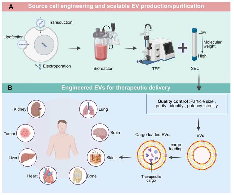
In the field of ophthalmic diseases, drug delivery is particularly difficult due to the presence of multiple physiological barriers in the eyes. Because of its good penetrability and biocompatibility, exosomes provide new solutions for the treatment of ocular neovascular diseases such as age-related macular degeneration (AMD), diabetic retinopathy (DR) and glaucoma. Engineering exosomes can be used to deliver anti-VEGF drugs or related siRNA to inhibit pathological angiogenesis. EVs themselves can also regulate corneal angiogenesis, lymphangiogenesis, and nerve repair, playing a role in corneal injury repair[181, 182]. The combination of exosomes with inorganic nanomaterials or hydrogels can further enhance their targeting and sustained release effect in the eye[183]. In terms of metabolic diseases, plant-derived extracellular vesicle-like nanoparticles (PELNs) show potential in regulating glucose and lipid metabolism disorders, providing a natural and safe intervention means for the treatment of obesity, diabetes and other diseases[184]. For complications of diabetes, such as diabetic foot ulcers, Exo-gel complex system loaded with angiogenic factors (Exo gel) can significantly accelerate chronic wound healing by regulating macrophage polarization and promoting angiogenesis[185]. In reproductive medicine and gynecological diseases, EVs have been explored for enhancing fertility treatments and supporting embryonic development[186]. In terms of endometrial repair and prevention of intrauterine adhesion (IUA), combining exosomes with hydrogels can effectively reduce fibrosis and promote endometrial regeneration[187]. Based on the above discussion, engineered EV-based drug delivery can be viewed as an integrated translational pipeline, including source-cell selection and engineering, EV production, isolation and purification, cargo loading, surface functionalization, quality control, disease-specific application, and clinical translation (Figure 5).
Integrated translational pipeline for engineered EV-based drug delivery systems, from source-cell engineering to clinical application. A. Source cells can be engineered by lipofection, viral transduction, or electroporation to generate EVs with desired cargos or surface features. Engineered or parental cells are expanded using scalable culture systems such as bioreactors, followed by EV isolation and purification using tangential flow filtration (TFF), size-exclusion chromatography (SEC), or integrated workflows. B. Purified EVs require quality-control assessment, including particle size, purity, identity, potency, and sterility. EVs can be further engineered through cargo loading and surface functionalization to generate cargo-loaded or targeted EVs for disease-specific delivery to organs or tissues. Created in BioRender. lian, W. (2026) https://BioRender.com/a5hzzat.
Clinical translation and scalable manufacturing of engineered EV therapeutics
Despite rapid progress in engineered EV-based drug delivery, clinical translation remains constrained by interrelated challenges in scalable manufacturing, purification, product standardization, potency evaluation, regulatory classification, and long-term safety assessment. Unlike conventional synthetic nanoparticles, EVs are biologically derived and intrinsically heterogeneous. Their composition and function are strongly influenced by source-cell type, culture conditions, and engineering strategy. Accordingly, the development of engineered EV therapeutics requires a translational framework that integrates good manufacturing practice (GMP) compatible production, predefined critical quality attributes (CQAs), mechanism-relevant potency assays, and systematic nonclinical and clinical safety evaluation[58, 188].
Scalable and GMP-compliant production
Scalable production is a prerequisite for moving engineered EVs from proof-of-concept studies to clinical development. Conventional two-dimensional culture systems remain useful for mechanistic studies but are generally insufficient for clinical-scale EV manufacturing because of their limited surface area, labor-intensive operation, and batch-to-batch variability. Three-dimensional culture systems, hollow-fiber bioreactors, stirred-tank bioreactors, and perfusion based platforms provide more suitable options for large scale EV production because they enable higher cell density, continuous harvest of conditioned medium, and improved control of oxygen tension, pH, nutrient supply, shear stress, and harvesting intervals[140, 189]. For example, large-scale bioreactor culture of mesenchymal stromal cells has been shown to markedly increase EV yield while allowing the assessment of sterility, potency, and functional activity, illustrating the feasibility of translation oriented EV manufacturing[140]. Suspension-adapted producer cells, such as HEK293-derived cells, may further support reproducible manufacturing when combined with serum-free or chemically defined media[77].
Manufacturing processes must preserve EVs batch consistency. Culture supplements, particularly fetal bovine serum, may introduce exogenous vesicles and undefined biological contaminants; therefore, EVs-depleted, serum-free, or chemically defined media are preferable for clinical-grade production. For engineered EVs, stable producer cell lines may offer better reproducibility than transient transfection, but genomic stability, transgene expression, residual host-cell DNA, and long-term culture performance should be monitored throughout cell banking and manufacturing[139, 190].
Downstream processing is another determinant of clinical feasibility. Ultracentrifugation remains widely used in basic research but is difficult to scale and may cause vesicle aggregation, and variable recovery. In contrast, tangential flow filtration (TFF), size-exclusion chromatography (SEC), ion-exchange chromatography, affinity capture, and integrated filtration-chromatography workflows are more compatible with GMP production. Comparative studies have shown that TFF combined with SEC can improve reproducibility, reduce processing time, and enhance cost-effectiveness compared with ultracentrifugation-based workflows[60]. For surface-engineered EVs, TFF-SEC workflows may also help remove free proteins or unbound components while exerting less stress on EVs than ultracentrifugation[191].
Standardization
The absence of unified quality criteria is a major barrier to the clinical development of engineered EV therapeutics. According to the MISEV2023 recommendations, EV studies should provide transparent information on EV nomenclature, source materials, pre-processing variables, separation methods, characterization, and functional validation[58]. For therapeutic products, these reporting requirements should be expanded into release specifications and CQAs. Relevant CQAs may include particle size distribution, particle concentration, morphology, protein-to-particle ratio, lipid composition, nucleic acid content, cargo-loading level, surface-marker profile, sterility, endotoxin level, mycoplasma status, storage stability, and potency-related functional readouts[58, 139].
Identity testing should not rely solely on classical EV-associated markers. Markers such as CD9, CD63, CD81, TSG101, and ALIX are useful for EV characterization, but they do not by themselves define product identity or therapeutic activity. For engineered EVs, identity testing should also confirm engineering-specific attributes, including targeting ligands, scaffold proteins, antibody fragments, bioorthogonal conjugation moieties, hybrid membrane components, or therapeutic cargos. In parallel, negative markers, such as endoplasmic reticulum or nuclear contaminants, should be assessed to exclude contamination by non-vesicular components[58, 139].
Potency assays are particularly challenging because engineered EVs may act through multiple mechanisms, including target-cell binding, cargo release, immune modulation, anti-inflammatory activity, cytotoxicity. Therefore, potency should be linked to the proposed mechanism of action rather than to particle number or total protein concentration alone. For nucleic acid-loaded EVs, potency assays may evaluate cargo abundance together with functional gene regulation. For immunomodulatory EVs, cytokine production, T-cell activation or suppression may be relevant readouts. For regenerative applications, angiogenesis, cell migration, or tissue-specific repair markers may be incorporated into potency testing[192].
Regulatory considerations and long-term safety
The regulatory classification of engineered EV products remains complex. EV products may be evaluated as biological products, cell-derived products, drug delivery systems, gene therapy related products. Surface-engineered EVs displaying targeting ligands or antibody fragments require additional evaluation of binding specificity and immunogenicity. Hybrid systems, such as EV-liposome or EV-lipid nanoparticle formulations, may further complicate regulatory evaluation because they combine biological and synthetic components[190].
Regulatory expectations should also consider the source of EVs. Autologous EVs may reduce immunogenicity but are difficult to standardize and scale. Allogeneic EVs are more suitable for off-the-shelf manufacturing but require donor screening, cell banking, traceability, and viral safety evaluation. Therefore, master cell banks and working cell banks should be established under GMP conditions, and release testing should include safety, purity, identity, and potency-related attributes[139, 190]. Dose definition also remains unresolved, because EV dose has been reported using particle number, total protein amount, lipid content, source-cell number, or active cargo amount. A combined dose description, including particle number, total protein, and active cargo amount, may provide a more informative basis for preclinical-to-clinical translation.
Although EVs are often considered biocompatible and less immunogenic than synthetic nanoparticles, engineered EVs should not be assumed to be inherently safe. Their biological effects depend on both native vesicle components and engineered modifications. Repeated administration may induce immune responses, unexpected tissue accumulation. Surface modification, PEGylation, ligand display, membrane fusion, and EV-lipid nanoparticle hybridization may alter biodistribution and clearance patterns, thereby requiring dedicated pharmacokinetic and biodistribution studies[139, 190]. Source-cell-related risks should also be carefully assessed. Tumor-derived EVs may possess tumor-homing properties but may carry oncogenic proteins, nucleic acids, or immunosuppressive signals. For EVs delivering gene-regulatory cargos, mRNA, or genome-editing systems, off-target genetic effects and prolonged biological activity require particular attention. Guidance for cellular and gene therapy products emphasizes science- and risk-based potency assurance and long-term follow-up for products with durable biological effects, which may provide useful regulatory principles for engineered EV products carrying long-acting nucleic acid or gene-editing cargos[189]. Notably, late-stage development of allogeneic cardiosphere-derived cell therapies with EV-mediated paracrine effects, such as deramiocel (CAP-1002) for Duchenne muscular dystrophy-associated cardiomyopathy, illustrates how scalable manufacturing, quality-control, and regulatory frameworks for EV-related therapeutics are already being implemented in practice[145, 146].
Overall, the clinical translation of engineered EV therapeutics depends on the convergence of scalable GMP-compliant production, robust purification workflows, standardized CQAs, mechanism-relevant potency assays, rational regulatory classification, and long-term safety validation. Future studies should move beyond proof-of-concept efficacy and establish reproducible, quality-controlled, and clinically relevant development pipelines for engineered EV-based drug delivery systems.
Conclusion
In recent years, extracellular vesicles (EVs) have emerged as promising biological nanocarriers for drug delivery because of their endogenous origin, biocompatibility, low immunogenicity, intrinsic targeting capacity, cargo-protective ability, and unique advantages in crossing biological barriers[7, 28]. With rapid advances in source-cell engineering, surface functionalization, cargo loading, membrane fusion, and hybrid vesicle development, engineered EV-based platforms have shown increasing potential for delivering small-molecule drugs, proteins, peptides, nucleic acid therapeutics, and immunomodulators. These strategies have expanded the therapeutic applicability of EVs in tumors, neurodegenerative diseases, musculoskeletal disorders, inflammatory diseases, and other pathological conditions[37].
Despite these advances, engineered EV therapeutics remain largely at the preclinical or early clinical stage, and several barriers must be overcome before broad clinical translation can be achieved. Key challenges include low and variable EV yield, product heterogeneity, insufficient targeting specificity, incomplete understanding of biodistribution and clearance, batch-to-batch variability, and uncertainty regarding long-term safety[193]. Future studies should move beyond proof-of-concept efficacy and establish standardized, mechanism-driven, and clinically relevant development pipelines. In particular, scalable GMP-compatible manufacturing, robust purification workflows, well-defined critical quality attributes, reliable identity markers, cargo- and indication-specific potency assays, and systematic safety evaluation will be essential for advancing engineered EV-based drug delivery systems toward clinical application[194, 195]. With continued progress in scaffold engineering, bioorthogonal modification, bioreactor-based production, quality control, and regulatory standardization, engineered EVs may become a versatile and translatable platform for precision medicine.
Acknowledgements
This work was supported by the National Key Research and Development Program of China (2022YFA1105600, 2022YFA1105601) and the Technology Innovation Program Shenzhen-Hong Kong Joint Project (Type A) (SGDX20240115113100001).
Author contributions statement
Lian Wang: Literature search, Data curation, Writing original draft; Xiangyu Li: Literature search, Data curation, Writing original draft; Qionghui Chen: Visualization, Data curation, Resources; Xianchuan Xu: Visualization, Data curation, Resources; Liwen Zhang: Literature screening, Data classification, Investigation; Peng Zhang: Literature screening, Data classification, Investigation; Jing Jiang: Supervision, Conceptualization, Writing review & editing, Project administration; Qizhou Lian: Supervision, Conceptualization, Writing review & editing, Project administration, Funding acquisition. All authors have read and agreed to the published version of the manuscript.
Competing Interests
The authors have declared that no competing interest exists.
References
1. Greening DW, Xu R, Rai A, Suwakulsiri W, Chen M, Simpson RJ. Clinical relevance of extracellular vesicles in cancer - therapeutic and diagnostic potential. Nature reviews. Clinical oncology. 2025;22:924-952
2. Huang C, Li J, Xie Z, Hu X, Huang Y. Relationship between exosomes and cancer: formation, diagnosis, and treatment. International journal of biological sciences. 2025;21:40-62
3. Chen YF, Luh F, Ho YS, Yen Y. Exosomes: a review of biologic function, diagnostic and targeted therapy applications, and clinical trials. Journal of biomedical science. 2024;31:67
4. Li J, Wang J, Chen Z. Emerging role of exosomes in cancer therapy: progress and challenges. Molecular cancer. 2025;24:13
5. Xu C, Jiang C, Li Z, Gao H, Xian J, Guo W. et al. Exosome nanovesicles: biomarkers and new strategies for treatment of human diseases. MedComm. 2024;5:e660
6. Liu JJJ, Liu D, To SKY, Wong AST. Exosomes in cancer nanomedicine: biotechnological advancements and innovations. Molecular cancer. 2025;24:166
7. Moghassemi S, Dadashzadeh A, Sousa MJ, Vlieghe H, Yang J, León-Félix CM. et al. Extracellular vesicles in nanomedicine and regenerative medicine: A review over the last decade. Bioactive materials. 2024;36:126-156
8. Dave KM, Pinky PP, D SM. Molecular engineering of extracellular vesicles for drug delivery: Strategies, challenges, and perspectives. Journal of controlled release: official journal of the Controlled Release Society. 2025;386:114068
9. Liu J, Ren L, Li S, Li W, Zheng X, Yang Y. et al. The biology, function, and applications of exosomes in cancer. Acta pharmaceutica Sinica. B. 2021;11:2783-2797
10. Gong Z, Cheng C, Sun C, Cheng X. Harnessing engineered extracellular vesicles for enhanced therapeutic efficacy: advancements in cancer immunotherapy. Journal of experimental & clinical cancer research: CR. 2025;44:138
11. Yang Q, Li S, Ou H, Zhang Y, Zhu G, Li S. et al. Exosome-based delivery strategies for tumor therapy: an update on modification, loading, and clinical application. Journal of nanobiotechnology. 2024;22:41
12. Zhang Y, Liu Q, Zhang X, Huang H, Tang S, Chai Y. et al. Recent advances in exosome-mediated nucleic acid delivery for cancer therapy. Journal of nanobiotechnology. 2022;20:279
13. Liang Y, Duan L, Lu J, Xia J. Engineering exosomes for targeted drug delivery. Theranostics. 2021;11:3183-3195
14. Xiao Q, Tan M, Yan G, Peng L. Revolutionizing lung cancer treatment: harnessing exosomes as early diagnostic biomarkers, therapeutics and nano-delivery platforms. Journal of nanobiotechnology. 2025;23:232
15. Sun M, Qin F, Bu Q, Zhao Y, Yang X, Zhang D. et al. Exosome-Based Therapeutics: A Natural Solution to Overcoming the Blood-Brain Barrier in Neurodegenerative Diseases. MedComm. 2025;6:e70386
16. Fu PJ, Luo Y, Zheng SY, Ma ZR, Xiao WF, Li H. et al. The Role of Extracellular Vesicles in Musculoskeletal Diseases. Journal of extracellular vesicles. 2025;14:e70205
17. Mizenko RR, Feaver M, Bozkurt BT, Lowe N, Nguyen B, Huang KW. et al. A critical systematic review of extracellular vesicle clinical trials. Journal of extracellular vesicles. 2024;13:e12510
18. Yang D, Zhang W, Zhang H, Zhang F, Chen L, Ma L. et al. Progress, opportunity, and perspective on exosome isolation - efforts for efficient exosome-based theranostics. Theranostics. 2020;10:3684-3707
19. Johnstone RM, Adam M, Hammond JR, Orr L, Turbide C. Vesicle formation during reticulocyte maturation. Association of plasma membrane activities with released vesicles (exosomes). The Journal of biological chemistry. 1987;262:9412-9420
20. Yu Z, Teng Y, Yang J, Yang L. The role of exosomes in adult neurogenesis: implications for neurodegenerative diseases. Neural regeneration research. 2024;19:282-288
21. Toh WS, Lai RC, Zhang B, Lim SK. MSC exosome works through a protein-based mechanism of action. Biochemical Society transactions. 2018;46:843-853
22. Mathieu M, Névo N, Jouve M, Valenzuela JI, Maurin M, Verweij FJ. et al. Specificities of exosome versus small ectosome secretion revealed by live intracellular tracking of CD63 and CD9. Nature communications. 2021;12:4389
23. Zhang G, Liu Z, Ding H, Zhou Y, Doan HA, Sin KWT. et al. Tumor induces muscle wasting in mice through releasing extracellular Hsp70 and Hsp90. Nature communications. 2017;8:589
24. Reynolds DE, Galanis G, Wang Y, Ko J. Single Extracellular Vesicle Analysis Using Droplet Microfluidics. Methods in molecular biology (Clifton, N.J.). 2023;2689:211-220
25. Dai J, Su Y, Zhong S, Cong L, Liu B, Yang J. et al. Exosomes: key players in cancer and potential therapeutic strategy. Signal transduction and targeted therapy. 2020;5:145
26. Sidhom K, Obi PO, Saleem A. A Review of Exosomal Isolation Methods: Is Size Exclusion Chromatography the Best Option? International journal of molecular sciences. 2020;21:6466
27. van der Meel R, Fens MH, Vader P, van Solinge WW, Eniola-Adefeso O, Schiffelers RM. Extracellular vesicles as drug delivery systems: lessons from the liposome field. Journal of controlled release: official journal of the Controlled Release Society. 2014;195:72-85
28. Tenchov R, Sasso JM, Wang X, Liaw WS, Chen CA, Zhou QA. Exosomes─Nature's Lipid Nanoparticles, a Rising Star in Drug Delivery and Diagnostics. ACS nano. 2022;16:17802-17846
29. Li W, Li H, Fan L, Zhao R, Dong F, Jia X. et al. The Potential Application of Exosomes as Therapeutic Agents, Carriers, and Biomarkers in Skin Diseases. International journal of nanomedicine. 2025;20:12627-12658
30. Xia Y, Zhang J, Liu G, Wolfram J. Immunogenicity of Extracellular Vesicles. Advanced materials (Deerfield Beach, Fla.). 2024;36:e2403199
31. Parada N, Romero-Trujillo A, Georges N, Alcayaga-Miranda F. Camouflage strategies for therapeutic exosomes evasion from phagocytosis. Journal of advanced research. 2021;31:61-74
32. Terstappen GC, Meyer AH, Bell RD, Zhang W. Strategies for delivering therapeutics across the blood-brain barrier. Nature reviews. Drug discovery. 2021;20:362-383
33. Sanadgol N, Abedi M, Hashemzaei M, Kamran Z, Khalseh R, Beyer C. et al. Exosomes as nanocarriers for brain-targeted delivery of therapeutic nucleic acids: advances and challenges. Journal of nanobiotechnology. 2025;23:453
34. Mehdizadeh S, Mamaghani M, Hassanikia S, Pilehvar Y, Ertas YN. Exosome-powered neuropharmaceutics: unlocking the blood-brain barrier for next-gen therapies. Journal of nanobiotechnology. 2025;23:329
35. Nouri Z, Barfar A, Perseh S, Motasadizadeh H, Maghsoudian S, Fatahi Y. et al. Exosomes as therapeutic and drug delivery vehicle for neurodegenerative diseases. Journal of nanobiotechnology. 2024;22:463
36. Kandimalla R, Saeed M, Tyagi N, Gupta RC, Aqil F. Exosome-based approaches in the management of Alzheimer's disease. Neuroscience and biobehavioral reviews. 2023;144:104974
37. Kim HI, Park J, Zhu Y, Wang X, Han Y, Zhang D. Recent advances in extracellular vesicles for therapeutic cargo delivery. Experimental & molecular medicine. 2024;56:836-849
38. Kooijmans SAA, de Jong OG, Schiffelers RM. Exploring interactions between extracellular vesicles and cells for innovative drug delivery system design. Advanced drug delivery reviews. 2021;173:252-278
39. Lee YJ, Shin KJ, Chae YC. Regulation of cargo selection in exosome biogenesis and its biomedical applications in cancer. Experimental & molecular medicine. 2024;56:877-889
40. Ko H, Kim CH, Son S, Shin JM, Park JH. Unlocking the therapeutic potential of tumor-derived small extracellular vesicles in cancer immunotherapy: A multifaceted approach. Biomaterials. 2026;327:123733
41. Wang H, Wang Z, Li B, Sui Y, Wu M, Guo Z. et al. Direct Cytosolic Delivery of Nanovesicles via Gigahertz Acoustic Streaming. ACS nano. 2025;19:16032-16042
42. Zhao J, Zhu W, Mao Y, Li X, Ling G, Luo C. et al. Unignored intracellular journey and biomedical applications of extracellular vesicles. Advanced drug delivery reviews. 2024;212:115388
43. Gu Q, Xue H, Liu Z, Rao J, Zeng L, Zhang C. et al. Developing Biomaterial-Based mRNA Delivery System for Lung Disease Treatment. Advanced science (Weinheim, Baden-Wurttemberg, Germany). 2025;12:e05413
44. Bian X, Zhou L, Luo Z, Liu G, Hang Z, Li H. et al. Emerging Delivery Systems for Enabling Precision Nucleic Acid Therapeutics. ACS nano. 2025;19:4039-4083
45. Muskan M, Abeysinghe P, Cecchin R, Branscome H, Morris KV, Kashanchi F. Therapeutic potential of RNA-enriched extracellular vesicles: The next generation in RNA delivery via biogenic nanoparticles. Molecular therapy: the journal of the American Society of Gene Therapy. 2024;32:2939-2949
46. Kalluri R, LeBleu VS. The biology, function, and biomedical applications of exosomes. Science (New York, N.Y.). 2020;367:eaau6977
47. van Niel G, D'Angelo G, Raposo G. Shedding light on the cell biology of extracellular vesicles. Nature reviews. Molecular cell biology. 2018;19:213-228
48. Xu M, Ji J, Jin D, Wu Y, Wu T, Lin R. et al. The biogenesis and secretion of exosomes and multivesicular bodies (MVBs): Intercellular shuttles and implications in human diseases. Genes & diseases. 2023;10:1894-1907
49. Kahlert C, Kalluri R. Exosomes in tumor microenvironment influence cancer progression and metastasis. Journal of molecular medicine (Berlin, Germany). 2013;91:431-437
50. Trajkovic K, Hsu C, Chiantia S, Rajendran L, Wenzel D, Wieland F. et al. Ceramide triggers budding of exosome vesicles into multivesicular endosomes. Science (New York, N.Y.). 2008;319:1244-1247
51. Choezom D, Gross JC. Neutral sphingomyelinase 2 controls exosome secretion by counteracting V-ATPase-mediated endosome acidification. J. Cell Sci. 2022;135:jcs259324
52. Perez-Hernandez D, Gutiérrez-Vázquez C, Jorge I, López-Martín S, Ursa A, Sánchez-Madrid F. et al. The intracellular interactome of tetraspanin-enriched microdomains reveals their function as sorting machineries toward exosomes. J. Biol. Chem. 2013;288:11649-11661
53. Jia Y, Yu L, Ma T, Xu W, Qian H, Sun Y. et al. Small extracellular vesicles isolation and separation: Current techniques, pending questions and clinical applications. Theranostics. 2022;12:6548-6575
54. Shirejini SZ, Inci F. The Yin and Yang of exosome isolation methods: conventional practice, microfluidics, and commercial kits. Biotechnology advances. 2022;54:107814
55. Taylor DD, Shah S. Methods of isolating extracellular vesicles impact down-stream analyses of their cargoes. Methods (San Diego, Calif.). 2015;87:3-10
56. Rahmatinejad F, Kharat Z, Jalili H, Renani MK, Mobasheri H. Comparison of morphology, protein concentration, and size distribution of bone marrow and Wharton's jelly-derived mesenchymal stem cells exosomes isolated by ultracentrifugation and polymer-based precipitation techniques. Tissue & cell. 2024;88:102427
57. Shen DD, Pang JR, Bi YP, Zhao LF, Li YR, Zhao LJ. et al. LSD1 deletion decreases exosomal PD-L1 and restores T-cell response in gastric cancer. Molecular cancer. 2022;21:75
58. Welsh JA, Goberdhan DCI, O'Driscoll L, Buzas EI, Blenkiron C, Bussolati B. et al. Minimal information for studies of extracellular vesicles (MISEV2023): From basic to advanced approaches. Journal of extracellular vesicles. 2024;13:e12404
59. Inamdar S, Nitiyanandan R, Rege K. Emerging applications of exosomes in cancer therapeutics and diagnostics. Bioengineering & translational medicine. 2017;2:70-80
60. Visan KS, Lobb RJ, Ham S, Lima LG, Palma C, Edna CPZ. et al. Comparative analysis of tangential flow filtration and ultracentrifugation, both combined with subsequent size exclusion chromatography, for the isolation of small extracellular vesicles. Journal of extracellular vesicles. 2022;11:e12266
61. Zhang L, Wang H, Zhao G, Li N, Wang X, Li Y. et al. Anti-Tim4 Grafting Strongly Hydrophilic Metal-Organic Frameworks Immunoaffinity Flake for High-Efficiency Capture and Separation of Exosomes. Analytical chemistry. 2021;93:6534-6543
62. Liangsupree T, Multia E, Riekkola ML. Recent advances in modern extracellular vesicle isolation and separation techniques. Journal of chromatography. A. 2026;1767:466602
63. Cao F, Gao Y, Chu Q, Wu Q, Zhao L, Lan T. et al. Proteomics comparison of exosomes from serum and plasma between ultracentrifugation and polymer-based precipitation kit methods. Electrophoresis. 2019;40:3092-3098
64. Wang C, Qiu J, Liu M, Wang Y, Yu Y, Liu H. et al. Microfluidic Biochips for Single-Cell Isolation and Single-Cell Analysis of Multiomics and Exosomes. Advanced science (Weinheim, Baden-Wurttemberg, Germany). 2024;11:e2401263
65. Zhuang J, Xia L, Zou Z, Yin J, Lin N, Mu Y. Recent advances in integrated microfluidics for liquid biopsies and future directions. Biosensors & bioelectronics. 2022;217:114715
66. Auquière M, Muccioli GG, des Rieux A. Methods and Challenges in Purifying Drug-Loaded Extracellular Vesicles. Journal of extracellular vesicles. 2025;14:e70097
67. Ng JQ, Abu Yazid NA, Tan SC, Monif M, Wong TW, Lee SY. Extracellular vesicles as delivery vehicles and therapeutic agents for glioblastoma treatment: A systematic review of in vitro and in vivo preclinical studies. Asian journal of pharmaceutical sciences. 2025;20:101043
68. Altıntaş Ö, Saylan Y. Exploring the Versatility of Exosomes: A Review on Isolation, Characterization, Detection Methods, and Diverse Applications. Analytical chemistry. 2023;95:16029-16048
69. Xylaki M, Chopra A, Weber S, Bartl M, Outeiro TF, Mollenhauer B. Extracellular Vesicles for the Diagnosis of Parkinson's Disease: Systematic Review and Meta-Analysis. Movement disorders: official journal of the Movement Disorder Society. 2023;38:1585-1597
70. Dutta S, Hornung S, Taha HB, Bitan G. Biomarkers for parkinsonian disorders in CNS-originating EVs: promise and challenges. Acta neuropathologica. 2023;145:515-540
71. Simon L, Lapinte V, Morille M. Exploring the role of polymers to overcome ongoing challenges in the field of extracellular vesicles. Journal of extracellular vesicles. 2023;12:e12386
72. Nila IS, Sumsuzzman DM, Khan ZA, Jung JH, Kazema AS, Kim SJ. et al. Identification of exosomal biomarkers and its optimal isolation and detection method for the diagnosis of Parkinson's disease: A systematic review and meta-analysis. Ageing research reviews. 2022;82:101764
73. Hassanpour Tamrin S, Sanati Nezhad A, Sen A. Label-Free Isolation of Exosomes Using Microfluidic Technologies. ACS nano. 2021;15:17047-17079
74. Lu Q, Zhang Z, Ding X. Isolation Techniques of Micro/Nano-Scaled Species for Biomedical Applications. Advanced science (Weinheim, Baden-Wurttemberg, Germany). 2025;12:e2414109
75. Deng S, Cao H, Cui X, Fan Y, Wang Q, Zhang X. Optimization of exosome-based cell-free strategies to enhance endogenous cell functions in tissue regeneration. Acta biomaterialia. 2023;171:68-84
76. Rädler J, Gupta D, Zickler A, Andaloussi SE. Exploiting the biogenesis of extracellular vesicles for bioengineering and therapeutic cargo loading. Molecular therapy: the journal of the American Society of Gene Therapy. 2023;31:1231-1250
77. Vo N, Tran C, Tran NHB, Nguyen NT, Nguyen T, Ho DTK. et al. A novel multi-stage enrichment workflow and comprehensive characterization for HEK293F-derived extracellular vesicles. Journal of extracellular vesicles. 2024;13:e12454
78. Prakash M, Cortez CD, Jayaraman A, Hsu SY, Huang YC, Yeh CY. et al. Innovative gene engineering and drug delivery systems for dendritic cells in cancer immunotherapy. Journal of biomedical science. 2025;32:95
79. Zhang F, Guo J, Zhang Z, Qian Y, Wang G, Duan M. et al. Mesenchymal stem cell-derived exosome: A tumor regulator and carrier for targeted tumor therapy. Cancer letters. 2022;526:29-40
80. Zou J, Yang W, Cui W, Li C, Ma C, Ji X. et al. Therapeutic potential and mechanisms of mesenchymal stem cell-derived exosomes as bioactive materials in tendon-bone healing. Journal of nanobiotechnology. 2023;21:14
81. Jung I, Shin S, Baek MC, Yea K. Modification of immune cell-derived exosomes for enhanced cancer immunotherapy: current advances and therapeutic applications. Experimental & molecular medicine. 2024;56:19-31
82. Debbi L, Guo S, Safina D, Levenberg S. Boosting extracellular vesicle secretion. Biotechnology advances. 2022;59:107983
83. Ostrowski M, Carmo NB, Krumeich S, Fanget I, Raposo G, Savina A. et al. Rab27a and Rab27b control different steps of the exosome secretion pathway. Nat. Cell Biol. 2010;12:19-30
84. Haraszti RA, Miller R, Stoppato M, Sere YY, Coles A, Didiot MC. et al. Exosomes Produced from 3D Cultures of MSCs by Tangential Flow Filtration Show Higher Yield and Improved Activity. Mol. Ther. 2018;26:2838-2847
85. Jang SC, Kim OY, Yoon CM, Choi DS, Roh TY, Park J. et al. Bioinspired exosome-mimetic nanovesicles for targeted delivery of chemotherapeutics to malignant tumors. ACS nano. 2013;7:7698-7710
86. Zhou ZY, Li ZB, Shi NS, Feng SS, Han YR, Liu MT. et al. Metal-polyphenol network-engineered mesenchymal stem cell-derived exosome mimetics mediate inflammatory/immune regulation for enhanced periodontal tissue regeneration. Biomaterials. 2026;327:123696
87. Mi T, Liu J, Luo J, Kong X, Tan X, Jin L. et al. CD24-targeted cystine and glucose oxidase cascade catalytic nanosystem triggers disulfidptosis in neuroblastoma. Materials today. Bio. 2025;35:102496
88. Lathwal S, Yerneni SS, Boye S, Muza UL, Takahashi S, Sugimoto N. et al. Engineering exosome polymer hybrids by atom transfer radical polymerization. Proceedings of the National Academy of Sciences of the United States of America. 2021;118:e2020241118
89. Dooley K, McConnell RE, Xu K, Lewis ND, Haupt S, Youniss MR. et al. A versatile platform for generating engineered extracellular vesicles with defined therapeutic properties. Molecular therapy: the journal of the American Society of Gene Therapy. 2021;29:1729-1743
90. Obuchi W, Zargani-Piccardi A, Leandro K, Rufino-Ramos D, Di Lanni E, Frederick DM. et al. Engineering of CD63 Enables Selective Extracellular Vesicle Cargo Loading and Enhanced Payload Delivery. Journal of extracellular vesicles. 2025;14:e70094
91. Niu Z, Zhou H, Zheng W, Hayes OG, Hou VWQ, Görgens A. et al. Screening scaffold proteins for improved functional delivery of luminal proteins using engineered extracellular vesicles. Journal of controlled release: official journal of the Controlled Release Society. 2025;384:113882
92. Yuan R, Mu Z, Zhang H, Tian Y, Xin Q, Tu Q. et al. RDYH58 functional exosomes targeting myofibroblasts loaded with siFKBP10 for inhibition of collagen biosynthesis and secretion of IPF. Acta pharmaceutica Sinica. B. 2025;15:6681-6697
93. Zhong F, Chen S, Yao F, Liu J, Zhang J, Jiang J. et al. Machine learning-driven glycolytic subtyping and exosome-based PKM splicing modulation overcome drug resistance in hyper-glycolytic myeloid leukemia. NPJ digital medicine. 2025;9:18
94. Yu X, Ding P, Guo M, Tang X, Wang Z, Zhang Y. et al. Extracellular vesicle-mediated delivery of circp53 suppresses the progression of multiple cancers by activating the CypD/TRAP/HSP90 pathway. Experimental & molecular medicine. 2025;57:1711-1726
95. Alvarez-Erviti L, Seow Y, Yin H, Betts C, Lakhal S, Wood MJ. Delivery of siRNA to the mouse brain by systemic injection of targeted exosomes. Nature biotechnology. 2011;29:341-345
96. Roefs MT, Gamauf J, Kroenigsberger B, Brancolini A, Traxlmayr MW, Arcalis E. et al. Rapid extracellular vesicle surface decoration with targeting moieties based on a fluorescein binding single chain variable fragment snorkel. Journal of controlled release: official journal of the Controlled Release Society. 2026;390:114558
97. Zheng W, Rädler J, Sork H, Niu Z, Roudi S, Bost JP. et al. Identification of scaffold proteins for improved endogenous engineering of extracellular vesicles. Nature communications. 2023;14:4734
98. Guo Y, Wang H, Liu S, Zhang X, Zhu X, Huang L. et al. Engineered extracellular vesicles with DR5 agonistic scFvs simultaneously target tumor and immunosuppressive stromal cells. Science advances. 2025;11:eadp9009
99. Shie MY, Huang SW, Chen Y, Chen MC, Pan CM, Chen CY. et al. Engineering HLA-G-targeted extracellular vesicles nanoplatform for enhanced cancer therapy through precise cancer drug delivery. Nature communications. 2025;16:11308
100. Shie MY, Chen MC, Chen Y, Huang SW, Lin YH, Yu MH. et al. Engineered extracellular vesicles-mediated curcumin delivery in brain microenvironment modulating lysosomes, mitochondria, and microglia reprogram for parkinson's disease therapy. Journal of nanobiotechnology. 2025;24:46
101. Jia G, Han Y, An Y, Ding Y, He C, Wang X. et al. NRP-1 targeted and cargo-loaded exosomes facilitate simultaneous imaging and therapy of glioma in vitro and in vivo. Biomaterials. 2018;178:302-316
102. Lu YT, Chen TY, Lin HH, Chen YW, Lin YX, Le DC. et al. Small Extracellular Vesicles Engineered Using Click Chemistry to Express Chimeric Antigen Receptors Show Enhanced Efficacy in Acute Liver Failure. Journal of extracellular vesicles. 2025;14:e70044
103. Elsharkasy OM, Hegeman CV, Driedonks TAP, Liang X, Lansweers I, Cotugno OL. et al. A modular strategy for extracellular vesicle-mediated CRISPR-Cas9 delivery through aptamer-based loading and UV-activated cargo release. Nature communications. 2025;16:10309
104. Hegeman CV, Elsharkasy OM, Driedonks TAP, Friesen KRJ, Vader P, de Jong OG. Modulating binding affinity of aptamer-based loading constructs enhances extracellular vesicle-mediated CRISPR/Cas9 delivery. Journal of controlled release: official journal of the Controlled Release Society. 2025;384:113853
105. Guan X, Wang LL, Yang Z, Wang Y, Zhao Y, Wang D. pH-responsive 2D niobium carbide nanosheets for targeted circPUM1 siRNA delivery in ovarian cancer therapy. Materials today. Bio. 2025;35:102314
106. Gao Y, Yuan X, Gu R, Wang N, Ren H, Song R. et al. Affinity Modifications of Porous Microscaffolds Impact Bone Regeneration by Modulating the Delivery Kinetics of Small Extracellular Vesicles. ACS nano. 2025;19:17813-17823
107. Dong S, Liu X, Bi Y, Wang Y, Antony A, Lee D. et al. Adaptive design of mRNA-loaded extracellular vesicles for targeted immunotherapy of cancer. Nature communications. 2023;14:6610
108. Li Y, Zhang X, Li Y, Sun L, Hu N, Lui S. et al. Neutrophil-based delivery platforms: from natural mechanisms to engineered therapeutics. Theranostics. 2026;16:123-155
109. Tang Y, Shen Y, Zang X, Wu P, Li L, Qian H. et al. Neutrophil membrane engineered human umbilical cord MSC-derived sEVs enhance anti-tumor efficacy for gastric cancer via delivering pentraxin 3. Journal of controlled release: official journal of the Controlled Release Society. 2025;383:113828
110. Liu H, Zeng Y, Fan W, Lan X, Liu C, He Y. et al. Platelet-bioengineered hiPSC-sEVs achieve targeted repair of fibrotic sinoatrial node in preclinical SND models. Nature communications. 2025;16:10974
111. Han Z, Huang H, Li B, Zhao R, Wang Q, Liu H. et al. Engineering exosome membrane disguised thermal responsive system for targeted drug delivery and controlled release across the blood-brain barrier. Materials today. Bio. 2025;32:101656
112. Wang D, Clark K, Rouen KC, Baird G, Polackal TN, Kumar P. et al. Hybrid Extracellular Vesicles with Combined Functional Properties from Mesenchymal Stem Cells and Astrocytes for Targeted Neurodegenerative Disease Applications. Journal of extracellular vesicles. 2025;14:e70175
113. Wang X, Munson MJ, Friis K, Marzeda A, Silva AM, Kohl F. et al. Hybrid Extracellular Vesicles for Efficient Loading and Functional Delivery of mRNA. Journal of extracellular vesicles. 2025;14:e70201
114. Chen Z, Xiong M, Tian J, Song D, Duan S, Zhang L. Encapsulation and assessment of therapeutic cargo in engineered exosomes: a systematic review. Journal of nanobiotechnology. 2024;22:18
115. Kwak S, Song CL, Lee J, Kim S, Nam S, Park YJ. et al. Development of pluripotent stem cell-derived epidermal organoids that generate effective extracellular vesicles in skin regeneration. Biomaterials. 2024;307:122522
116. Cheng Q, Dai Z, Shi X, Duan X, Wang Y, Hou T. et al. Expanding the toolbox of exosome-based modulators of cell functions. Biomaterials. 2021;277:121129
117. Kostyusheva A, Romano E, Yan N, Lopus M, Zamyatnin AA Jr, Parodi A. Breaking barriers in targeted Therapy: Advancing exosome Isolation, Engineering, and imaging. Advanced drug delivery reviews. 2025;218:115522
118. Mehryab F, Rabbani S, Shahhosseini S, Shekari F, Fatahi Y, Baharvand H. et al. Exosomes as a next-generation drug delivery system: An update on drug loading approaches, characterization, and clinical application challenges. Acta biomaterialia. 2020;113:42-62
119. Herrmann IK, Wood MJA, Fuhrmann G. Extracellular vesicles as a next-generation drug delivery platform. Nature nanotechnology. 2021;16:748-759
120. Liu C, Su C. Design strategies and application progress of therapeutic exosomes. Theranostics. 2019;9:1015-1028
121. Kim MS, Haney MJ, Zhao Y, Mahajan V, Deygen I, Klyachko NL. et al. Development of exosome-encapsulated paclitaxel to overcome MDR in cancer cells. Nanomedicine: nanotechnology, biology, and medicine. 2016;12:655-664
122. Familtseva A, Jeremic N, Tyagi SC. Exosomes: cell-created drug delivery systems. Molecular and cellular biochemistry. 2019;459:1-6
123. Luan X, Sansanaphongpricha K, Myers I, Chen H, Yuan H, Sun D. Engineering exosomes as refined biological nanoplatforms for drug delivery. Acta pharmacologica Sinica. 2017;38:754-763
124. Poinsot V, Pizzinat N, Ong-Meang V. Engineered and Mimicked Extracellular Nanovesicles for Therapeutic Delivery. Nanomaterials (Basel, Switzerland). 2024;14:639
125. Susa F, Limongi T, Dumontel B, Vighetto V, Cauda V. Engineered Extracellular Vesicles as a Reliable Tool in Cancer Nanomedicine. Cancers. 2019;11:1979
126. Ortega A, Martinez-Arroyo O, Forner MJ, Cortes R. Exosomes as Drug Delivery Systems: Endogenous Nanovehicles for Treatment of Systemic Lupus Erythematosus. Pharmaceutics. 2020;13:3
127. Sato YT, Umezaki K, Sawada S, Mukai SA, Sasaki Y, Harada N. et al. Engineering hybrid exosomes by membrane fusion with liposomes. Scientific reports. 2016;6:21933
128. Fuhrmann G, Serio A, Mazo M, Nair R, Stevens MM. Active loading into extracellular vesicles significantly improves the cellular uptake and photodynamic effect of porphyrins. Journal of controlled release: official journal of the Controlled Release Society. 2015;205:35-44
129. Hong Y, Zhang H, Zeng L, Wang Y, You Y, Shen J. et al. Universal Microfluidic Platform for Multifunctional Surface Modification of Small Extracellular Vesicles. Journal of extracellular vesicles. 2025;14:e70215
130. Zhang H, Sun M, Chen J, Wang S, Liu J, Yang H. et al. Immunological conversion of triple-negative breast cancer by engineered extracellular vesicle-mediated pyroptosis. Journal of controlled release: official journal of the Controlled Release Society. 2026;389:114493
131. Saari H, Lázaro-Ibáñez E, Viitala T, Vuorimaa-Laukkanen E, Siljander P, Yliperttula M. Microvesicle- and exosome-mediated drug delivery enhances the cytotoxicity of Paclitaxel in autologous prostate cancer cells. Journal of controlled release: official journal of the Controlled Release Society. 2015;220:727-737
132. Oskouie MN, Aghili Moghaddam NS, Butler AE, Zamani P, Sahebkar A. Therapeutic use of curcumin-encapsulated and curcumin-primed exosomes. Journal of cellular physiology. 2019;234:8182-8191
133. Zeng H, Guo S, Ren X, Wu Z, Liu S, Yao X. Current Strategies for Exosome Cargo Loading and Targeting Delivery. Cells. 2023;12:1416
134. Lou G, Song X, Yang F, Wu S, Wang J, Chen Z. et al. Exosomes derived from miR-122-modified adipose tissue-derived MSCs increase chemosensitivity of hepatocellular carcinoma. Journal of hematology & oncology. 2015;8:122
135. Liang G, Zhu Y, Ali DJ, Tian T, Xu H, Si K. et al. Engineered exosomes for targeted co-delivery of miR-21 inhibitor and chemotherapeutics to reverse drug resistance in colon cancer. Journal of nanobiotechnology. 2020;18:10
136. Hadla M, Palazzolo S, Corona G, Caligiuri I, Canzonieri V, Toffoli G. et al. Exosomes increase the therapeutic index of doxorubicin in breast and ovarian cancer mouse models. Nanomedicine (London, England). 2016;11:2431-2441
137. Haney MJ, Klyachko NL, Zhao Y, Gupta R, Plotnikova EG, He Z. et al. Exosomes as drug delivery vehicles for Parkinson's disease therapy. Journal of controlled release: official journal of the Controlled Release Society. 2015;207:18-30
138. Kalani A, Chaturvedi P, Kamat PK, Maldonado C, Bauer P, Joshua IG. et al. Curcumin-loaded embryonic stem cell exosomes restored neurovascular unit following ischemia-reperfusion injury. The international journal of biochemistry & cell biology. 2016;79:360-369
139. Takakura Y, Hanayama R, Akiyoshi K, Futaki S, Hida K, Ichiki T. et al. Quality and Safety Considerations for Therapeutic Products Based on Extracellular Vesicles. Pharmaceutical research. 2024;41:1573-1594
140. Kink JA, Bellio MA, Forsberg MH, Lobo A, Thickens AS, Lewis BM. et al. Large-scale bioreactor production of extracellular vesicles from mesenchymal stromal cells for treatment of acute radiation syndrome. Stem cell research & therapy. 2024;15:72
141. Thakur A, Rai D. Global requirements for manufacturing and validation of clinical grade extracellular vesicles. The journal of liquid biopsy. 2024;6:100278
142. Gao F, Chiu SM, Motan DA, Zhang Z, Chen L, Ji HL. et al. Mesenchymal stem cells and immunomodulation: current status and future prospects. Cell death & disease. 2016;7:e2062
143. Siu CW, Liao SY, Liu Y, Lian Q, Tse HF. Stem cells for myocardial repair. Thrombosis and haemostasis. 2010;104:6-12
144. Kou M, Huang L, Yang J, Chiang Z, Chen S, Liu J. et al. Mesenchymal stem cell-derived extracellular vesicles for immunomodulation and regeneration: a next generation therapeutic tool? Cell death & disease. 2022;13:580
145. McDonald CM, Marbán E, Hendrix S, Hogan N, Ruckdeschel Smith R, Eagle M. et al. Repeated intravenous cardiosphere-derived cell therapy in late-stage Duchenne muscular dystrophy (HOPE-2): a multicentre, randomised, double-blind, placebo-controlled, phase 2 trial. Lancet. 2022;399:1049-1058
146. Taylor M, Jefferies J, Byrne B, Lima J, Ambale-Venkatesh B, Ostovaneh MR. et al. Cardiac and skeletal muscle effects in the randomized HOPE-Duchenne trial. Neurology. 2019;92:e866-e878
147. Zhao P, Wang M, Liu Z, Zhang M, Cai Z, Zhang B. et al. Advances in biomimetic nano delivery systems for pulmonary hypertension therapy. Acta biomaterialia. 2026;211:266-287
148. Raguraman R, Bhavsar D, Kim D, Ren X, Sikavitsas V, Munshi A. et al. Tumor-targeted exosomes for delivery of anticancer drugs. Cancer letters. 2023;558:216093
149. Xu Y, Feng K, Zhao H, Di L, Wang L, Wang R. Tumor-derived extracellular vesicles as messengers of natural products in cancer treatment. Theranostics. 2022;12:1683-1714
150. Fu S, Gao C, Yu Q, Gong R, Yang Z, Zang X. et al. Design of nano-delivery systems for pancreatic cancer immunotherapy. Cancer letters. 2025;635:218098
151. Pereira-Silva M, Veiga F, Paiva-Santos AC, Concheiro A, Alvarez-Lorenzo C. Biomimetic nanosystems for pancreatic cancer therapy: A review. Journal of controlled release: official journal of the Controlled Release Society. 2025;383:113824
152. Wu X, Ban C, Deng W, Bao X, Tang N, Wu Y. et al. Unveiling the PDK4-centered rituximab-resistant mechanism in DLBCL: the potential of the "Smart" exosome nanoparticle therapy. Molecular cancer. 2024;23:144
153. Liu T, Yao W, Sun W, Yuan Y, Liu C, Liu X. et al. Components, Formulations, Deliveries, and Combinations of Tumor Vaccines. ACS nano. 2024;18:18801-18833
154. Guo J, Zhao W, Xiao X, Liu S, Liu L, Zhang L. et al. Reprogramming exosomes for immunity-remodeled photodynamic therapy against non-small cell lung cancer. Bioactive materials. 2024;39:206-223
155. Xue X, Wang X, Pang M, Yu L, Qian J, Li X. et al. An exosomal strategy for targeting cancer-associated fibroblasts mediated tumors desmoplastic microenvironments. Journal of nanobiotechnology. 2024;22:196
156. Zhou X, Liu Q, Wang X, Yao X, Zhang B, Wu J. et al. Exosomal ncRNAs facilitate interactive 'dialogue' between tumor cells and tumor-associated macrophages. Cancer letters. 2023;552:215975
157. Jiang Y, Xu X, Fan D, Liu P, Zhou M, Cheng M. et al. Advancing Tumor-Targeted Chemo-Immunotherapy: Development of the CAR-M-derived Exosome-Drug Conjugate. Journal of medicinal chemistry. 2024;67:13959-13974
158. Macedo-Pereira A, Martins C, Lima J, Sarmento B. Digging the intercellular crosstalk via extracellular vesicles: May exosomes be the drug delivery solution for target glioblastoma? Journal of controlled release: official journal of the Controlled Release Society. 2023;358:98-115
159. Wang J, Tang W, Yang M, Yin Y, Li H, Hu F. et al. Inflammatory tumor microenvironment responsive neutrophil exosomes-based drug delivery system for targeted glioma therapy. Biomaterials. 2021;273:120784
160. Jiang J, Lu Y, Chu J, Zhang X, Xu C, Liu S. et al. Anti-EGFR ScFv functionalized exosomes delivering LPCAT1 specific siRNAs for inhibition of lung cancer brain metastases. Journal of nanobiotechnology. 2024;22:159
161. Batista IA, Machado JC, Melo SA. Advances in exosomes utilization for clinical applications in cancer. Trends in cancer. 2024;10:947-968
162. Liang Y, Iqbal Z, Lu J, Wang J, Zhang H, Chen X. et al. Cell-derived nanovesicle-mediated drug delivery to the brain: Principles and strategies for vesicle engineering. Molecular therapy: the journal of the American Society of Gene Therapy. 2023;31:1207-1224
163. Rehman FU, Liu Y, Zheng M, Shi B. Exosomes based strategies for brain drug delivery. Biomaterials. 2023;293:121949
164. Si Q, Wu L, Pang D, Jiang P. Exosomes in brain diseases: Pathogenesis and therapeutic targets. MedComm. 2023;4:e287
165. Yang L, Li S, Hou C, Zhang Y, Cheng L, Wang Z. et al. Biomimetic Nanoregulators Mediated Tyrosine Hydroxylase mRNA and Stimulator of Interferon Genes Antagonist Codelivery for Synergistic Therapy on Parkinson's Disease. ACS nano. 2025;19:33734-33748
166. Pang C, Zhang J, Gu Y, Zhang Q, Zhao Y. The biological roles of exosome-encapsulated traditional Chinese medicine monomers in neuronal disorders. Journal of pharmaceutical analysis. 2025;15:101131
167. Jang YJ, Kang SJ, Park HS, Lee DH, Kim JH, Kim JE. et al. Drug delivery strategies with lipid-based nanoparticles for Alzheimer's disease treatment. Journal of nanobiotechnology. 2025;23:99
168. Yin T, Liu Y, He B, Gong B, Chu J, Gao C. et al. Cell primitive-based biomimetic nanomaterials for Alzheimer's disease targeting and therapy. Materials today. Bio. 2023;22:100789
169. Xia X, Wang Y, Zheng JC. Extracellular vesicles, from the pathogenesis to the therapy of neurodegenerative diseases. Translational neurodegeneration. 2022;11:53
170. Liu X, Shen L, Wan M, Xie H, Wang Z. Peripheral extracellular vesicles in neurodegeneration: pathogenic influencers and therapeutic vehicles. Journal of nanobiotechnology. 2024;22:170
171. Zhu Y, Xi Q, Liu Y, Zhou Y, Liao J, Wu Q. Recent advances in exosome-based nanodelivery systems for Parkinson's disease. Biomaterials. 2026;325:123548
172. Lv Z, Xu J, Cai X, Wang Y, Yang X, Wang H. et al. Exosome-Based Therapeutics for Musculoskeletal Disorders: Advances in Engineering, Targeting, and Biomaterial Integration. ACS nano. 2025;19:33681-33716
173. Wu M, Chen Z, Song B, Wang X, Liang W. Current status and future perspectives of research on intra-articular drug delivery systems for osteoarthritis therapy. Acta biomaterialia. 2025;203:59-77
174. Zhang M, Hu W, Cai C, Wu Y, Li J, Dong S. Advanced application of stimuli-responsive drug delivery system for inflammatory arthritis treatment. Materials today. Bio. 2022;14:100223
175. Zhang J, Li C, Liu H, Wang Q, Xu AY, Shah K. et al. Hydrogel delivery systems in intervertebral disc degeneration: Current status and future perspectives. Journal of controlled release: official journal of the Controlled Release Society. 2025;386:114066
176. Chen J, Gan X, Su S, Jiao S, Gong Z, Liu Z. et al. Conditional sequential delivery of ginkgetin and rapamycin orchestrates inflammation and autophagy to alleviate intervertebral disc degeneration. Journal of controlled release: official journal of the Controlled Release Society. 2025;381:113556
177. Deng D, Li X, Zhang JJ, Yin Y, Tian Y, Gan D. et al. Biotin-Avidin System-Based Delivery Enhances the Therapeutic Performance of MSC-Derived Exosomes. ACS nano. 2023;17:8530-8550
178. Wang C, Xu M, Fan Q, Li C, Zhou X. Therapeutic potential of exosome-based personalized delivery platform in chronic inflammatory diseases. Asian journal of pharmaceutical sciences. 2023;18:100772
179. Tariq H, Bukhari SZ, An R, Dong J, Ihsan A, Younis MR. Stem cell-derived exosome delivery systems for treating atherosclerosis: The new frontier of stem cell therapy. Materials today. Bio. 2025;30:101440
180. Oh MK, Park HS, Chae DH, Yu A, Park JH, Heo J. et al. Engineered extracellular vesicles reprogram T cells by targeting PD-1 and PHB1 signaling in inflammatory bowel disease. Signal transduction and targeted therapy. 2025;10:418
181. Guan J, Meng F, Wang C, Zhang B, Chen J, Han J. Recent advances in engineered exosome-based therapies for ocular vascular disease. Journal of nanobiotechnology. 2025;23:526
182. Pedersen C, Chen VT, Herbst P, Zhang R, Elfert A, Krishan A. et al. Target specification and therapeutic potential of extracellular vesicles for regulating corneal angiogenesis, lymphangiogenesis, and nerve repair. The ocular surface. 2024;34:459-476
183. Geng Y, Zhang J, Song S, Xie M, Bi H, Min H. et al. Inorganic nanomaterials as promising therapeutic agents for ocular neovascularization: Progress and perspectives. Materials today. Bio. 2025;33:102051
184. Liu R, Zhang F, He X, Huang K. Plant Derived Exosome-Like Nanoparticles and Their Therapeutic Applications in Glucolipid Metabolism Diseases. Journal of agricultural and food chemistry. 2025;73:6385-6399
185. Meng Y, Sun J, Ma Y, Kong C. Exo-hydrogel therapy: a revolutionary approach to managing diabetic complications. Journal of nanobiotechnology. 2025;23:558
186. Wang Y, Sun H, Ye F, Li Z, Fan Z, Fu X. et al. Systematic review on the extracellular vesicles in reproductive medicine and gamete union. Journal of pharmaceutical analysis. 2025;15:101261
187. Sun W, Xu W, Xiao M, Zhang X, Chen J, Zhang J. et al. Enhancing exosome stability and delivery with natural polymers to prevent intrauterine adhesions and promote endometrial regeneration: a review. Journal of nanobiotechnology. 2025;23:529
188. Ma CY, Zhai Y, Li CT, Liu J, Xu X, Chen H. et al. Translating mesenchymal stem cell and their exosome research into GMP compliant advanced therapy products: Promises, problems and prospects. Medicinal research reviews. 2024;44:919-938
189. Terai S, Asonuma M, Hoshino A, Kino-Oka M, Ochiya T, Okada K. et al. Guidance on the clinical application of extracellular vesicles. Regenerative therapy. 2025;29:43-50
190. Wang CK, Tsai TH, Lee CH. Regulation of exosomes as biologic medicines: Regulatory challenges faced in exosome development and manufacturing processes. Clinical and translational science. 2024;17:e13904
191. Kawai-Harada Y, Nimmagadda V, Harada M. Scalable isolation of surface-engineered extracellular vesicles and separation of free proteins via tangential flow filtration and size exclusion chromatography (TFF-SEC). BMC Methods. 2024;1:9
192. Gimona M, Brizzi MF, Choo ABH, Dominici M, Davidson SM, Grillari J. et al. Critical considerations for the development of potency tests for therapeutic applications of mesenchymal stromal cell-derived small extracellular vesicles. Cytotherapy. 2021;23:373-380
193. Song J, Song B, Yuan L, Yang G. Multiplexed strategies toward clinical translation of extracellular vesicles. Theranostics. 2022;12:6740-6761
194. Piffoux M, Volatron J, Cherukula K, Aubertin K, Wilhelm C, Silva AKA. et al. Engineering and loading therapeutic extracellular vesicles for clinical translation: A data reporting frame for comparability. Advanced drug delivery reviews. 2021;178:113972
195. Brezgin S, Parodi A, Kostyusheva A, Ponomareva N, Lukashev A, Sokolova D. et al. Technological aspects of manufacturing and analytical control of biological nanoparticles. Biotechnology advances. 2023;64:108122
Author contact
Corresponding authors: Qizhou Lian, Email: lianqizhouedu.cn; Jing Jiang, Email: jiangjing3210com.