Impact Factor ISSN: 1449-2288




Int J Biol Sci 2027; 23(2):753-773. doi:10.7150/ijbs.141459 This issue Cite
Review
Short-Chain Fatty Acids in Liver Diseases: Pathogenesis and Therapies
Shanshan Ge1,*, Haoyu Yu1,*, Ruoyu Yang2,*, Xinyu Lin1, Pei Pan1  , Wenyong Wu3,4 , Tao Xu1
1. Inflammation and Immune Mediated Diseases Laboratory of Anhui Province; School of Pharmaceutical Sciences, Anhui Medical University, 81 Meishan Road, Hefei, Anhui 230032, China.
2. Department of Orthopedics & Spine Surgery, The First Affiliated Hospital of Anhui Medical University, 218 Jixi Road, Hefei, Anhui 230022, China.
3. Department of General Surgery, Anhui No.2 Provincial People's Hospital, 1868 Dangshan Road, Hefei, Anhui 230041, China.
4. Anhui Institute of Medicine, 632 Furong Road, Hefei, Anhui 230601, China.
*These authors contributed equally: Shanshan Ge, Haoyu Yu, Ruoyu Yang.
Received 2026-7-24; Accepted 2026-10-9; Published 2027-1-1
Abstract
Liver diseases are the major causes of disease burden, with their prevalence continuing to rise and their pathogenesis being closely linked to the gut-liver axis. Short-chain fatty acids (SCFAs), mainly acetate, propionate, and butyrate, are primarily produced by the fermentation of dietary fiber by gut microbiota and by secretion from probiotics. SCFAs can modulate liver disease progression by regulating intestinal barrier integrity and permeability, inflammatory signaling, metabolism, liver fibrosis, and hepatocarcinogenesis. This review briefly discusses the functions of acetate, propionate, and butyrate, and summarizes their roles in metabolic dysfunction-associated steatotic liver disease (MASLD), alcohol-associated liver disease (ALD), metabolic dysfunction and alcohol-associated liver disease (MetALD), liver cirrhosis, and HCC by, integrating the related adenosine monophosphate-activated protein kinase, G protein-coupled receptor, and peroxisome proliferator-activated receptor signaling pathways. This review also compares SCFAs derived from commensal microbiota- or probiotic-associated fermentation, exogenous dietary intake, and endogenous cellular production, emphasizing that microbiota-derived SCFAs may provide more site-specific and sustained gut-liver axis signaling. In addition, acetate is the final product of alcohol metabolism, and its role in ALD and MetALD is elaborated. Finally, this review evaluates translational strategies, including prebiotics, probiotics, synbiotics, microencapsulation, and targeted delivery, to clarify their potential clinical applications in liver diseases.
Keywords: Liver diseases, commensal gut microbiota, short-chain fatty acids, gut-liver axis, clinical treatment
1. Introduction
Liver diseases are a group of disorders characterized by liver injury, inflammatory cell infiltration, and hepatic stellate cell (HSC) activation, processes that cumulatively impair liver function and disrupt liver architecture [1]. HSC activation is a key event in the development of hepatic fibrosis [2] , and short-chain fatty acid (SCFA)-related signaling has been reported to influence HSC proliferation, apoptosis, and profibrogenic activity through metabolic and inflammatory pathways. Approximately 2 million people die from liver diseases worldwide each year, accounting for about 4% of global deaths [3]. Acute liver diseases are often caused by hepatitis virus infection, whereas chronic liver diseases are usually associated with alcohol consumption, HBV infection, HCV infection, and related factors [4]. Beyond these well recognized etiologies, accumulating evidence indicated that the gut-liver axis serves as a core bidirectional anatomical and functional axis governing the pathological mechanisms of various liver diseases [5-7]. Anatomically, the liver is directly connected to the intestinal tract via the portal venous system, which transports massive gut-derived microbial metabolites, bacterial components, and inflammatory mediators to hepatic tissues [7]. Unlike anatomical principles, under physiological conditions, a complete intestinal barrier and balanced gut microbiota maintain gut-liver homeostasis and protect the liver from harmful stimuli. However, under pathological conditions, intestinal dysbiosis, intestinal permeability elevation, and intestinal barrier dysfunction trigger the translocation of endotoxins into the portal circulation, further inducing persistent hepatic inflammation and lipid metabolic disorders [5]. Taken together, such pathological processes link multiple causative stimuli, all of which funnel into the same shared pathway driving liver injury and repair, controlling the progression of liver disease from early damage to established pathological manifestations. Pathological changes in early liver disease are usually mild. For example, early metabolic dysfunction-associated steatotic liver disease (MASLD), alcohol-associated liver disease (ALD), and metabolic dysfunction and alcohol-associated liver disease (MetALD) may present with steatosis without overt inflammation [8]. Pathological changes are more pronounced in severe stages, such as marked bridging fibrosis and early nodule formation in advanced chronic liver disease [9]. Based on pathogenesis and etiology, liver diseases can be classified into MASLD, ALD, MetALD, drug-induced liver injury (DILI), cirrhosis, and hepatocellular carcinoma (HCC) [10-13]. Given the core role of the gut-liver axis in liver disease progression, gut microbiota-derived microbial metabolites such as SCFAs have attracted increasing attention as critical regulators of hepatic pathological processes.
SCFAs are metabolic byproducts generated by dietary fiber fermentation [14], and can also arise from exogenous dietary intake and endogenous cellular metabolism in humans [15, 16]. In the human body, the dominant source of SCFAs is the anaerobic fermentation of indigestible carbohydrates by commensal gut microbiota [17]. Notably, beneficial microorganisms within the commensal gut microbiota are live microorganisms that confer health benefits to the host by stabilizing gastrointestinal function when ingested in adequate amounts [18]. Therefore, probiotic-associated SCFA production should be regarded as a regulatory component within the commensal microbial ecosystem rather than as the sole source of these metabolites. In addition, SCFAs regulate lipid, cholesterol, and glucose metabolism, exert anti-inflammatory effects, and maintain intestinal barrier integrity [19]. These processes directly link SCFA biology with gut-related diseases, systemic inflammation, and liver disease progression [20, 21].
This review summarized studies on SCFAs and liver diseases based on three correlations between them. First, the liver receives gut-derived metabolites and microbial products through the portal vein [6, 7], and is therefore highly sensitive to changes in the gut microbiota and intestinal barrier function [1]. Second, SCFAs are not only energy substrates but also signaling molecules that act through receptor-mediated and epigenetic mechanisms, including free fatty acid receptor (FFAR)/G protein-coupled receptor (GPR), adenosine monophosphate-activated protein kinase (AMPK), peroxisome proliferator-activated receptor (PPAR), and HDAC-related signaling pathways [22, 23]. Third, liver diseases such as MASLD, ALD, cirrhosis, and HCC share overlapping pathological processes, including lipid metabolic disorders, endotoxin-driven inflammation, oxidative stress, and fibrosis. Therefore, this review summarized the molecular mechanisms of SCFAs in major liver diseases, compares SCFAs from different sources, and evaluates emerging translational strategies, while highlighting evidence gaps and the context-dependent effects of microbiota-derived SCFAs.
2. Methods
This study conducted a structured narrative search across 4 core academic databases, including Scopus, PubMed, Springer Nature, and Web of Science. The search encompassed all studies published up to August 2026 to ensure the inclusion of the latest research findings. For the PubMed online database, a comprehensive literature search was performed using its "Advanced search" function to optimize retrieval accuracy. The core search strings, concatenated by Boolean operators "OR" and "AND" to cover key concepts and avoid omissions, were constructed as follows: ("short-chain fatty acids" OR "SCFAs" OR "acetate" OR "propionate" OR "butyrate") AND ("liver diseases" OR "hepatic diseases" OR "metabolic dysfunction-associated steatotic liver disease" OR "MASLD" OR "non-alcoholic steatohepatitis" OR "MASH" OR "alcohol-associated liver disease" OR "ALD" OR "liver cirrhosis" OR "hepatocellular carcinoma" OR "HCC" OR "hepatic fibrosis" OR "metabolic dysfunction and alcohol-associated liver disease" OR "MetALD" OR "gut-liver axis"). No pre-set filters (e.g., publication type, language) were applied during the initial PubMed search to maximize the breadth of literature retrieval.
3. Overview of SCFAs
3.1 Basic Characteristics and Functions of SCFAs: Acetate, Propionate, and Butyrate
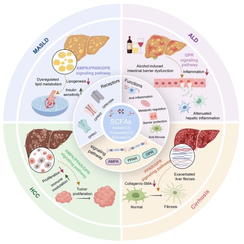
SCFAs are saturated fatty acids with a carbon chain length of 1-6 carbon atoms [24]. SCFAs are generated via glycolytic fermentation by anaerobic commensal microbes from complex carbohydrates that resist digestion in the upper gastrointestinal tract in the colon. This process converts carbohydrates into pyruvate and then generates acetate, propionate, and butyrate through microbial metabolic pathways dependent on acetyl-CoA, succinate, acrylate, and butyryl-CoA, respectively [22, 25-28]. In addition, SCFAs are mainly produced in the cecum and proximal colon, and their concentrations gradually decrease from the proximal to the distal colon as fermentation substrates are depleted [26]. Substrate source, fermentation site, gut microbiota composition, intestinal transit time, and colonic pH all influence SCFA generation [27, 28]. Butyrate is an important energy source for colonocytes, and therefore only a smaller fraction reaches the portal circulation. By contrast, acetate and propionate that are not metabolized by colonocytes can enter the liver through the portal vein [22]. Although only limited quantities of microbiota-derived butyrate transit via the portal vein under physiological conditions, butyrate can still exert marked hepatoprotective effects via dual mechanisms. On the one hand, a major proportion of butyrate acts locally within the colonic lumen and epithelium to preserve intestinal barrier integrity, regulate mucosal immune homeostasis, and restrain translocation of microbial-derived pro-inflammatory mediators, thereby mitigating hepatic injury indirectly through gut-liver axis crosstalk, independent of high portal-vein butyrate concentrations [29]. On the other hand, butyrate functions as a potent HDAC inhibitor with high intrinsic biological activity; even low portal concentrations are sufficient to trigger intracellular signaling responses in hepatocytes. Notably, many preclinical studies demonstrated that butyrate-mediated liver protection employs exogenous pharmacological butyrate supplementation, which elevates portal butyrate exposure beyond physiological microbial fermentation levels [30]. Thus, SCFAs are important mediators linking the microbiota with host physiology, and their reduced production or redistribution is associated with metabolic diseases [31, 32]. It is also worth noting that SCFAs have been investigated as biomarkers of disease-related metabolic alterations [33], including liver diseases (Figure 1).
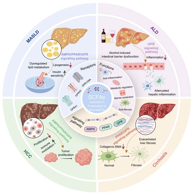
The regulatory roles of short-chain fatty acids (SCFAs, including acetate, propionate and butyrate) in the progression of chronic liver diseases. SCFAs bind to G protein-coupled receptor (GPR)41, GPR43 and GPR109A receptors and activate downstream AMPK, PPAR and PI3K/AKT signaling pathways, thereby exerting anti-inflammatory, metabolic regulation, barrier protection, anti-tumor and anti-fibrotic effects. In ALD, SCFAs alleviate alcohol-induced intestinal barrier damage and subsequent hepatic inflammatory responses. In MASLD, SCFAs restore disordered hepatic lipid metabolism. However, the effects of SCFAs are context-dependent: they suppress tumor proliferation in HCC, while they may aggravate liver fibrosis during cirrhosis progression.
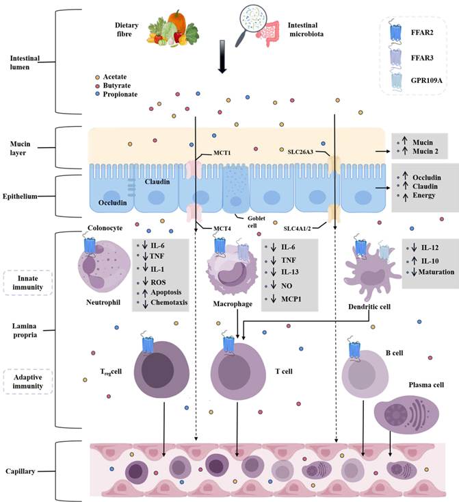
The major functions of SCFAs involve GPRs located on different cell types, particularly FFAR2 and FFAR3[34]. In addition, acetate, propionate, and butyrate differ in receptor affinity and tissue exposure. Therefore, their biological effects depend on concentration and anatomical site. In addition, cell experiments have shown that SCFAs promote intestinal barrier integrity by fueling colonocytes, increasing tight junction proteins such as occludin and claudin, stimulating mucus production, and limiting LPS production [35, 36]. This barrier regulatory effect helps explain how colonic SCFA production influences hepatic inflammation. In terms of metabolic regulation, mechanistic findings obtained from controlled in-vitro cell experiments have shown that propionate inhibits hepatic gluconeogenesis through the GPR43/AMPK related signaling pathway [37], whereas acetate and butyrate reduce lipogenesis and regulate leptin secretion [38-40]. Leptin links adipose endocrine activity, obesity-related insulin resistance, and hepatic lipid accumulation with MASLD. SCFAs can also activate AMPK, thereby connecting energy sensing with glucose and lipid metabolism. In mouse models of steatohepatitis, SCFA administration reduces hepatic steatosis [41, 42]. Overall, SCFAs regulate liver-related physiological processes by integrating intestinal barrier repair, immune modulation, and metabolic signaling (Figure 2).
Schematic of short-chain fatty acids (SCFAs)-mediated regulation of intestinal barrier and immune homeostasis. Dietary fiber fermentation by gut microbiota generates SCFAs (acetate, butyrate, propionate), which strengthen intestinal barrier function by upregulating tight junction proteins (occludin, claudin) and mucins, and fuel colonocytes. SCFAs are transported across the epithelium via MCT1/4, SLC26A3, and SLC4A1/2, and act on immune cells in the lamina propria through free fatty acid receptor (FFAR)2/3 and GPR109A. SCFAs suppress pro-inflammatory cytokine production in innate immune cells (neutrophils, macrophages, dendritic cells) and promote anti-inflammatory responses, while modulating adaptive immune cells (Treg, T, B cells) to maintain gut immune homeostasis, with systemic effects via capillary circulation.
Because SCFA generation is determined not only by substrate availability but also by host lifestyle and microbial ecology, exercise-related metabolic adaptation may interact with gut microbial ecology, intestinal permeability, energy expenditure, and insulin sensitivity, thereby indirectly affecting SCFA exposure and downstream metabolic signaling [43-48]. Mechanistically, exercise remodels multiple gastrointestinal physiological parameters to modify SCFA production. Regular moderate intensity exercise alters intestinal motility and gastrointestinal transit time, optimizes splanchnic intestinal blood perfusion (preventing the up to 50 % reduction of splanchnic blood flow induced by strenuous exercise), maintains intestinal barrier integrity, and reshapes gut microbiota composition by enriching SCFA-producing microbial taxa, collectively facilitating microbial fermentation and SCFA synthesis [49, 50]. By contrast, excessive or high intensity exercise (≥60 %-70 % V̇O2max) can trigger splanchnic hypoperfusion mediated intestinal ischemia-reperfusion injury, which reduces splanchnic blood flow by up to 50 %, acutely elevates intestinal permeability, and disrupts microbial community homeostasis, which may conversely impair gut derived SCFA generation [49]. The bidirectional splanchnic and intestinal physiological changes driven by physical activity constitute the intermediate links connecting exercise status to SCFA-mediated hepatic metabolic regulation.
3.2 Source Differences of SCFAs and Their Effects on Liver Diseases
In humans, SCFAs can broadly be divided into three sources: fermentation of carbohydrates mediated by commensal gut microbiota, exogenous dietary intake, and endogenous cellular production (Table 1). Commensal gut microbiota-derived SCFAs are mainly acetate, propionate, and butyrate [5]. These metabolites are produced locally in the colon, forming high luminal concentrations and acting on intestinal epithelial cells and immune cells before entering the portal circulation. Endogenous SCFAs are generated by host metabolic pathways and mainly participate in cellular energy and intermediary metabolism [15, 16]. Exogenous dietary SCFAs are derived from fermented foods or oral supplements, but their concentrations, absorption sites, and hepatic first-pass exposure differ from SCFAs generated by colonic fermentation [51]. Therefore, SCFAs from different sources differ in concentration, exposure site, and capacity to participate in gut-liver axis regulation [22, 52].
Summary of major SCFA sources and liver-relevant features.
| Source category | Representative origin | Main SCFAs | Site/exposure feature | Liver-relevant implication | References |
|---|---|---|---|---|---|
| Commensal gut microbiota-mediated fermentation | Anaerobic fermentation of dietary fiber in cecum/proximal colon | Acetate, propionate, butyrate | High colonic concentration; portal delivery after epithelial uptake | Barrier support, immune modulation, metabolic signaling through gut-liver axis | [6][22] [23][24]39] |
| Probiotic-associated production | Administered beneficial strains within the resident microbial ecosystem | Strain-dependent acetate/propionate/butyrate patterns | Depends on strain, colonization, diet, and formulation | May combine SCFAs generation with microbial immunomodulation; efficacy is context-dependent | [42][43] [45] |
| Exogenous dietary intake | Fermented foods or oral SCFA supplements | Variable; often acetate-rich or formulation-dependent | Absorption and hepatic first-pass exposure differ from colonic fermentation | Potential supplementation route but may lack sustained local colonic signaling | [41] |
| Endogenous cellular production | Host intermediary metabolism and cellular pathways | Small intracellular pools | Local cellular energy/intermediary metabolism | Supports energy metabolism; less directly linked to microbiota-mediated gut-liver signaling | [14][15] [46] |
SCFAs from different sources may have distinct effects on liver diseases. Commensal microbiota- or probiotic-associated SCFA production has potential advantages because different microbial strains have distinct SCFA-generating capacities and preferences [25]. For example, Bacteroidetes mainly produce acetate and propionate, whereas Firmicutes are important producers of butyrate [53]. Different microbial strains and community-specificity can shape the concentration and temporal pattern of local intestinal SCFA exposure. Additionally, clinical studies have shown that probiotic-associated SCFAs may also synergize with the immunomodulatory effects of the administered microorganisms themselves, which may explain why some probiotic or synbiotic strategies show effects beyond those of SCFA supplementation alone [54, 55]. Nevertheless, the proposed site specific and sustained advantages of microbiota-derived SCFAs remain largely mechanistic inference rather than empirically validated conclusions in liver disease relevant models. For example, pharmacokinetic studies have demonstrated that orally supplemented free SCFAs undergo rapid absorption in the upper small intestine, with limited fractions reaching the colonic lumen, whereas microbiota-driven fermentation generates SCFAs locally within the colon, establishing persistent luminal concentration gradients along the large intestine [56]. Accordingly, gradients form the hypothetical basis for spatial and temporal superiority of microbial SCFAs. However, head-to-head pharmacokinetic, pharmacodynamic, or side by side therapeutic comparison studies between microbiota produced and exogenously supplemented SCFAs are still scarce in liver disease relevant animal models. Although the advantage remains unproven under liver disease conditions, the hypothesis is widely invoked to explain divergent therapeutic outcomes observed between microbial intervention and direct SCFA supplementation, and thus merits discussion. In addition, microbial species, colonization efficiency, diet, disease stage, and delivery formulation may all influence therapeutic efficacy. Mechanistically, cell studies have shown that SCFAs link glucose and lipid metabolism: intestinal and hepatic SCFA signaling can influence gluconeogenesis, insulin sensitivity, fatty acid oxidation, de novo lipogenesis, cholesterol handling, thermogenesis, and fat storage through FFAR/GPR, AMPK, PPAR, and HDAC-related mechanisms [43, 57, 58]. These metabolic effects intersect with inflammatory and barrier pathways, providing a rationale for SCFA-targeted strategies in MASLD, ALD, cirrhosis, and HCC.
4. Functions of SCFAs in Liver Diseases
SCFAs influence liver diseases through shared pathological processes. At the intestinal level, they support epithelial energy supply, tight junction integrity, mucus production, and immune tolerance. At the portal venous and hepatic levels, they regulate LPS exposure, cytokine production, glucose and lipid metabolism, oxidative stress, HSC activation, and tumor cell survival. This section summarized the functions and effects of SCFAs in MASLD, ALD, cirrhosis, and HCC, while also integrate the background, SCFA types, mechanisms, and effects in major liver diseases (Table 2).
Classification and Mechanisms of Short-Chain Fatty Acids Regulated SCFA Production by Probiotics
| Target Liver Disease | Type of SCFA (Number of carbon atoms) | Key effects | Key findings | Key target | Study type | References |
|---|---|---|---|---|---|---|
| MASLD | Acetate (C2) | Reduce BMI, visceral fat and serum triglycerides | Reduce BMI and blood lipids | FFAR2 | In vivo mice | [54] |
| Propionate (C3) | Improve insulin resistance, regulate lipid metabolism | Improve MASLD | FFAR3 | In vitro cellular | [34] | |
| Butyrate (C4) | Improve liver steatosis, reduce cholesterol and gamma-glutamyl transferase | Delay MASLD progression | GPR109A | Clinical trial | [45] [50] | |
| MASH | Propionate (C3) | Improve liver steatosis and enhance the expression of lipoxin-related proteins | Suppress lipid synthesis genes | FFAR3 | In vitro cellular | [32] [53] |
| Butyrate (C4) | Reduce liver inflammatory infiltration and lower liver triglyceride levels | Reduce MASH inflammation | PPARα | Clinical trial | [50] [101] | |
| ALD | Acetate (C2) | Reduce endotoxin translocation and lower plasma LPS levels | Strengthen the intestinal barrier | GPR43 | Clinical trial | [59] [83] |
| Butyrate (C4) | Reduce liver injury by downregulating cleaved GSDMD and IL-1β | Improve liver injury and inflammation | HDAC | In vivo mice | [69] | |
| Mixture SCFAs (C2+C3+C4) | Improv alcohol-induced liver Steatosis and Inflammation | Improve alcoholic liver steatosis | NF-κB | Clinical trial | [61] [98] | |
| AH | Butyrate (C4) | Reduce TNF-α and IL-6 levels to alleviate liver damage | Reduce AH inflammation | PPARγ | In vivo mice | [104] [69] |
| Mixture SCFAs (C2+C3+C4) | Reduce the accumulation of triglycerides (TG) and free fatty acids (FFA) in the liver, thereby alleviating fatty degeneration | Increase liver NAD+, reduce liver fat accumulation | PPARα | In vivo mice | [103] [68] | |
| HF | Acetate (C2) | Reduce liver fibrosis and decrease α-SMA expression | Reduce the HF-induced liver fibrosis | PPARγ | In vivo mice | [100] |
| Butyrate (C4) | Reduce collagen deposition and induce apoptosis of activated HSCs | Suppress HSC activity | AMPK | In vivo mice | [78] | |
| Mixture SCFAs (C2+C3+C4) | Delay the progression of fibrosis and downregulate the release of pro-inflammatory cytokine | Delay HF progression | Intestinal Barrier-related Targets | In vivo mice | [52] [83] | |
| HCC | Butyrate (C4) | Delay disease progression and induce apoptosis in liver cancer cells | Delay HCC progression | AMPK | In vivo mice | [70] |
| Mixture SCFAs (C2+C3+C4) | Suppress tumorigenesis and improve the inflammatory tumor microenvironment | Suppress HCC development | GPR43 | In vitro cellular | [89] [100] | |
| Cirrhosis | Mixed SCFAs (C2+C3+C4) | Potentially reduce gut-derived inflammatory signaling | Fecal acetate, propionate and butyrate are reduced in cirrhosis | Gut-liver axis | Clinical trial | [106] |
| Butyrate (C4) | May mitigate barrier dysfunction and LPS translocation | Mechanistic rationale requires disease-specific clinical validation | Intestinal barrier-related targets | Clinical trial | [27][28] [106] |
4.1 SCFAs in MASLD
MASLD is a spectrum of diseases caused by the accumulation of lipids, such as triglycerides, in the liver. It can progress from metabolic dysfunction-associated steatotic liver disease to MASH and eventually develop into cirrhosis and HCC [58]. Currently, approximately 38% of adults and 7% of children and adolescents worldwide have MASLD, and the prevalence of adult MASLD is expected to exceed 55% by 2040[59]. MASLD has substantially increased the global disease burden owing to complex etiologies, including environmental factors and lifestyle. MASLD encompasses lipid accumulation and insulin resistance as its major pathophysiological mechanisms [60, 61]. Gut microbiota dysbiosis is also an important mechanism because it can alter SCFA production, intestinal permeability, bile acid metabolism, and endotoxin exposure. Studies show that gut microbiota dysbiosis is common in patients with MASLD [62].
SCFAs are involved in the pathogenesis of MASLD and also have potential therapeutic value. In particular, findings derived from noninterventional observational cohort studies revealed that circulating SCFA levels are lower in patients with early-stage MASLD relative to healthy individuals [63, 64], elevated plasma SCFA can be detected in patients with advanced MASLD-cirrhosis by metabolic disturbance, a phenomenon that is not primarily driven by boosted intestinal microbial synthesis of SCFAs [65]. Instead, it is largely attributed to cirrhotic liver injury that compromises hepatic capacity for SCFA uptake and metabolic clearance. Concurrent portosystemic shunting further enables gut-derived SCFAs to bypass hepatic first-pass metabolism and directly enter systemic blood circulation, consequently raising measurable plasma SCFA concentrations [65]. Patients with MASLD usually present metabolic abnormalities such as elevated plasma triglycerides and insulin resistance [66]. Accordingly, a finding generated from a human interventional trial demonstrate that supplementation with butyrate preparations can improve liver related indicators, including triglycerides, cholesterol, and gamma glutamyl transferase among participants presenting with hepatic steatosis and metabolic syndrome [67]. This interventional clinical evidence reflects therapeutic outcomes under controlled human trial conditions. Mechanistically, sodium butyrate can downregulate lipid synthesis-related proteins such as SREBP-1c and ACC1 and improve lipid metabolism by upregulating lipoxin-related proteins [68]. SCFAs can also regulate hepatic metabolism by inhibiting hepatic gluconeogenesis and lipogenesis, enhancing hepatic cholesterol uptake, and regulating leptin secretion [69-71]. Leptin participates in appetite, energy expenditure, and insulin sensitivity, thereby affecting metabolic disorder and the risk of MASLD. Studies conducted through animal experiments have shown that butyrate and acetate can also maintain intestinal barrier homeostasis by enhancing tight junction protein expression, alleviating high-fat diet-induced mucosal injury, and limiting LPS entry into the liver via the portal vein [72, 73]. Overall, SCFAs influence MASLD by integrating lipid metabolism, insulin resistance, intestinal permeability, and inflammatory responses [42, 74].
4.2 SCFAs in ALD
ALD is a spectrum of liver injuries induced by chronic excessive alcohol consumption, including AH, AC, and acute-on-chronic liver failure presenting as an acute episode [75]. Currently, approximately 2.4 billion people worldwide drink alcohol, making ALD an important global health problem. Approximately 2 million people die from liver disease each year, nearly half of whom are associated with alcohol-induced cirrhosis [76]. ALD pathogenesis involves multiple interrelated mechanisms. Alcohol and its metabolic byproducts, such as acetaldehyde, can directly cause hepatocellular injury, and liver injury can also be perpetuated by DAMPs. Meanwhile, alcohol affects the gut microbiota and intestinal permeability, allowing bacterial components such as LPS to enter the portal circulation, stimulate hepatic immune responses, and aggravate liver injury [77].
Studies showed that fecal SCFA concentrations and the abundance of SCFA-producing bacteria are both reduced in patients with ALD [78, 79]. Chronic alcohol intake increases plasma LPS levels and hepatic expression of pro-inflammatory cytokines such as IL-1β, IL-8, and TNF-α, whereas SCFA administration decreases the mRNA levels of these pro-inflammatory cytokines compared with alcohol exposure alone [80]. Furthermore, preclinical in-vivo evidence generated from ethanol-fed mouse animal-model experiments shows that administration of probiotic strains such as Pediococcus pentosaceus can counteract intestinal butyrate depletion, reduce circulating endotoxin levels, and ameliorate hepatic inflammation as well as alcohol-induced hepatic steatosis [81]. This mechanistic evidence is obtained under controlled experimental animal conditions. Butyrate can also indirectly inhibit alcohol-induced hepatocyte pyroptosis by reducing intestinal permeability and plasma LPS levels. After butyrate treatment, pyroptosis-related proteins such as cleaved GSDMD and cleaved caspase-1 are downregulated in mice liver tissue [82]. These findings suggested that SCFAs improve ALD mainly by enhancing the intestinal barrier, reducing endotoxin entry into the blood and portal vein, and suppressing hepatic inflammatory signaling.
4.3 MetALD
MetALD is a category within the spectrum of steatotic liver disease, characterized by hepatic steatosis, at least one cardiometabolic risk factor, and weekly alcohol consumption of 140-350 g in women and 210-420 g in men [83]. Notably, alcohol intake above the upper threshold shifts disease classification toward predominantly ALD. Importantly, the concept of MetALD emphasizes that metabolic dysfunction and alcohol exposure are not independent processes, which may jointly drive the progression of liver injury [84]. Of note, an epidemiological meta-analysis of 19 studies from 9 countries, involving 4,543,341 adults with steatotic liver disease was conducted. The meta-analysis estimated that MetALD accounts for approximately 10% of this population, and with MetALD being more common in men [85]. Beyond these epidemiological findings, the pathogenesis of MetALD relies on multiple interacting processes, including lipid overload, oxidative stress, endoplasmic reticulum stress, gut dysbiosis, intestinal barrier disruption, inflammation, and fibrogenesis.
The effects of SCFAs in MetALD depend on the molecular species, site of action, and the combined milieu created by alcohol exposure and metabolic dysfunction. First, chronic ethanol exposure reduces butyrate-producing bacteria that use the acetyl-CoA pathway, and the resulting butyrate deficiency may compromise energy supply to the colonic epithelium and protection of tight junctions [86]. On the contrary, butyrate originates exclusively from gut microbial metabolism, while acetate can be produced by gut bacteria and can also enter the circulation as an end product of ethanol oxidation. Therefore, elevated acetate may reflect either microbial fermentation or host ethanol metabolic flux [87]. Furthermore, increased microbiota-derived acetate can coincide with attenuated intestinal and hepatic injury under specific dietary interventions, suggesting that the effects of acetate are source dependent [88]. In addition, the propionate of SCFAs can suppress hepatocyte gluconeogenesis through the GPR43/AMPK signaling pathway, linking it more directly to the regulation of hepatic glucose metabolism and attenuation of MetALD progression.
4.4 Acetate in ALD and MetALD
In ALD, acetate is a major end product of ethanol clearance. Ethanol is converted to acetaldehyde through pathways involving alcohol dehydrogenase and CYP2E1 and is subsequently oxidized to acetate by ALDH2 [89]. Of note, ethanol oxidation increases the hepatic NADH/NAD+ ratio, and may be accompanied by elevated reactive oxygen species and impaired fatty acid oxidation. In turn, elevated circulating acetate often occurs in parallel with a high ethanol metabolic burden [89]. Additionally, stable-isotope tracing has shown that ethanol-derived carbon can be incorporated into acetate, acetyl-CoA, and histone acetyl groups, indicating that acetate can link ethanol metabolism to epigenetic regulation [90]. Meanwhile, within hepatocytes, ACSS2 activates acetate to acetyl-CoA and participates in acetylation-dependent regulation of lipogenesis-related genes, thereby further promoting acute alcohol-induced steatosis [91]. Nevertheless, acetate does not invariably exacerbate liver injury. For example, dietary interventions that promote Bifidobacterium-associated acetate production can ameliorate intestinal and liver injury in binge-drinking models, further indicating that microbiota derived and ethanol derived acetate have distinct biological implications [88]. Thus, the net effect of acetate in ALD depends on its source, tissue compartment, and hepatocellular utilization [92]. For example, recent studies show that the gut and liver play an important role in metabolizing acetaldehyde via the gut and liver ALDH2, generating acetate in the gut and circulation, respectively [93]. Specifically, definition of such divergent actions is essential for defining context dependent acetate functions when assessing SCFA targeted therapeutic strategies for ALD.
In MetALD, acetate is embedded in a metabolic milieu distinct from that of ALD alone, with the alcohol-derived acetate load superimposed on insulin resistance, enhanced adipose-tissue lipolysis, and hepatic lipid overload. Accordingly, acetate more closely represents a convergence point for alcohol-derived carbon flux, dietary substrates, and microbial metabolism [84]. For example, fructose can be converted to acetate by the gut microbiota, after which hepatic ACSS2 activates acetate and channels it into lipogenic pathways, indicating that diet-derived carbon flux can bypass part of the canonical pathways of acetyl-CoA generation [94]. However, acetate supply itself does not determine the level of de novo lipo-genesis. For example, in obesity, even when hepatic acetyl-CoA levels decline, lipogenic enzyme expression and compensatory pathways may still sustain lipid synthesis [95]. In parallel, acetate generated in the intestinal lumen can regulate immune and barrier functions through FFAR2 related signaling pathway and microbiota barrier interactions [34]. Overall, acetate in MetALD may amplify preexisting lipogenic and epigenetic programs while retaining local regulatory effects in the gut, the ultimate direction of acetate's action depends on the metabolic context and tissue exposure [96]. Accordingly, careful stratification of metabolic context is required to predict acetate mediated outcomes in MetALD.
Taken together, the main difference between ALD and MetALD is not the acetate molecule itself, but the metabolic context in which acetate is produced and utilized. In ALD, the acetate pool is more directly influenced by ethanol dose and drinking pattern, and its significance is primarily associated with ethanol oxidation, redox imbalance, and alcohol-induced remodeling of the gut microbiota [87]. By contrast, in MetALD, acetate reaches a liver already affected by insulin resistance and lipid accumulation. Furthermore, dietary and microbiota-derived carbon may further expand the hepatic acetate pool. Acetate is therefore more likely to function as a metabolic convergence point in MetALD than in ALD alone.
4.5 SCFAs in Cirrhosis
Cirrhosis is a common terminal stage of chronic liver disease, characterized by diffuse fibrosis, regenerative nodules, portal hypertension, intestinal barrier dysfunction, and systemic inflammation. Because cirrhosis is accompanied by marked gut microbiota dysbiosis and bacterial product translocation, SCFA disturbance is biologically closely related to disease progression and complications. Indeed, data collected from noninterventional observational patient cohorts indicated that fecal SCFA concentrations, especially acetate, propionate, and butyrate, are significantly reduced in patients with cirrhosis [97]. This observational patient data describes phenotypic correlation between diminished fecal SCFA abundance and cirrhotic disease status without experimental intervention.
Mechanistically, SCFAs may influence cirrhosis through three main pathways. First, butyrate supports colonocyte energy metabolism and barrier maintenance, thereby limiting gut-derived endotoxin exposure. Second, acetate and propionate entering the portal vein may regulate hepatic metabolism and inflammatory responses through GPR/FFAR, AMPK, and PPAR signaling. Third, SCFA related regulation of immune cytokines may influence the inflammatory microenvironment that drives fibrosis and cirrhotic decompensation. Nevertheless, current evidence more strongly supports association and mechanistic plausibility than proven clinical efficacy. Therefore, future studies should determine whether restoring SCFA levels through diet, prebiotics, probiotics, synbiotics, or targeted delivery can improve cirrhosis-related outcomes, including hepatic encephalopathy, infection risk, and portal inflammatory signaling.
4.6 SCFAs in HCC
HCC is the major type of primary liver cancer, accounting for approximately 90% of primary liver cancer cases. Primary liver cancer is the third leading cause of cancer-related death worldwide [98]. The global prevalence of HCC is approximately 15-30 cases per 100,000 people, with more than 70% of cases occurring in Asia [99]. HCC development is a multifactorial, multistep, and polygenic process, at the core of which are gene mutations and abnormal epigenetic modifications in hepatocytes under internal and external triggers. This process is accompanied by an imbalance between cell proliferation and apoptosis, abnormal angiogenesis, and microenvironmental remodeling, ultimately leading to malignant transformation [100]. Established risk factors for HCC include chronic HBV and HCV infection, cirrhosis, and MASLD [101, 102].
Recent evidence suggested that SCFAs exert context-dependent effects in HCC [103, 104]. SCFAs exert anti-tumor effects mainly through reinforcing intestinal barrier function, attenuating inflammatory signaling, and directly regulating the metabolism and apoptosis of tumor cells. For example, SCFA intake can promote CSF2 expression in HBx transgenic mouse models, reduce precancerous lesions and tumor nodules, inhibit the activity of HBx-positive HCC cells, and downregulate multiple HBx-activated oncogenic pathways [105]. SCFAs can also enhance intestinal barrier integrity [106], thereby limiting inflammation-related drivers of hepatocarcinogenesis, such as TNF-α and IL-6 signaling. In Huh7 cells, SCFAs have been reported to induce ROS production and mitochondrial membrane dysfunction. This mechanism is more consistent with inducing apoptosis or reducing cell viability than with enhancing tumor cell survival [107]. Therefore, potential pro-tumor or disease-promoting associations should be discussed separately. Increased SCFAs or their derivatives in the feces and serum of patients with MASLD-HCC may reflect altered microbial ecology, impaired hepatic metabolism, or tumor-associated metabolic reprogramming. Thus, SCFAs in HCC should not be regarded as absolutely beneficial or harmful, their effects depend on tumor stage, concentration, source, microbial environment, and host metabolic status.
5. Regulatory Mechanisms of SCFAs in Liver Diseases
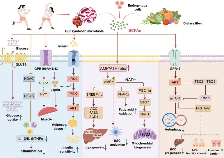
As liver disease research continues to advance, SCFAs are regarded as signaling metabolites that connect intestinal ecology with hepatic metabolism, immunity, and fibrosis. This section summarized signaling pathways associated with SCFAs and liver diseases, including AMPK, PPAR, and GPR signaling pathways (Figure 3).
The metabolic regulatory mechanism of dietary fiber-derived short-chain fatty acids (SCFAs) by intestinal microbiota. Exercise reshapes the composition of gut commensal microbiota, while dietary fiber is fermented by intestinal bacteria to produce SCFAs, which together with endogenous SCFAs exert multiple biological effects. SCFAs bind to GPR41, GPR43 and GPR109A receptors, then improve insulin sensitivity in muscle and adipose tissue by elevating GLUT4-mediated glucose uptake, promoting the secretion of GLP-1 and PYY, and inhibiting NF-κB-related inflammatory responses. In hepatocytes, elevated AMP/ATP ratio activates AMPK signaling, which suppresses lipogenic Srebp-1c/ACC/FASN pathway and enhances PPARα-dependent fatty acid oxidation. Meanwhile, AMPK upregulates the SIRT1/PGC-1α axis to boost mitochondrial biogenesis and inhibits hepatic stellate cell (HSC) activation. In addition, SCFA-triggered GPR43/AKT/mTOR cascade promotes autophagy, restrains HCC progression, reduces LPS translocation, and maintains the integrity of the intestinal barrier.
5.1 AMPK Signaling Pathway
AMPK is a key regulator of energy metabolism, consisting of a catalytic α subunit and regulatory β and γ subunits, and is expressed in the liver [108]. The activation state of AMPK is regulated by the intracellular AMP/ATP ratio. ATP inhibits AMPK, whereas AMP activates AMPK. SCFAs can promote activation of the AMPK signaling pathway by altering energy status and increasing the hepatic AMP/ATP ratio [43]. In addition, SCFAs can also indirectly regulate AMPK. For example, butyrate can inhibit HDAC, thereby upregulating AMPK-α expression [109]. In liver diseases such as MASLD-related fibrosis and HCC, AMPK activation is associated with improvement in metabolic disorders, inflammation, and fibrosis [110].
SCFA-induced activation of AMPK signaling pathway can reduce downstream lipid synthesis regulators such as SREBP-1c, FASN, ACC, and SCD1 in MASLD, thereby regulating fatty acid and cholesterol production and ameliorating MASLD-related metabolic stress [111, 112]. During hepatic fibrosis, SCFAs may inhibit HSC activation by suppressing HSC proliferation and promoting apoptosis of activated HSCs through AMPK signaling pathway [113]. In HCC models, SCFAs can activate AMPK and inhibit mTORC1, thereby reducing proliferation of HCC cells such as Huh7 cells [114]. These results suggested that AMPK links SCFA exposure with energy balance, lipid metabolism, fibrosis, and tumor cell proliferation. Of note, autophagy is a downstream cellular process associated with the AMPK-mTORC1 axis [115-117]. For example, in cells under energy stress, AMPK activation usually antagonizes mTORC1 signaling, and mTORC1 is a central regulator of autophagy related cellular homeostasis [116, 117]. In the current context of liver diseases, SCFA related AMPK activation has been associated with mTORC1 inhibition, reduced HCC cell proliferation, and apoptosis of Huh7 cells [114, 118]. Thus, through the AMPK-mTORC1 axis, SCFAs may therefore extend their regulatory influence from metabolic reprogramming to autophagy dependent cellular control, a way increasingly implicated in liver disease progression.
More specifically, the SCFA-AMPK-mTORC1-autophagy axis can be organized as a sequential regulatory module. SCFA-induced changes in cellular energy status and butyrate-mediated HDAC inhibition favor AMPK activation [42,107]. Activated AMPK suppresses mTORC1. Because active mTORC1 inhibits the autophagy-initiating kinase ULK1 under nutrient-replete conditions, AMPK mediated inhibition of mTORC1 removes this brake. In parallel, AMPK directly phosphorylates ULK1 at activating sites, thereby initiating autophagy during energy stress [113-115]. Additionally, autophagy converges with AMPK dependent suppression of HSC activation in fibrosis [111]. Meanwhile, in HCC, butyrate induced autophagy through Akt/mTOR signaling pathway has been demonstrated in HCC cells [112], while butyrate induced apoptosis has also been reported independently [116]. Thus, AMPK-mTORC1-ULK1 axis provides a mechanistic bridge from SCFA sensing to autophagy related regulation of fibro genic and tumor cell phenotypes, although direct evidence that this complete axis mediates SCFA effects in vivo remains limited.
5.2 GPR Signaling Pathway
GPCRs are membrane proteins with a seven-transmembrane helix structure and are the starting point of cellular signal transduction [119]. GPCRs can influence multiple disease categories, including cardiovascular diseases, neurological diseases, metabolic diseases, and cancer [120]. SCFA-related GPCRs include GPR43, GPR41, and GPR109A [121]. GPR43 and GPR41 are widely distributed in the liver, adipose tissue, intestinal epithelium, and immune cells, whereas GPR109A is mainly expressed in myeloid cells and hepatocytes [122]. Different SCFAs show different affinities for these GPCRs [123], allowing SCFAs to influence liver diseases through tissue-specific receptor signaling.
SCFAs can trigger GPR43 signaling, promote potassium efflux and membrane hyperpolarization, and influence NLRP3 inflammasome activity [69]. Studies conducted through animal experiments have shown that SCFAs can also promote intestinal gluconeogenesis through the GPR41-mediated gut-brain neural axis, thereby improving insulin resistance in MASLD [124][104]. Butyrate reduces hepatic inflammatory cytokines by activating GPR109A signaling pathway, thereby alleviating MASLD-related symptoms [125]. In ALD, alcohol-induced intestinal barrier disruption and endotoxemia can be alleviated by SCFAs through GPR43-mediated enhancement of barrier function [126] and GPR109A-mediated reduction of endotoxin translocation. In HCC, SCFAs can stimulate GLP-1 release by activating GPR43[127], whereas GLP-1-related signaling is associated with reduced hepatic lipid content and inhibition of HCC development [128].
5.3 PPAR Signaling Pathway
PPARs are ligand-dependent nuclear transcription factors. The PPAR family comprises three subtypes: PPARα, PPARγ, and PPARβ/δ. Each subtype has specific biological activity according to its ligands and tissue localization [129-131], and regulates fatty acid metabolism, cell differentiation, cell migration, and inflammatory responses [132].
SCFAs can act as natural ligands or indirect regulators of PPARs and influence liver diseases by activating PPAR signaling pathway [133]. In MASLD, SCFAs promote fatty acid β-oxidation by activating PPARα, thereby reducing hepatic triglyceride synthesis and accumulation. In a high-fat diet-induced MASLD model, butyrate supplementation can increase hepatic PPARα expression, increase fatty acid oxidation-related genes, reduce hepatic triglyceride content, and improve hepatic steatosis [134]. Additionally, SCFAs can also suppress hepatic inflammatory cytokines such as TNF-α by activating PPARγ signaling pathway, thereby alleviating hepatic inflammatory injury [135]. In ALD, SCFAs reduce TG and FFA levels by activating PPARα signaling pathway, promote fatty acid metabolism, and reduce hepatic lipid accumulation. PPARα activation can increase hepatic NAD+ levels and accelerate alcohol clearance [136]. SCFAs can also reduce TNF-α and IL-6 release through PPARγ signaling pathway, thereby suppressing inflammatory responses and alleviating AH and liver injury [137]. In fibrosis and HCC-related contexts, PPARγ activation may inhibit HSC activation and proliferation, increase aquaporin 3 expression, reduce AP-1 levels, and suppress collagen synthesis [138].
Overall, the AMPK, GPR, and PPAR signaling pathway operate through interlinked mechanisms. GPR activation at the intestinal and immune cell levels can alter endocrine signaling, barrier integrity, and inflammatory status. AMPK senses cellular energy stress and coordinates glucose and lipid metabolism. PPARs translate metabolic signals into transcriptional programs that regulate fatty acid oxidation, inflammation, and fibrosis. Butyrate-mediated inhibition of HDACs further links SCFAs with epigenetic regulation. These signaling pathway converge on several liver-related endpoints, including reduced LPS exposure, improved lipid handling, decreased inflammatory cytokine production, and regulation of HSC and tumor cell behavior. Collectively, this integrated perspective helps explain why the same SCFA may exhibit beneficial, neutral, or context-dependent effects depending on disease stage, tissue compartment, and delivery strategy.
6. Clinical Applications of SCFAs in Liver Diseases
As key metabolites of the gut microbiota, SCFAs have received broad attention in recent years. Changes in SCFA levels and composition are closely associated with multiple diseases, supporting their value as potential noninvasive biomarkers [23]. However, interpretation of SCFAs requires consideration of sample type and disease stage. Altered SCFA generation can reflect gut microbiota dysbiosis, whereas elevated circulating SCFAs in liver diseases may also indicate impaired hepatic metabolism or altered clearance and do not necessarily represent increased colonic production. Plasma SCFA analysis in patients with MASLD shows significant increases in several SCFAs, including propionate, formate, valerate, and α-methyl butyrate, which are positively correlated with disease severity [65]. By contrast, fecal SCFAs, especially acetate, propionate, and butyrate, are significantly reduced in patients with cirrhosis [97]. Such inconsistent findings between plasma and fecal compartments underscore that SCFA measurements are highly sample dependent, rather than universally comparable across difference sample types. Fecal and circulating SCFA concentrations may display divergent or even opposing associations with liver-related phenotypes, which arises from the combined effects of microbial production, intestinal absorption, host tissue utilization, and hepatic clearance [139]. Fecal SCFA concentrations largely reflect net residual luminal output after colonic microbial fermentation, and are influenced by microbiota composition, fermentation substrates, and intestinal transit time; hence they serve as a partial proxy for in situ colonic SCFA production [140]. Following microbial production within the gut lumen, SCFAs are absorbed across the intestinal epithelium via monocarboxylate transporters, and absorption efficiency is modulated by gut barrier integrity and transporter-protein expression status [141]. A large proportion of absorbed butyrate is rapidly utilized as the primary energy fuel for colonocytes before entering portal venous circulation. Under physiological conditions, the liver performs substantial first-pass extraction and metabolic degradation of portal-delivered SCFAs and restricts their spill-over into the systemic bloodstream. In advanced liver disease such as MASLD-related cirrhosis, hepatocyte dysfunction together with portosystemic shunting markedly impairs hepatic SCFA-clearing capacity [65]. Consequently, circulating SCFA concentrations can increase even when colonic microbial SCFA production (reflected by faecal measurements) declines, creating this paradoxical biomarker discrepancy. Collectively, these findings suggest that SCFAs may serve as useful candidate biomarkers, but fecal, portal venous, hepatic, and systemic circulatory compartments should not be interpreted interchangeably. In addition, SCFAs are also associated with obesity [44] and T2DM [142], further supporting their connection with metabolic liver disease.
Targeted regulation of SCFAs can improve the progression of multiple diseases. For example, sodium butyrate has been shown to increase clinical remission rates in patients with ulcerative colitis and has good safety [143]. Meanwhile, results derived from a human interventional trial indicated that oral microencapsulated sodium butyrate (3 capsules/day, 1800 mg/day) can improve quality of life in patients with IBD. Compared with conventional oral sodium butyrate formulations, this microencapsulated delivery strategy achieves enhanced bioavailability in human subjects [144]. From the perspective of clinical adherence, microencapsulation may also improve tolerability by reducing odor, taste, rapid absorption, and gastrointestinal discomfort, although liver disease-specific adherence data remain limited. Targeted SCFA regulation also shows promise in MASLD/MASH; for example, self-assembling polymeric SCFA prodrugs can alleviate steatohepatitis and hepatic fibrosis [145]. Liposome-encapsulated acetate nanoparticles used for colorectal cancer can significantly reduce tumor volume (LITA-CAN: 106.8 ± 113.3 mm²; control: 269.7 ± 87.5 mm²; P < 0.05) [146]. Nevertheless, it is worth noting that despite promising advances in formulation design, several liver patient specific safety concerns still warrant careful consideration. Beyond the gastrointestinal tolerability issues described above, sodium salt based SCFA supplements impose extra sodium load, which carries particular clinical relevance for decompensated cirrhosis patients requiring sodium restricted diets [139, 147]. Moreover, pronounced inter-individual microbiome variability modulates endogenous SCFA-producing capacity, contributing to heterogeneous therapeutic responses to microbiota-targeted interventions [139]. Importantly, human safety data for exogenous SCFA supplementation in advanced cirrhosis and HCC remain scarce, given context dependent bioactivity demonstrated in pre-clinical studies, favorable outcomes observed in mild liver disease cannot be simply generalized to late-stage liver disorders.
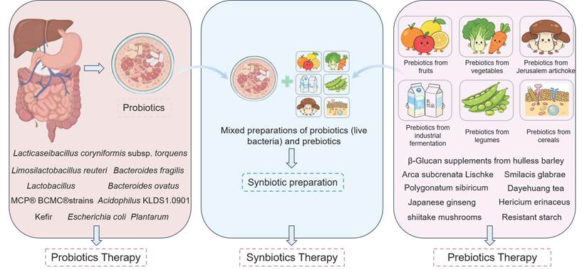
At present, SCFAs are not yet widely used in clinical practice for liver diseases. Existing strategies mainly include prebiotics such as inulin and fructo-oligosaccharides, high-fiber diets, probiotic or synbiotic modulation, and oral SCFA supplementation. Synbiotic therapy refers to the combined use of probiotics and prebiotics and has been widely explored as a strategy to restore gut-liver axis function (Figure 4). Even with promising findings derived from IBD and T2DM research, substantial practical limitations remain for the direct extrapolation of these results to liver disease related investigations. Specifically, chief limitations include poor oral bioavailability and rapid splanchnic clearance of free SCFAs: human stable isotope data show only 36 % of colonic acetate, 9.2 % of propionate, and merely 2.4 % of butyrate can reach systemic circulation, with extensive first pass hepatic extraction especially for butyrate, orally administered free SCFAs are rapidly metabolized in the upper gastrointestinal tract, and circulating concentrations return to baseline within approximately 120 min after oral dosing [22]. Additionally, there is a lack of validated compartment specific biomarkers [65, 74]. Thus, to overcome the limitations and advance clinical application of SCFAs in liver diseases, further clinical translation requires standardization of dose, formulation, patient selection, disease stage stratification, biomarker monitoring, and long-term safety assessment.
The microecological regulation plan for the liver and intestines. Specifically presents the strains and related products corresponding to the probiotic therapy, various substances corresponding to the prebiotic (polysaccharide) therapy, and the combined therapy as the three types of intervention methods.
7. Differences and Similarities of Acetate, Propionate, and Butyrate-mediated Functions in Liver Diseases
Acetate, propionate, and butyrate share several roles in host energy metabolism, microbial metabolite sensing, and gut-liver communication. However, the three SCFAs differ in absorption, tissue exposure, receptor preference, and metabolic fate. These differences prevent acetate, propionate, and butyrate from being treated as equivalent metabolites. Within this section, the distinct features of the three SCFAs are first described, followed by the shared biological properties of these metabolites, to clarify their respective roles in liver disease (Table 3).
Comparison of acetate, propionate, and butyrate: principal sources, exposure patterns, mechanistic features, and evidence relevant to liver disease.
| SCFA species | Major source | Exposure pattern | Key feature | Liver disease relevance | References |
|---|---|---|---|---|---|
| Acetate(C2) | Microbial fermentation; Ethanol oxidation | High portal and systemic exposure | ACSS2-linked acetyl-CoA and acetylation | Source-dependent; key to ALD/MetALD carbon flux and lipogenesis | [22][91] [92] |
| Propionate(C3) | Microbial fermentation | Marked hepatic first pass uptake | Predominantly hepatic metabolic signal | Closely linked to hepatic glucose/lipid regulation; limited human evidence | [28][37] [151] |
| Butyrate(C4) | Microbial fermentation | Colonocyte use; Low portal exposure | Colonocyte fuel; barrier and immune regulation | Gut-liver protection; Preclinical support in MASLD/ALD | [37][57] [88][154] |
7.1 Differences among Acetate, Propionate, and Butyrate
Acetate, propionate, and butyrate differ substantially in their tissue exposure patterns, receptor preferences, and predominant functions in liver disease. Acetate is generally the most abundant SCFA in the colon, propionate is extensively taken up by the liver after entering the portal vein, and most butyrate is oxidized by colonocytes before reaching the portal circulation [148]. Consequently, acetate is more likely to serve as a systemic circulating carbon source, propionate produces a more prominent hepatic first-pass metabolic signal, and butyrate acts predominantly at the local intestinal level [28]. Furthermore, receptor preferences also differ: acetate and propionate generally activate FFAR2/GPR43, whereas propionate and butyrate signal through FFAR3/GPR41.
Of note, butyrate can also activate GPR109A [34]. Among the three SCFAs, acetate is distinguished by its diverse origins and its ability to enter acetyl-CoA metabolism directly through ACSS2, and may therefore provide an additional carbon source for lipid synthesis during alcohol exposure or excessive fructose intake [94]. Nevertheless, acetate does not necessarily promote steatosis, because its net effect is also shaped by energy balance, tissue uptake, and compensatory metabolic pathways [95]. By contrast, acetate displays lower hepatic first-pass uptake than propionate, which is therefore more closely linked to the regulation of hepatic glucose and lipid metabolism, with relatively limited systemic exposure [148]. Specifically, propionate can suppress hepatocyte gluconeogenesis through the GPR43/CaMKKβ/AMPK signaling pathway, although evidence remains insufficient to establish that propionate supplementation consistently reduces liver fat in humans [37]. In comparison, butyrate is an important oxidative substrate for colonocytes and is more strongly associated with maintenance of the intestinal barrier, mucosal immunity, and epigenetic regulation [149]. Moreover, because colonocytes consume most butyrate, the proportion reaching the liver under physiological conditions is lower than that of acetate and propionate, meaning its benefits in liver disease are thought to be largely indirectly mediated through the gut-liver axis [150]. Besides, in obesity or MASLD animal models, butyrate can improve hepatic mitochondrial function and steatosis [151]. Of note, in ALD, chronic ethanol exposure reduces butyrate producing bacteria and the associated biosynthetic pathways. Consequently, butyrate deficiency initially manifests as impaired protection of the intestinal barrier [152]. Butyrate supplementation can reduce endotoxemia and inflammatory pyroptosis, indicating that its principal site of action lies along the intestinal barrier inflammation axis [82]. By contrast, butyrate primarily targets intestinal barrier and inflammatory pathways, whereas acetate more prominently conveys the carbon flux and acetylation signals generated by ethanol metabolism and participates in alcohol-induced remodeling of the gut microbiota [90]. In MetALD, the barrier protective effects of butyrate may complement the metabolic effects of acetate and propionate.
Overall, acetate, propionate, and butyrate exhibit critical differences and should not be regarded as interchangeable therapeutic agents or biomarkers. Specifically, principal differences among these three SCFAs lie in systemic carbon flux, hepatic first-pass metabolism, and local intestinal barrier and immune regulation. Among acetate, propionate, and butyrate, acetate exhibits the greatest metabolic plasticity, propionate is more closely aligned with the regulation of hepatic glucose and lipid metabolism, and butyrate is more strongly oriented toward protection of the gut-liver axis. Accordingly, evaluation of these three SCFAs should distinguish fecal abundance, portal-vein exposure, hepatic uptake, and systemic circulating levels rather than inferring overall function from the concentration measured in a single compartment. Future comparative studies should also standardize dose and delivery methods and stratify analyses according to liver disease etiology and stage.
7.2 Similarities among Acetate, Propionate, and Butyrate
Acetate, propionate, and butyrate exhibit distinct biological characteristics, as outlined above, yet these three SCFA species share multiple biological features relevant to liver disease. Acetate, propionate, and butyrate can be generated by colonic microbial fermentation of undigested carbohydrates and subsequently absorbed and metabolized by the host. Of note, a stable isotope study in healthy humans demonstrated that each colonically administered SCFA underwent systemic recovery and oxidation, indicating that all three SCFAs participate in host carbon and energy metabolism despite differences in systemic availability [56]. In addition, acetate, propionate, and butyrate can activate FFAR2/GPR43, providing a shared receptor-mediated route for sensing microbial metabolites, although their potencies and additional receptor preferences differ [153]. Notably, a scientific study indicated that all three types of SCFAs may support intestinal regulatory immune responses: individual administration of acetate, propionate, or butyrate increased colonic Foxp3-positive regulatory T-cell numbers in germ-free mice, demonstrating a common capacity to regulate mucosal immune homeostasis [154]. Mucosal immune homeostasis documented in germ free mice provides a mechanistic rationale for investigating shared effects along the gut-liver axis, yet cannot confirm equivalent suppression of hepatic inflammation by all three SCFAs, and these immune findings do not fully represent their metabolic effects. In detail, metabolic effects can converge on reduced hepatic lipid accumulation. For example, a study has shown that in high-fat-diet-fed mice, dietary supplementation with these SCFAs promoted a PPAR-γ-dependent shift from lipogenesis toward fatty acid oxidation, involving increased UCP2 expression, a higher AMP/ATP ratio, and AMPK activation in liver and adipose tissue [155]. Thus, the similarities extend beyond their chemical classification to overlapping receptor signaling, intestinal immune regulation, and metabolic pathways relevant to steatotic liver disease.
8. SCFAs other than Acetate, Propionate, and Butyrate in liver disease
While acetate, propionate, and butyrate represent over 95% of the SCFAs in the human body, "minor" SCFAs and branched-chain fatty acids (BCFAs) play distinct, highly significant roles in chronic liver conditions like MASLD and cirrhosis. Minor SCFAs differ from acetate, propionate, and butyrate in both carbon skeleton and metabolic origin. Common straight-chain minor SCFAs include valerate and hexanoate, whereas BCFAs include isobutyrate, isovalerate, and 2-methylbutyrate [14]. In addition, BCFAs are generated primarily through microbial degradation of valine, leucine, and isoleucine and therefore reflect the extent of proteolytic fermentation more strongly than straight-chain SCFAs.
Interpretation of minor SCFAs is highly dependent on the biological compartment sampled. Specifically, fecal concentrations of minor SCFAs primarily reflect luminal substrate availability, microbial synthesis, epithelial uptake, and intestinal transport, whereas plasma concentrations additionally integrate portal absorption, hepatic clearance, host synthesis, and renal excretion. Notably, between feces and plasma, differences in minor SCFA concentrations are particularly important in advanced liver disease, where reduced hepatic uptake and portosystemic shunting can alter circulating SCFA concentrations. Therefore, elevated plasma concentrations of minor SCFAs cannot be directly equated with enhanced beneficial fermentation [156]. Accordingly, simultaneous comparison of fecal, portal venous, liver tissue, and systemic circulation samples is more likely to clarify the sources, organ clearance, and direction of action of minor SCFAs [65].
Among minor straight-chain SCFAs, valerate currently has relatively direct mechanistic evidence in the liver. In preclinical HCC models related to NAFLD/MASLD, valerate suppresses oncogenic Rho-GTPase signaling pathway via GPR41/FFAR3 and GPR43/FFAR2 expressed in HCC cells while improving intestinal barrier integrity [157]. Remarkably, for minor SCFAs, hexanoate has not yet shown the same protective effect as propionate in diet-induced steatohepatitis models [158]. Accordingly, hexanoate is currently better regarded as a metabolic or microbial signature, and its role in liver disease requires further validation.
Although BCFAs are less abundant, they may connect proteolytic fermentation with hepatic metabolic dysregulation. For instance, a study has shown that diverse dietary proteins can increase microbiota-derived BCFAs and exacerbate hepatic insulin resistance through mTORC1 signaling pathway [159]. Moreover, the BCFAs, isobutyrate and isovalerate, can exert metabolic effects by alter glucose and lipid metabolism in adipocytes, suggesting that they may influence hepatic lipid input through adipose-liver crosstalk [160]. However, the metabolic effects are also influenced by diet, obesity status, microbiota composition, and dose, and no clinical trial has yet demonstrated that BCFAs can directly treat MASLD or MetALD [161]. Additionally, elevated circulating isobutyrate and isovalerate may also reflect reduced hepatic clearance, portosystemic shunting, or altered proteolytic fermentation in cirrhosis.
Overall, minor SCFAs and BCFAs differ substantially in their origins, receptor preferences, and tissue exposure and therefore cannot be interpreted using a uniform framework. Specifically, for minor SCFAs, valerate more closely resembles a candidate mediator with receptor-signaling potential, whereas hexanoate and BCFAs are currently better regarded as indicators of the metabolic milieu or microbial function. Furthermore, hepatic uptake, portosystemic shunting, and renal excretion can alter circulating SCFA concentrations in advanced liver disease. Thus, the concentrations of minor SCFAs and BCFAs will be an important consideration in the treatment of these diseases.
9. Future Perspectives
As research continues to advance, the role of SCFAs in liver diseases has received increasing attention. As signaling molecules mediating interactions between the gut microbiota and the host, SCFAs have been shown to regulate metabolic, inflammatory, barrier, and fibrotic processes. This section discussed the potential of SCFA biotherapies, traditional Chinese medicine-derived active components that regulate SCFA generation, and targeted delivery strategies.
In recent years, research on SCFA-related biotherapies has continued to increase. A recent study showed that oral administration of an SCFA-producing biotherapy increased SCFA levels in adults with T1D, altered the gut microbiota, and induced immunomodulatory changes, thereby delaying T1D progression [162]. This study used modified polysaccharide biomolecules to deliver acetate and butyrate (HAMSAB). Although this study was not a liver disease trial, its biological rationale may be relevant to liver diseases because diabetes, gut microbiota dysbiosis, LPS release, and portal inflammatory signaling are shared components of the gut-liver axis [163]. SCFAs can counteract LPS-induced inflammation and tissue injury by enhancing intestinal barrier integrity, regulating the gut microbiota, and suppressing inflammatory pathways [164]. Therefore, SCFA biotherapy should be regarded more as a hypothesis-generating direction for future liver disease research than as an established clinical treatment. Similarly, oral APS can increase the abundance of SCFA-producing gut bacteria and elevate SCFA levels in an adenine-induced kidney injury model [165]. The traditional Chinese medicine formula JCM-16021 can enrich butyrate-producing commensal bacteria and alleviate IBS-D [166]. These non-hepatic studies support a shared mechanism involving gut microbiota remodeling, SCFA restoration, epithelial barrier repair, and inflammation control. Because the liver and gut are connected through the gut-liver axis [7], future studies should test whether traditional Chinese medicine-derived active components capable of regulating SCFAs can be translated into liver disease-specific interventions. Targeted delivery is also an important direction. Polyethylene glycol-poly (vinyl ester) copolymers conjugated with propionate or butyrate can form self-assembled nanomicelles (NanoPA/NanoBA), maintain activity in the liver for more than 72 h after oral administration, and exhibit greater stability than free SCFAs [145]. Butyrate-loaded chitosan/hyaluronan nanoparticles can inhibit neutrophil ROS release and show sustained anti-inflammatory effects in IBD models [167]. These polymers modified SCFA systems may protect SCFA activity and prolong duration of action, but their liver specific efficacy, safety, and pharmacokinetics require further validation. Of note, based on the promising preclinical formulation findings, future translational exploration for liver diseases should fully exploit the diversity of gut-liver-axis-oriented interventions, while accounting for practical constraints facing clinical translation. While observations from IBD and T1D illustrated the conceptual promise of SCFA centered therapeutic strategies, pathophysiological differences across disease contexts caution against straightforward extrapolation to hepatic disorders. In addition, apart from optimized delivery platforms, combinatorial regimens merging formulation-based SCFA intervention with microbiota-modulating approaches deserve greater attention. Meanwhile, consideration of inter-individual variability, including baseline gut microbial profiles and distinct liver disease phenotypes, will also help unpack heterogeneous treatment responses and guide rational intervention design.
Based on the content of this section, SCFAs show important potential in maintaining liver health. Future work may develop SCFA-targeted delivery systems, screen traditional Chinese medicine-derived components that can stably regulate SCFA-producing microbiota, and design formulations capable of delivering SCFAs to intestinal or hepatic compartments with controlled kinetics. Collectively, these technical and pharmacological advances may open new avenues for comprehensive liver-disease interventions, yet real-world clinical benefits remain to be validated in dedicated investigations.
10. Conclusions
This review focuses on SCFAs such as acetate, propionate, and butyrate, and systematically summarizes their mechanisms, functions, and application prospects in liver diseases. SCFAs exert regulatory effects on MASLD, ALD, cirrhosis, and HCC through interrelated mechanisms involving intestinal barrier integrity, endotoxin translocation, inflammatory responses, glucose and lipid metabolism, HSC activity, and tumor cell proliferation or apoptosis. AMPK, GPR, and PPAR signaling pathways form a core regulatory network rather than independent mechanisms. Compared with exogenous dietary intake or endogenous cellular production, commensal microbiota- or probiotic-associated SCFA generation may provide sustained local exposure and coordinated microbiota-host signaling, but this advantage is context-dependent and should not be interpreted as universally superior. SCFAs also show potential as biomarkers and therapeutic targets. Future studies should focus on standardizing SCFA measurement in different biological compartments, targeted delivery systems, rational probiotic/prebiotic/synbiotic design, and clinical trials that define which patients, disease stages, and formulations are most likely to benefit.
Abbreviations
ACC1: acetyl-CoA carboxylase 1; AH: alcoholic hepatitis; ALD: alcohol-associated liver disease; AMPK: adenosine monophosphate-activated protein kinase; BMI: body mass index; FFAR: free fatty acid receptor; FFA: free fatty acid; GPR: G protein-coupled receptor; GSDMD: gasdermin D; HCC: hepatocellular carcinoma; HDAC: histone deacetylase; HF: hepatic fibrosis; HSC: hepatic stellate cell; IL: interleukin; LPS: lipopolysaccharide; MASLD: metabolic dysfunction-associated steatotic liver disease; MetALD: metabolic dysfunction and alcohol-associated liver disease; MASH: metabolic dysfunction-associated steatohepatitis; mTORC1: mammalian target of rapamycin complex 1; NF-κB: nuclear factor kappa B; PPAR: peroxisome proliferator-activated receptor; ROS: reactive oxygen species; SREBP-1c: sterol regulatory element-binding protein 1c; TG: triglyceride; TNF-α: tumor necrosis factor-α.
Acknowledgements
This work was supported by the National Natural Science Foundation of China (82373932); Natural Science Foundation of Anhui Province (2408085QH242, 2508085J050); Anhui Outstanding Young Teachers Cultivation Program (YQZD2023023); Anhui Province University Outstanding Youth Research Project (2024AH020006); Open Fund of the Joint Research Center for Occupational Medicine and Health, Institute of Major Health at the National Science Center for Comprehensive Science in Hefei (OMH-2023-07); Anhui Provincial Higher Education Institution Key Project of Natural Science Research (2023AH40391,2023AH10084); School-Level Tutoring & Ideological and Political Studio (0601067124); School-Level College Students' Innovative Training Program (0601067120, 0601067122); 2024 Provincial Quality Project - School-Enterprise Cooperative Practical Education Base (0601067123).
Author Contributions
Shanshan Ge: Conceptualization; writing - original draft; writing - review and editing; visualization; Haoyu Yu: Conceptualization; writing - original draft; writing - review and editing; Ruoyu Yang: Conceptualization; writing - original draft; writing - review and editing; Xinyu Lin: Conceptualization; writing - original draft; writing - review and editing; Pei Pan: Conceptualization; writing - original draft; writing - review and editing; Wenyong Wu: Conceptualization; writing - original draft; writing - review and editing; Tao Xu: Conceptualization; supervision; Funding acquisition; writing - original draft; writing - review and editing.
Competing Interests
The authors have declared that no competing interest exists.
References
1. Han H, Desert R, Das S, Song Z, Athavale D, Ge X. et al. Danger signals in liver injury and restoration of homeostasis. Journal of hepatology. 2020;73:933-51
2. Friedman SL. Mechanisms of hepatic fibrogenesis. Gastroenterology. 2008;134:1655-69
3. Devarbhavi H, Asrani SK, Arab JP, Nartey YA, Pose E, Kamath PS. Global burden of liver disease: 2023 update. Journal of hepatology. 2023;79:516-37
4. Wong MCS, Huang JLW, George J, Huang J, Leung C, Eslam M. et al. The changing epidemiology of liver diseases in the Asia-Pacific region. Nature reviews Gastroenterology & hepatology. 2019;16:57-73
5. Hsu CL, Schnabl B. The gut-liver axis and gut microbiota in health and liver disease. Nature reviews Microbiology. 2023;21:719-33
6. Tripathi A, Debelius J, Brenner DA, Karin M, Loomba R, Schnabl B. et al. The gut-liver axis and the intersection with the microbiome. Nature reviews Gastroenterology & hepatology. 2018;15:397-411
7. Albillos A, de Gottardi A, Rescigno M. The gut-liver axis in liver disease: Pathophysiological basis for therapy. Journal of hepatology. 2020;72:558-77
8. Seitz HK, Bataller R, Cortez-Pinto H, Gao B, Gual A, Lackner C. et al. Alcoholic liver disease. Nature reviews Disease primers. 2018;4:17
9. De Boer YS, Van Nieuwkerk CM, Witte BI, Mulder CJ, Bouma G, Bloemena E. Assessment of the histopathological key features in autoimmune hepatitis. Histopathology. 2015;66:351-62
10. Ali SMJ, Lai M. Metabolic Dysfunction-Associated Steatotic Liver Disease. Annals of internal medicine. 2025;178:Itc1-itc16
11. Gao B, Arab JP, Liangpunsakul S, Ding WX, Szabo G, Mehal W. et al. Metabolic dysfunction and alcohol-associated liver disease (MetALD). eGastroenterology. 2025;3:e100319
12. Horn P, Tacke F. Key takeaways from the updated multidisciplinary European MASLD guidelines. eGastroenterology. 2025;3:e100196
13. Sun YK, Zhang YF, Xie L, Rong F, Zhu XY, Xie J. et al. Progress in the treatment of drug-induced liver injury with natural products. Pharmacological research. 2022;183:106361
14. Rios-Covian D, González S, Nogacka AM, Arboleya S, Salazar N, Gueimonde M. et al. An Overview on Fecal Branched Short-Chain Fatty Acids Along Human Life and as Related With Body Mass Index: Associated Dietary and Anthropometric Factors. Frontiers in microbiology. 2020;11:973
15. Xi M, Yan Y, Duan S, Li T, Szeto IM, Zhao A. Short-chain fatty acids in breast milk and their relationship with the infant gut microbiota. Frontiers in microbiology. 2024;15:1356462
16. Bellanti F, Lo Buglio A, Vendemiale G. Hepatic Mitochondria-Gut Microbiota Interactions in Metabolism-Associated Fatty Liver Disease. Metabolites. 2023;13:322
17. Facchin S, Bertin L, Bonazzi E, Lorenzon G, De Barba C, Barberio B. et al. Short-Chain Fatty Acids and Human Health: From Metabolic Pathways to Current Therapeutic Implications. Life (Basel, Switzerland). 2024;14:559
18. Aziz T, Naveed M, Sarwar A, Makhdoom SI, Mughal MS, Ali U. et al. Functional Annotation of Lactiplantibacillus plantarum 13-3 as a Potential Starter Probiotic Involved in the Food Safety of Fermented Products. Molecules (Basel, Switzerland). 2022;27:5399
19. Ramos Meyers G, Samouda H, Bohn T. Short Chain Fatty Acid Metabolism in Relation to Gut Microbiota and Genetic Variability. Nutrients. 2022;14:5361
20. Nami Y, Shaghaghi Ranjbar M, Modarres Aval M, Haghshenas B. Harnessing Lactobacillus-derived SCFAs for food and health: Pathways, genes, and functional implications. Current research in microbial sciences. 2025;9:100496
21. Yan M, Pan D, Chen L, Pang J, Shao Y, Cheng Q. et al. Role of intestinal SCFAs homeostasis in the hepatoprotective effect of Clostridium butyricum in T2DM. NPJ biofilms and microbiomes. 2025;11:206
22. Koh A, De Vadder F, Kovatcheva-Datchary P, Bäckhed F. From Dietary Fiber to Host Physiology: Short-Chain Fatty Acids as Key Bacterial Metabolites. Cell. 2016;165:1332-45
23. Mann ER, Lam YK, Uhlig HH. Short-chain fatty acids: linking diet, the microbiome and immunity. Nature reviews Immunology. 2024;24:577-95
24. van der Hee B, Wells JM. Microbial Regulation of Host Physiology by Short-chain Fatty Acids. Trends in microbiology. 2021;29:700-12
25. Deleu S, Machiels K, Raes J, Verbeke K, Vermeire S. Short chain fatty acids and its producing organisms: An overlooked therapy for IBD? EBioMedicine. 2021;66:103293
26. Hanus M, Parada-Venegas D, Landskron G, Wielandt AM, Hurtado C, Alvarez K. et al. Immune System, Microbiota, and Microbial Metabolites: The Unresolved Triad in Colorectal Cancer Microenvironment. Frontiers in immunology. 2021;12:612826
27. McLoughlin RF, Berthon BS, Jensen ME, Baines KJ, Wood LG. Short-chain fatty acids, prebiotics, synbiotics, and systemic inflammation: a systematic review and meta-analysis. The American journal of clinical nutrition. 2017;106:930-45
28. Wong JM, de Souza R, Kendall CW, Emam A, Jenkins DJ. Colonic health: fermentation and short chain fatty acids. Journal of clinical gastroenterology. 2006;40:235-43
29. Canfora EE, Meex RCR, Venema K, Blaak EE. Gut microbial metabolites in obesity, NAFLD and T2DM. Nature reviews Endocrinology. 2019;15:261-73
30. Amiri P, Arefhosseini S, Bakhshimoghaddam F, Jamshidi Gurvan H, Hosseini SA. Mechanistic insights into the pleiotropic effects of butyrate as a potential therapeutic agent on NAFLD management: A systematic review. Frontiers in nutrition. 2022;9:1037696
31. Qin J, Li Y, Cai Z, Li S, Zhu J, Zhang F. et al. A metagenome-wide association study of gut microbiota in type 2 diabetes. Nature. 2012;490:55-60
32. Forslund K, Hildebrand F, Nielsen T, Falony G, Le Chatelier E, Sunagawa S. et al. Disentangling type 2 diabetes and metformin treatment signatures in the human gut microbiota. Nature. 2015;528:262-266
33. Du Y, He C, An Y, Huang Y, Zhang H, Fu W. et al. The Role of Short Chain Fatty Acids in Inflammation and Body Health. International journal of molecular sciences. 2024;25:7379
34. Kimura I, Ichimura A, Ohue-Kitano R, Igarashi M. Free Fatty Acid Receptors in Health and Disease. Physiological reviews. 2020;100:171-210
35. Nagpal R, Wang S, Ahmadi S, Hayes J, Gagliano J, Subashchandrabose S. et al. Human-origin probiotic cocktail increases short-chain fatty acid production via modulation of mice and human gut microbiome. Scientific reports. 2018;8:12649
36. Liu P, Wang Y, Yang G, Zhang Q, Meng L, Xin Y. et al. The role of short-chain fatty acids in intestinal barrier function, inflammation, oxidative stress, and colonic carcinogenesis. Pharmacological research. 2021;165:105420
37. Yoshida H, Ishii M, Akagawa M. Propionate suppresses hepatic gluconeogenesis via GPR43/AMPK signaling pathway. Archives of biochemistry and biophysics. 2019;672:108057
38. Xiong Y, Miyamoto N, Shibata K, Valasek MA, Motoike T, Kedzierski RM. et al. Short-chain fatty acids stimulate leptin production in adipocytes through the G protein-coupled receptor GPR41. Proceedings of the National Academy of Sciences of the United States of America. 2004;101:1045-50
39. Soliman MM, Ahmed MM, Salah-Eldin AE, Abdel-Aal AA. Butyrate regulates leptin expression through different signaling pathways in adipocytes. Journal of veterinary science. 2011;12:319-23
40. Weitkunat K, Schumann S, Nickel D, Kappo KA, Petzke KJ, Kipp AP. et al. Importance of propionate for the repression of hepatic lipogenesis and improvement of insulin sensitivity in high-fat diet-induced obesity. Molecular nutrition & food research. 2016;60:2611-21
41. Deng M, Qu F, Chen L, Liu C, Zhang M, Ren F. et al. SCFAs alleviated steatosis and inflammation in mice with NASH induced by MCD. The Journal of endocrinology. 2020;245:425-37
42. Shimizu H, Masujima Y, Ushiroda C, Mizushima R, Taira S, Ohue-Kitano R. et al. Dietary short-chain fatty acid intake improves the hepatic metabolic condition via FFAR3. Scientific reports. 2019;9:16574
43. den Besten G, van Eunen K, Groen AK, Venema K, Reijngoud DJ, Bakker BM. The role of short-chain fatty acids in the interplay between diet, gut microbiota, and host energy metabolism. Journal of lipid research. 2013;54:2325-40
44. Koutoukidis DA, Jebb SA, Zimmerman M, Otunla A, Henry JA, Ferrey A. et al. The association of weight loss with changes in the gut microbiota diversity, composition, and intestinal permeability: a systematic review and meta-analysis. Gut microbes. 2022;14:2020068
45. Mailing LJ, Allen JM, Buford TW, Fields CJ, Woods JA. Exercise and the Gut Microbiome: A Review of the Evidence, Potential Mechanisms, and Implications for Human Health. Exercise and sport sciences reviews. 2019;47:75-85
46. Allen JM, Mailing LJ, Niemiro GM, Moore R, Cook MD, White BA. et al. Exercise Alters Gut Microbiota Composition and Function in Lean and Obese Humans. Medicine and science in sports and exercise. 2018;50:747-57
47. Clarke SF, Murphy EF, O'Sullivan O, Lucey AJ, Humphreys M, Hogan A. et al. Exercise and associated dietary extremes impact on gut microbial diversity. Gut. 2014;63:1913-20
48. Monda V, Villano I, Messina A, Valenzano A, Esposito T, Moscatelli F. et al. Exercise Modifies the Gut Microbiota with Positive Health Effects. Oxidative medicine and cellular longevity. 2017;2017:3831972
49. Mohr AE, Mach N, Pugh J, Grosicki GJ, Allen JM, Karl JP. et al. Mechanisms underlying alterations of the gut microbiota by exercise and their role in shaping ecological resilience. FEMS microbiology reviews. 2025;49:fuaf037
50. Gao C, Wei J, Lu C, Wang L, Dong D, Sun M. A new perspective in intestinal microecology: lifting the veil of exercise regulation of cardiometabolic diseases. Gut microbes. 2024;16:2404141
51. Zhao Y, Chen J, Qin Y, Yuan J, Yu Z, Ma R. et al. Linking Short-Chain Fatty Acids to Systemic Homeostasis: Mechanisms, Therapeutic Potential, and Future Directions. Journal of nutrition and metabolism. 2025;2025:8870958
52. Ríos-Covián D, Ruas-Madiedo P, Margolles A, Gueimonde M, de Los Reyes-Gavilán CG, Salazar N. Intestinal Short Chain Fatty Acids and their Link with Diet and Human Health. Frontiers in microbiology. 2016;7:185
53. Ashaolu TJ, Ashaolu JO, Adeyeye SAO. Fermentation of prebiotics by human colonic microbiota in vitro and short-chain fatty acids production: a critical review. Journal of applied microbiology. 2021;130:677-87
54. Ragavan ML, Hemalatha S. The functional roles of short chain fatty acids as postbiotics in human gut: future perspectives. Food science and biotechnology. 2024;33:275-85
55. Baba Y, Tsuge D, Aoki R. Enhancement of carbohydrate metabolism by probiotic and prebiotic intake promotes short-chain fatty acid production in the gut microbiome: a randomized, double-blind, placebo-controlled crossover trial. Bioscience, biotechnology, and biochemistry. 2025;89:1191-202
56. Boets E, Gomand SV, Deroover L, Preston T, Vermeulen K, De Preter V. et al. Systemic availability and metabolism of colonic-derived short-chain fatty acids in healthy subjects: a stable isotope study. The Journal of physiology. 2017;595:541-55
57. Canfora EE, Jocken JW, Blaak EE. Short-chain fatty acids in control of body weight and insulin sensitivity. Nature reviews Endocrinology. 2015;11:577-91
58. Kong L, Lu Y, Zhang S, Nan Y, Qiao L. Role of nutrition, gene polymorphism, and gut microbiota in non-alcoholic fatty liver disease. Discovery medicine. 2017;24:95-106
59. Younossi ZM, Kalligeros M, Henry L. Epidemiology of metabolic dysfunction-associated steatotic liver disease. Clinical and molecular hepatology. 2025;31:S32-S50
60. Robinson KE, Shah VH. Pathogenesis and pathways: nonalcoholic fatty liver disease & alcoholic liver disease. Translational gastroenterology and hepatology. 2020;5:49
61. Kim KH, Lee MS. Pathogenesis of Nonalcoholic Steatohepatitis and Hormone-Based Therapeutic Approaches. Frontiers in endocrinology. 2018;9:485
62. Yang C, Wu J, Yang L, Hu Q, Li L, Yang Y. et al. Altered gut microbial profile accompanied by abnormal short chain fatty acid metabolism exacerbates nonalcoholic fatty liver disease progression. Scientific reports. 2024;14:22385
63. Tsai HJ, Hung WC, Hung WW, Lee YJ, Chen YC, Lee CY. et al. Circulating Short-Chain Fatty Acids and Non-Alcoholic Fatty Liver Disease Severity in Patients with Type 2 Diabetes Mellitus. Nutrients. 2023;15:1712
64. Dai X, Hou H, Zhang W, Liu T, Li Y, Wang S. et al. Microbial Metabolites: Critical Regulators in NAFLD. Frontiers in microbiology. 2020;11:567654
65. Thing M, Werge MP, Kimer N, Hetland LE, Rashu EB, Nabilou P. et al. Targeted metabolomics reveals plasma short-chain fatty acids are associated with metabolic dysfunction-associated steatotic liver disease. BMC gastroenterology. 2024;24:43
66. Sellmann C, Priebs J, Landmann M, Degen C, Engstler AJ, Jin CJ. et al. Diets rich in fructose, fat or fructose and fat alter intestinal barrier function and lead to the development of nonalcoholic fatty liver disease over time. The Journal of nutritional biochemistry. 2015;26:1183-92
67. Fogacci F, Giovannini M, Di Micoli V, Grandi E, Borghi C, Cicero AFG. Effect of Supplementation of a Butyrate-Based Formula in Individuals with Liver Steatosis and Metabolic Syndrome: A Randomized Double-Blind Placebo-Controlled Clinical Trial. Nutrients. 2024;16:2454
68. Zheng M, Yang X, Wu Q, Gong Y, Pang N, Ge X. et al. Butyrate Attenuates Hepatic Steatosis Induced by a High-Fat and Fiber-Deficient Diet via the Hepatic GPR41/43-CaMKII/HDAC1-CREB Pathway. Molecular nutrition & food research. 2023;67:e2200597
69. Li X, He M, Yi X, Lu X, Zhu M, Xue M. et al. Short-chain fatty acids in nonalcoholic fatty liver disease: New prospects for short-chain fatty acids as therapeutic targets. Heliyon. 2024;10:e26991
70. Nguyen TD, Prykhodko O, Hållenius FF, Nyman M. Monobutyrin Reduces Liver Cholesterol and Improves Intestinal Barrier Function in Rats Fed High-Fat Diets. Nutrients. 2019;11:308
71. Zhao Y, Liu J, Hao W, Zhu H, Liang N, He Z. et al. Structure-Specific Effects of Short-Chain Fatty Acids on Plasma Cholesterol Concentration in Male Syrian Hamsters. Journal of agricultural and food chemistry. 2017;65:10984-92
72. Zhou D, Pan Q, Xin FZ, Zhang RN, He CX, Chen GY. et al. Sodium butyrate attenuates high-fat diet-induced steatohepatitis in mice by improving gut microbiota and gastrointestinal barrier. World journal of gastroenterology. 2017;23:60-75
73. Beisner J, Filipe Rosa L, Kaden-Volynets V, Stolzer I, Günther C, Bischoff SC. Prebiotic Inulin and Sodium Butyrate Attenuate Obesity-Induced Intestinal Barrier Dysfunction by Induction of Antimicrobial Peptides. Frontiers in immunology. 2021;12:678360
74. Pohl K, Moodley P, Dhanda A. The effect of increasing intestinal short-chain fatty acid concentration on gut permeability and liver injury in the context of liver disease: A systematic review. Journal of gastroenterology and hepatology. 2022;37:1498-506
75. Crabb DW, Im GY, Szabo G, Mellinger JL, Lucey MR. Diagnosis and Treatment of Alcohol-Associated Liver Diseases: 2019 Practice Guidance From the American Association for the Study of Liver Diseases. Hepatology (Baltimore, Md). 2020;71:306-33
76. Rehm J, Samokhvalov AV, Shield KD. Global burden of alcoholic liver diseases. Journal of hepatology. 2013;59:160-8
77. Dunn W, Shah VH. Pathogenesis of Alcoholic Liver Disease. Clinics in liver disease. 2016;20:445-56
78. Couch RD, Dailey A, Zaidi F, Navarro K, Forsyth CB, Mutlu E. et al. Alcohol induced alterations to the human fecal VOC metabolome. PloS one. 2015;10:e0119362
79. Smirnova E, Puri P, Muthiah MD, Daitya K, Brown R, Chalasani N. et al. Fecal Microbiome Distinguishes Alcohol Consumption From Alcoholic Hepatitis But Does Not Discriminate Disease Severity. Hepatology (Baltimore, Md). 2020;72:271-86
80. Quintanilla ME, Santapau D, Diaz E, Valenzuela Martinez I, Medina N, Landskron G. et al. Intragastric administration of short chain fatty acids greatly reduces voluntary ethanol intake in rats. Scientific reports. 2024;14:29260
81. Jiang XW, Li YT, Ye JZ, Lv LX, Yang LY, Bian XY. et al. New strain of Pediococcus pentosaceus alleviates ethanol-induced liver injury by modulating the gut microbiota and short-chain fatty acid metabolism. World journal of gastroenterology. 2020;26:6224-40
82. Zhang T, Li J, Liu CP, Guo M, Gao CL, Zhou LP. et al. Butyrate ameliorates alcoholic fatty liver disease via reducing endotoxemia and inhibiting liver gasdermin D-mediated pyroptosis. Annals of translational medicine. 2021;9:873
83. Rinella ME, Lazarus JV, Ratziu V, Francque SM, Sanyal AJ, Kanwal F. et al. A multisociety Delphi consensus statement on new fatty liver disease nomenclature. Journal of hepatology. 2023;79:1542-56
84. Gratacós-Ginès J, Ariño S, Sancho-Bru P, Bataller R, Pose E. MetALD: Clinical aspects, pathophysiology and treatment. JHEP reports: innovation in hepatology. 2025;7:101250
85. Tampaki M, Tsochatzis E, Lekakis V, Cholongitas E. Prevalence, characteristics and outcomes of patients with metabolic and alcohol related/associated liver disease (MetALD): a systematic review and meta-analysis. Metabolism: clinical and experimental. 2025;163:156101
86. Peng L, Li ZR, Green RS, Holzman IR, Lin J. Butyrate enhances the intestinal barrier by facilitating tight junction assembly via activation of AMP-activated protein kinase in Caco-2 cell monolayers. The Journal of nutrition. 2009;139:1619-25
87. Martino C, Zaramela LS, Gao B, Embree M, Tarasova J, Parker SJ. et al. Acetate reprograms gut microbiota during alcohol consumption. Nature communications. 2022;13:4630
88. Morishima S, Abe A, Okamoto S, Kapoor MP, Osumi M, Oda M. et al. Partially hydrolyzed guar gum suppresses binge alcohol-induced liver fat accumulation via gut environment modulation in mice. Journal of gastroenterology and hepatology. 2024;39:2700-8
89. Zakhari S. Overview: how is alcohol metabolized by the body? Alcohol research & health: the journal of the National Institute on Alcohol Abuse and Alcoholism. 2006;29:245-54
90. Kriss CL, Gregory-Lott E, Storey AJ, Tackett AJ, Wahls WP, Stevens SM Jr. In Vivo Metabolic Tracing Demonstrates the Site-Specific Contribution of Hepatic Ethanol Metabolism to Histone Acetylation. Alcoholism, clinical and experimental research. 2018;42:1909-23
91. Lu Y, Chen Y, Hu W, Wang M, Wen X, Yang J. Inhibition of ACSS2 attenuates alcoholic liver steatosis via epigenetically regulating de novo lipogenesis. Liver international: official journal of the International Association for the Study of the Liver. 2023;43:1729-40
92. Wang M, Wen X, Feng Z, Choubey M, Chen S, Pan R. et al. ACSS2 protects against alcohol-induced hepatocyte ferroptosis through regulation of hepcidin expression. Nature communications. 2025;16:5491
93. Fu Y, Mackowiak B, Lin YH, Maccioni L, Lehner T, Pan H. et al. Coordinated action of a gut-liver pathway drives alcohol detoxification and consumption. Nature metabolism. 2024;6:1380-96
94. Zhao S, Jang C, Liu J, Uehara K, Gilbert M, Izzo L. et al. Dietary fructose feeds hepatic lipogenesis via microbiota-derived acetate. Nature. 2020;579:586-91
95. Yenilmez B, Kelly M, Zhang GF, Wetoska N, Ilkayeva OR, Min K. et al. Paradoxical activation of transcription factor SREBP1c and de novo lipogenesis by hepatocyte-selective ATP-citrate lyase depletion in obese mice. The Journal of biological chemistry. 2022;298:102401
96. Ayares G, Diaz LA, Idalsoaga F, Alkhouri N, Noureddin M, Bataller R. et al. MetALD: New Perspectives on an Old Overlooked Disease. Liver international: official journal of the International Association for the Study of the Liver. 2025;45:e70017
97. Bloom PP, Luévano JM Jr, Miller KJ, Chung RT. Deep stool microbiome analysis in cirrhosis reveals an association between short-chain fatty acids and hepatic encephalopathy. Annals of hepatology. 2021;25:100333
98. Bray F, Laversanne M, Sung H, Ferlay J, Siegel RL, Soerjomataram I. et al. Global cancer statistics 2022: GLOBOCAN estimates of incidence and mortality worldwide for 36 cancers in 185 countries. CA: a cancer journal for clinicians. 2024;74:229-63
99. Zhang CH, Cheng Y, Zhang S, Fan J, Gao Q. Changing epidemiology of hepatocellular carcinoma in Asia. Liver international: official journal of the International Association for the Study of the Liver. 2022;42:2029-41
100. Seyhan D, Allaire M, Fu Y, Conti F, Wang XW, Gao B. et al. Immune microenvironment in hepatocellular carcinoma: from pathogenesis to immunotherapy. Cellular & molecular immunology. 2025;22:1132-58
101. Llovet JM, Kelley RK, Villanueva A, Singal AG, Pikarsky E, Roayaie S. et al. Hepatocellular carcinoma. Nature reviews Disease primers. 2021;7:6
102. Yang Z, Liu Z, Liu W, Dong XC. Steatotic liver disease and cancer: from pathogenesis to therapeutic targets. eGastroenterology. 2025;3:e100218
103. Zhang M, Huang X, Zhang Y, Yu M, Yuan X, Xu Y. et al. Gut microbial metabolite butyrate suppresses hepatocellular carcinoma growth via CXCL11-dependent enhancement of natural killer cell infiltration. Gut microbes. 2025;17:2519706
104. Che Y, Chen G, Guo Q, Duan Y, Feng H, Xia Q. Gut microbial metabolite butyrate improves anticancer therapy by regulating intracellular calcium homeostasis. Hepatology (Baltimore, Md). 2023;78:88-102
105. McBrearty N, Arzumanyan A, Bichenkov E, Merali S, Merali C, Feitelson M. Short chain fatty acids delay the development of hepatocellular carcinoma in HBx transgenic mice. Neoplasia (New York, NY). 2021;23:529-38
106. Shen S, Khatiwada S, Behary J, Kim R, Zekry A. Modulation of the Gut Microbiome to Improve Clinical Outcomes in Hepatocellular Carcinoma. Cancers. 2022;14:2099
107. Pant K, Yadav AK, Gupta P, Islam R, Saraya A, Venugopal SK. Butyrate induces ROS-mediated apoptosis by modulating miR-22/SIRT-1 pathway in hepatic cancer cells. Redox biology. 2017;12:340-9
108. Wu J, Puppala D, Feng X, Monetti M, Lapworth AL, Geoghegan KF. Chemoproteomic analysis of intertissue and interspecies isoform diversity of AMP-activated protein kinase (AMPK). The Journal of biological chemistry. 2013;288:35904-12
109. Li Z, Yi CX, Katiraei S, Kooijman S, Zhou E, Chung CK. et al. Butyrate reduces appetite and activates brown adipose tissue via the gut-brain neural circuit. Gut. 2018;67:1269-79
110. Yadav P, Singh SK, Weiskirchen R, Navik U. Sclareol mitigates steatosis, inflammation, and fibrosis through the regulation of AMPK/SREBP1/NF-κB/TGF-β pathways in metabolic dysfunction-associated steatohepatitis. Biomedicine & pharmacotherapy = Biomedecine & pharmacotherapie. 2025;191:118512
111. Gao Z, Yin J, Zhang J, Ward RE, Martin RJ, Lefevre M. et al. Butyrate improves insulin sensitivity and increases energy expenditure in mice. Diabetes. 2009;58:1509-17
112. Kondo T, Kishi M, Fushimi T, Kaga T. Acetic acid upregulates the expression of genes for fatty acid oxidation enzymes in liver to suppress body fat accumulation. Journal of agricultural and food chemistry. 2009;57:5982-6
113. Li W, Deng M, Gong J, Zhang X, Ge S, Zhao L. Sodium Acetate Inhibit TGF-β1-Induced Activation of Hepatic Stellate Cells by Restoring AMPK or c-Jun Signaling. Frontiers in nutrition. 2021;8:729583
114. Pant K, Saraya A, Venugopal SK. Oxidative stress plays a key role in butyrate-mediated autophagy via Akt/mTOR pathway in hepatoma cells. Chemico-biological interactions. 2017;273:99-106
115. Donohoe DR, Garge N, Zhang X, Sun W, O'Connell TM, Bunger MK. et al. The microbiome and butyrate regulate energy metabolism and autophagy in the mammalian colon. Cell metabolism. 2011;13:517-26
116. Kim J, Kundu M, Viollet B, Guan KL. AMPK and mTOR regulate autophagy through direct phosphorylation of Ulk1. Nature cell biology. 2011;13:132-41
117. Ueno T, Komatsu M. Autophagy in the liver: functions in health and disease. Nature reviews Gastroenterology & hepatology. 2017;14:170-84
118. Emanuele S, D'Anneo A, Bellavia G, Vassallo B, Lauricella M, De Blasio A. et al. Sodium butyrate induces apoptosis in human hepatoma cells by a mitochondria/caspase pathway, associated with degradation of beta-catenin, pRb and Bcl-XL. European journal of cancer (Oxford, England: 1990). 2004;40:1441-52
119. Lagerström MC, Schiöth HB. Structural diversity of G protein-coupled receptors and significance for drug discovery. Nature reviews Drug discovery. 2008;7:339-57
120. Overington JP, Al-Lazikani B, Hopkins AL. How many drug targets are there? Nature reviews Drug discovery. 2006;5:993-6
121. Kimura I, Ozawa K, Inoue D, Imamura T, Kimura K, Maeda T. et al. The gut microbiota suppresses insulin-mediated fat accumulation via the short-chain fatty acid receptor GPR43. Nature communications. 2013;4:1829
122. Dong L, Xu Z, Huang G, Zhang R, Deng M, Huang F. et al. Lychee Pulp-Derived Dietary Fiber-Bound Phenolic Complex Upregulates the SCFAs-GPRs-ENS Pathway and Aquaporins in Loperamide-Induced Constipated Mice by Reshaping Gut Microbiome. Journal of agricultural and food chemistry. 2023;71:15087-96
123. Milligan G, Stoddart LA, Smith NJ. Agonism and allosterism: the pharmacology of the free fatty acid receptors FFA2 and FFA3. British journal of pharmacology. 2009;158:146-53
124. Li X, Shimizu Y, Kimura I. Gut microbial metabolite short-chain fatty acids and obesity. Bioscience of microbiota, food and health. 2017;36:135-40
125. Singh N, Thangaraju M, Prasad PD, Martin PM, Lambert NA, Boettger T. et al. Blockade of dendritic cell development by bacterial fermentation products butyrate and propionate through a transporter (Slc5a8)-dependent inhibition of histone deacetylases. The Journal of biological chemistry. 2010;285:27601-8
126. Zhao Y, Chen F, Wu W, Sun M, Bilotta AJ, Yao S. et al. GPR43 mediates microbiota metabolite SCFA regulation of antimicrobial peptide expression in intestinal epithelial cells via activation of mTOR and STAT3. Mucosal immunology. 2018;11:752-62
127. Tolhurst G, Heffron H, Lam YS, Parker HE, Habib AM, Diakogiannaki E. et al. Short-chain fatty acids stimulate glucagon-like peptide-1 secretion via the G-protein-coupled receptor FFAR2. Diabetes. 2012;61:364-71
128. Mio K, Ogawa R, Tadenuma N, Aoe S. Arabinoxylan as well as β-glucan in barley promotes GLP-1 secretion by increasing short-chain fatty acids production. Biochemistry and biophysics reports. 2022;32:101343
129. Duszka K, Gregor A, Guillou H, König J, Wahli W. Peroxisome Proliferator-Activated Receptors and Caloric Restriction-Common Pathways Affecting Metabolism, Health, and Longevity. Cells. 2020;9:1708
130. Xi Y, Zhang Y, Zhu S, Luo Y, Xu P, Huang Z. PPAR-Mediated Toxicology and Applied Pharmacology. Cells. 2020;9:352
131. Gamdzyk M, Doycheva DM, Malaguit J, Enkhjargal B, Tang J, Zhang JH. Role of PPAR-β/δ/miR-17/TXNIP pathway in neuronal apoptosis after neonatal hypoxic-ischemic injury in rats. Neuropharmacology. 2018;140:150-61
132. Song W, Zhong C, Yuan Y, Zhu Q, Wang Y, Yin H. et al. Peroxisome proliferator-activated receptor-coactivator 1-beta (PGC-1β) modulates the expression of genes involved in adipogenesis during preadipocyte differentiation in chicken. Gene. 2020;741:144516
133. Hammarstedt A, Gogg S, Hedjazifar S, Nerstedt A, Smith U. Impaired Adipogenesis and Dysfunctional Adipose Tissue in Human Hypertrophic Obesity. Physiological reviews. 2018;98:1911-41
134. Kamata S, Honda A, Ishii I. Current Clinical Trial Status and Future Prospects of PPAR-Targeted Drugs for Treating Nonalcoholic Fatty Liver Disease. Biomolecules. 2023;13:1264
135. Xiong Z, Chan SL, Zhou J, Vong JSL, Kwong TT, Zeng X. et al. Targeting PPAR-gamma counteracts tumour adaptation to immune-checkpoint blockade in hepatocellular carcinoma. Gut. 2023;72:1758-73
136. Yue R, Chen GY, Xie G, Hao L, Guo W, Sun X. et al. Activation of PPARα-catalase pathway reverses alcoholic liver injury via upregulating NAD synthesis and accelerating alcohol clearance. Free radical biology & medicine. 2021;174:249-63
137. Borbath I, Horsmans Y. The Role of PPARgamma in Hepatocellular Carcinoma. PPAR research. 2008;2008:209520
138. Singh S, Kumar A, Gupta S, Agrawal R. Curative role of natural PPARγ agonist in non-alcoholic fatty liver disease (NAFLD). Tissue barriers. 2024;12:2289830
139. Morrison DJ, Preston T. Formation of short chain fatty acids by the gut microbiota and their impact on human metabolism. Gut microbes. 2016;7:189-200
140. Louis P, Flint HJ. Formation of propionate and butyrate by the human colonic microbiota. Environmental microbiology. 2017;19:29-41
141. Parada Venegas D, De la Fuente MK, Landskron G, González MJ, Quera R, Dijkstra G. et al. Short Chain Fatty Acids (SCFAs)-Mediated Gut Epithelial and Immune Regulation and Its Relevance for Inflammatory Bowel Diseases. Frontiers in immunology. 2019;10:277
142. Wang S, Lin H, Jia X, Lin Y, Hu C, Li M. et al. Circulating short-chain and branched short-chain fatty acids and the risk of incident type 2 diabetes: findings from the 4C study. Life metabolism. 2025;4:loaf001
143. Vernero M, De Blasio F, Ribaldone DG, Bugianesi E, Pellicano R, Saracco GM. et al. The Usefulness of Microencapsulated Sodium Butyrate Add-On Therapy in Maintaining Remission in Patients with Ulcerative Colitis: A Prospective Observational Study. Journal of clinical medicine. 2020;9:3941
144. Facchin S, Vitulo N, Calgaro M, Buda A, Romualdi C, Pohl D. et al. Microbiota changes induced by microencapsulated sodium butyrate in patients with inflammatory bowel disease. Neurogastroenterology and motility. 2020;32:e13914
145. Shashni B, Tajika Y, Ikeda Y, Nishikawa Y, Nagasaki Y. Self-assembling polymer-based short chain fatty acid prodrugs ameliorate non-alcoholic steatohepatitis and liver fibrosis. Biomaterials. 2023;295:122047
146. Brody LP, Sahuri-Arisoylu M, Parkinson JR, Parkes HG, So PW, Hajji N. et al. Cationic lipid-based nanoparticles mediate functional delivery of acetate to tumor cells in vivo leading to significant anticancer effects. International journal of nanomedicine. 2017;12:6677-85
147. EASL Clinical Practice Guidelines for the management of patients with decompensated cirrhosis. Journal of hepatology. 2018; 69: 406-60.
148. Cummings JH, Pomare EW, Branch WJ, Naylor CP, Macfarlane GT. Short chain fatty acids in human large intestine, portal, hepatic and venous blood. Gut. 1987;28:1221-7
149. Hodgkinson K, El Abbar F, Dobranowski P, Manoogian J, Butcher J, Figeys D. et al. Butyrate's role in human health and the current progress towards its clinical application to treat gastrointestinal disease. Clinical nutrition (Edinburgh, Scotland). 2023;42:61-75
150. Pant K, Venugopal SK, Lorenzo Pisarello MJ, Gradilone SA. The Role of Gut Microbiome-Derived Short-Chain Fatty Acid Butyrate in Hepatobiliary Diseases. The American journal of pathology. 2023;193:1455-67
151. Mollica MP, Mattace Raso G, Cavaliere G, Trinchese G, De Filippo C, Aceto S. et al. Butyrate Regulates Liver Mitochondrial Function, Efficiency, and Dynamics in Insulin-Resistant Obese Mice. Diabetes. 2017;66:1405-18
152. Singhal R, Donde H, Ghare S, Stocke K, Zhang J, Vadhanam M. et al. Decrease in acetyl-CoA pathway utilizing butyrate-producing bacteria is a key pathogenic feature of alcohol-induced functional gut microbial dysbiosis and development of liver disease in mice. Gut microbes. 2021;13:1946367
153. Brown AJ, Goldsworthy SM, Barnes AA, Eilert MM, Tcheang L, Daniels D. et al. The Orphan G protein-coupled receptors GPR41 and GPR43 are activated by propionate and other short chain carboxylic acids. The Journal of biological chemistry. 2003;278:11312-9
154. Smith PM, Howitt MR, Panikov N, Michaud M, Gallini CA, Bohlooly YM. et al. The microbial metabolites, short-chain fatty acids, regulate colonic Treg cell homeostasis. Science (New York, NY). 2013;341:569-73
155. den Besten G, Bleeker A, Gerding A, van Eunen K, Havinga R, van Dijk TH. et al. Short-Chain Fatty Acids Protect Against High-Fat Diet-Induced Obesity via a PPARγ-Dependent Switch From Lipogenesis to Fat Oxidation. Diabetes. 2015;64:2398-408
156. Bloemen JG, Olde Damink SW, Venema K, Buurman WA, Jalan R, Dejong CH. Short chain fatty acids exchange: Is the cirrhotic, dysfunctional liver still able to clear them? Clinical nutrition (Edinburgh, Scotland). 2010;29:365-9
157. Lau HC, Zhang X, Ji F, Lin Y, Liang W, Li Q. et al. Lactobacillus acidophilus suppresses non-alcoholic fatty liver disease-associated hepatocellular carcinoma through producing valeric acid. EBioMedicine. 2024;100:104952
158. Tengeler AC, Gart E, Wiesmann M, Arnoldussen IAC, van Duyvenvoorde W, Hoogstad M. et al. Propionic acid and not caproic acid, attenuates nonalcoholic steatohepatitis and improves (cerebro) vascular functions in obese Ldlr(-/-).Leiden mice. FASEB journal: official publication of the Federation of American Societies for Experimental Biology. 2020;34:9575-93
159. Choi JE, Sebastian C, Ferrer CM, Lewis CA, Sade-Feldman M, LaSalle T. et al. A unique subset of glycolytic tumour-propagating cells drives squamous cell carcinoma. Nature metabolism. 2021;3:182-95
160. Heimann E, Nyman M, Pålbrink AK, Lindkvist-Petersson K, Degerman E. Branched short-chain fatty acids modulate glucose and lipid metabolism in primary adipocytes. Adipocyte. 2016;5:359-68
161. Yehia NA, Lai KZH, Semnani-Azad Z, Blanco Mejia S, Bazinet RP, Beaudry JL. et al. Association of branched chain fatty acids with cardiometabolic disorders in humans: a systematic review. Nutrition reviews. 2023;81:180-90
162. Tillett BJ, Dwiyanto J, Secombe KR, George T, Zhang V, Anderson D. et al. SCFA biotherapy delays diabetes in humanized gnotobiotic mice by remodeling mucosal homeostasis and metabolome. Nature communications. 2025;16:2893
163. Abdalla MMI. Gut-liver axis in diabetes: Mechanisms and therapeutic opportunities. World journal of gastroenterology. 2025;31:109090
164. Martin JLA, Cartwright NM, Hutchinson AL, Robinson LE, Ma DWL, Monk JM. Differential Effects of Short-Chain Fatty Acids on L6 Myotube Inflammatory Mediator Production in Response to Lipopolysaccharide- or Palmitic Acid-Stimulation. Nutrients. 2022;14:2826
165. Liu W, Zhang Y, Hu D, Huang L, Liu X, Lu Z. Oral Astragalus polysaccharide alleviates adenine-induced kidney injury by regulating gut microbiota-short-chain fatty acids-kidney G protein-coupled receptors axis. Renal failure. 2024;46:2429693
166. Zhai L, Zheng Y, Lo CW, Xu S, Jiang X, Liu Q. et al. Butyrate-producing commensal bacteria mediates the efficacy of herbal medicine JCM-16021 on abdominal pain in diarrhea-predominant irritable bowel syndrome: a randomized clinical trial. Phytomedicine: international journal of phytotherapy and phytopharmacology. 2025;145:157040
167. Sacco P, Decleva E, Tentor F, Menegazzi R, Borgogna M, Paoletti S. et al. Butyrate-Loaded Chitosan/Hyaluronan Nanoparticles: A Suitable Tool for Sustained Inhibition of ROS Release by Activated Neutrophils. Macromolecular bioscience. 2017 17
Author contact
Corresponding author: Tao Xu, Inflammation and Immune Mediated Diseases Laboratory of Anhui Province; School of Pharmaceutical Sciences, Anhui Medical University, 81 Meishan Road, Hefei, Anhui 230032, China. Email: xutaoedu.cn. Wenyong Wu, Department of General Surgery, Anhui No.2 Provincial People's Hospital, 1868 Dangshan Road, Hefei, Anhui 230041, China. Anhui Institute of Medicine, 632 Furong Road, Hefei, Anhui 230601, China. Email: wwyedu.cn. Pei Pan, Inflammation and Immune Mediated Diseases Laboratory of Anhui Province; School of Pharmaceutical Sciences, Anhui Medical University, 81 Meishan Road, Hefei, Anhui 230032, China. Email: peipanedu.cn.